. 2018 Sep 15;27(18):3233-3245.
doi: 10.1093/hmg/ddy218.

Pathogenic variant in EPHB4 results in central conducting lymphatic anomaly

Affiliations

Pathogenic variant in EPHB4 results in central conducting lymphatic anomaly

Dong Li et al. Hum Mol Genet. .

Abstract

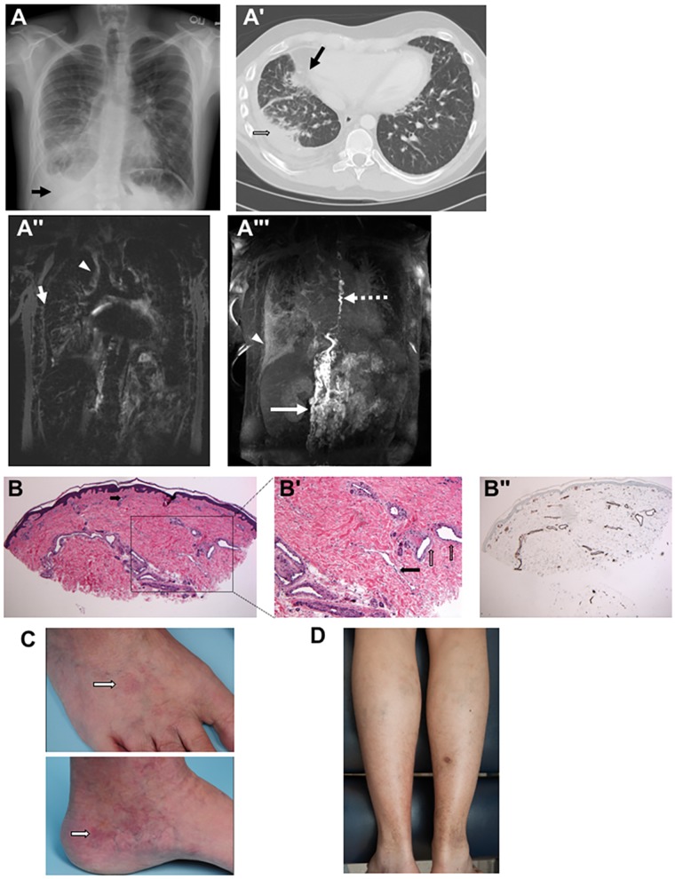
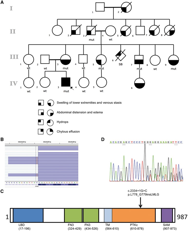
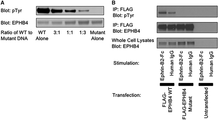
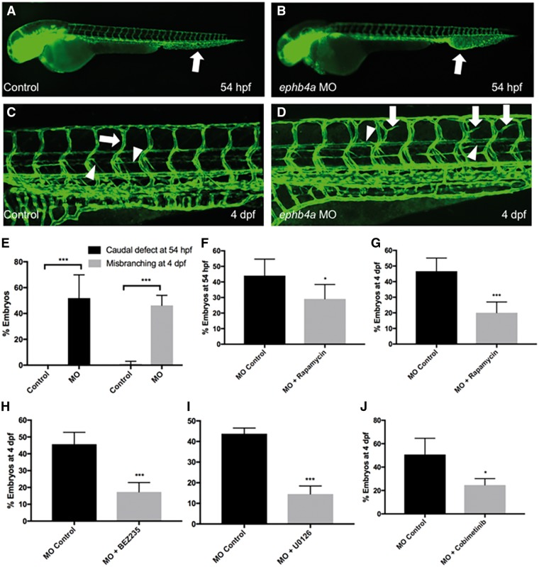
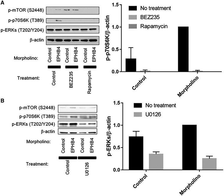
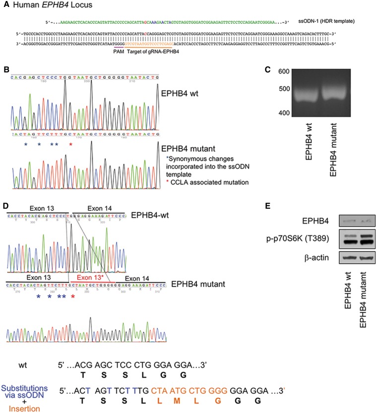
Central conducting lymphatic anomaly (CCLA) is one of the complex lymphatic anomalies characterized by dilated lymphatic channels, lymphatic channel dysmotility and distal obstruction affecting lymphatic drainage. We performed whole exome sequencing (WES) of DNA from a four-generation pedigree and examined the consequences of the variant by transfection of mammalian cells and morpholino and rescue studies in zebrafish. WES revealed a heterozygous mutation in EPHB4 (RefSeq NM_004444.4; c.2334 + 1G>C) and RNA-Seq demonstrated that the EPHB4 mutation destroys the normal donor site, which leads to the use of a cryptic splice donor that results in retention of the intervening 12-bp intron sequence. Transient co-expression of the wild-type and mutant EPHB4 proteins showed reduced phosphorylation of tyrosine, consistent with a loss-of-function effect. Zebrafish ephb4a morpholino resulted in vessel misbranching and deformities in the lymphatic vessel development, indicative of possible differentiation defects in lymphatic vessels, mimicking the lymphatic presentations of the patients. Immunoblot analysis using zebrafish lysates demonstrated over-activation of mTORC1 as a consequence of reduced EPHB4 signaling. Strikingly, drugs that inhibit mTOR signaling or RAS-MAPK signaling effectively rescued the misbranching phenotype in a comparable manner. Moreover, knock-in of EPHB4 mutation in HEK293T cells also induced mTORC1 activity. Our data demonstrate the pathogenicity of the identified EPHB4 mutation as a novel cause of CCLA and suggesting that ERK inhibitors may have therapeutic benefits in such patients with complex lymphatic anomalies.


Figures

Figure 1.
Figure 1.
 Clinical images and skin histopathology in patients with lymphatic anomalies. (A) Chest radiograph of the proband shows prominent interstitial lung markings with a right-sided pleural effusion (arrow) at 24 years of age. (A′) Chest CT scan of the proband shows right-sided pleural effusion (open arrow) and ground glass opacifications (black arrow) at 23 years old. (A″) T2-weighted MRI demonstrating high peribronchial (arrow) and pulmonary interstitial signal (arrowhead). (A′″) MIP of DCMRL showing retroperitoneal masses (arrow) dilated and tortuous thoracic duct (TD) (dashed arrow) with retrograde perfusion of the right lung (arrow head). (B and B′) Skin histopathology of mother of proband demonstrates a markedly increased number of dermal vascular structures. Both dilated lymphatic channels and venules/capillaries are noted (B, hematoxylin and eosin, 40×, black arrow indicates capillary in the superficial reticular dermis; B′, hematoxylin and eosin, 100×, black arrow is pointing at a dilated lymphatic channel and open vertical arrows are pointing at dilated venules). (B″) The dilation and thickness of venolymphatic channels stains positive for CD31 (40×), with a subset also positive for D2–40 (data not shown). (C) Faint, vaguely reticulated matted telangiectatic patch on the medial left foot of mother of proband, along with scattered varicosities. The dorsal foot shows a similar faint telangiectatic patch. White arrow is pointing at a telangiectatic patch in both clinical images. (D) Photograph of posterior calves of proband demonstrating discoloration and stasis dermatitis suggestive of longstanding venous insufficiency. Of note, the lesion on the right posterior calf is partially healed biopsy site. The biopsy site took several weeks longer to heal completely than would be anticipated, further supportive of venous insufficiency.
Figure 2.
Figure 2.
 Identification of mutation in EPHB4. (A) Pedigree of studied family and cosegregating pattern of the EPHB4 mutation. The open circles denote unaffected females and the open squares denote unaffected males; the solid figures indicate affected subjects. EPHB4 genotypes are noted beneath the symbol for each subject from whom DNA was available for testing. (B) RNA-Seq demonstrates EPHB4 splice-altering mutation destroys the normal donor site, which leads to the use of a cryptic splice donor (the coding strand of EPHB4 corresponds to the chromosome’s reverse strand) that causes the retention of the intervening 12 bp of the intron. (C) Schematic of EPHB4 protein showing conserved domains and variant identified in the affected individuals. Abbreviations are as follows: LBD, ligand binding domain; FN3, fibronectin type 3 domain; TM, transmembrane domain; PTKc, catalytic domain of the protein tyrosine kinases; SAM, sterile alpha motif. (D) The in-frame insertion was confirmed by Sanger sequencing of cDNA from patient.
Figure 3.
Figure 3.
 LoF mutation in EPHB4. (A) HEK293T cells were transiently transfected with wild-type EPHB4 alone, mutant EPHB4 alone or the mixtures of the wild-type and mutant in the indicated ratios. (B) A375 cells were transfected with wild-type EPHB4, mutant EPHB4 or left untransfected. Cells were stimulated with either plate-bound Ephrin-B2-Fc or human IgG1 as a control. Cells were lysed, and transfected proteins were immunoprecipitated. IPs were blotted for phosphotyrosine (top) or EPHB4 (middle). Whole cell lysates were blotted for EPHB4 to demonstrate equal expression (bottom).
Figure 4.
Figure 4.
  ephb4a MOs induces mTORC1 signaling-dependent expansion of the caudal vascular plexus and miss-guided vessels in the intersomitic vasculature. (A and B) Injection of ephb4a MO (800 um) in the tg(fli1: EGFP) zebrafish line (in which endothelial cells is marked by EGFP expression) induces expansion and fusion of the caudal vasculature at 54 hpf (arrows). (C) At 4 dpf the zebrafish vascular system consists of intersomitic blood vessels that project in dorso-ventral direction (arrow) as well as thinner lymphatic vessels (arrowheads). The lymphatic parachordal line runs horizontally along the trunk (downward arrowhead) while the intersegmental lymphatic vessels project downwards (upward arrowhead). (D) ephb4a MO induces misguided vessels that resemble blood vessels (arrows) and lymphatic vessels (arrowheads). (Images of C and D are merged from two neighboring confocal scans) (E) The defects in the caudal plexus are detected in 52% of ephb4a MO-injected larvae at 54 hpf, misguided vasculature is present in 46% on day 4 (***P <0.001). (F and G) Rapamycin can significantly reduce the number of animals with caudal defect and misbranching at 54 hpf (F, *P <0.05) and 4 dpf (G, ***P <0.001), respectively. (HJ) BEZ235 (H, ***P <0.001), U0126 (I, ***P <0.001) and Cobimetibib (J, *P <0.05) similarly rescue the branching defects on day 4.
Figure 5.
Figure 5.
 Perturbation attributed to ephb4a MO in developing zebrafish upregulate mTORC1 signaling and activation of mTORC1 can be inhibited by treatment of mTORC1 inhibitors. (A and B) Lysates from zebrafish larvae treated with either ephb4a MO or control MO, and untreated, or treated with Rapamycin or BEZ235 (A) or with U0126 (a MEK inhibitor) (B) were separated by SDS-PAGE and blotted for phospho-mTOR S2448, phospho-p70S6K T389 and phospho-ERKs. Blotting for β-actin was used as a loading control. Normalized quantification of p-p70S6K and p-ERKs against β-actin was performed from three independent experiments. Data are presented as mean ± SD.
Figure 6.
Figure 6.
 Knock-in of EPHB4 mutation in HEK293T cells results in increased mTORC1 activity. (A) Schematic drawing of targeted genome editing at EPHB4 locus using CRISPR/Cas9. (B) Sanger sequence traces of genomic DNA after the knock-in. (C) RT-PCR analysis suggested a small insertion in the knock-in cells compare with the wild-type cells. (D) The four-amino-acid insertion induced by CRISPR/Cas9 confirmed by standard Sanger sequencing of RT-PCR product. (E) Western blot of the wild-type HEK293T cells and EPHB4 mutant cells by gene editing. Cells containing the EPHB4 splice-altering mutation displayed higher p-p70S6K levels than wild-type cells.

References

    1. Alitalo K., Tammela T., Petrova T.V. (2005) Lymphangiogenesis in development and human disease. Nature, 438, 946–953. - PubMed
    1. Adams R.H., Alitalo K. (2007) Molecular regulation of angiogenesis and lymphangiogenesis. Nat. Rev. Mol. Cell. Biol., 8, 464–478. - PubMed
    1. Tammela T., Alitalo K. (2010) Lymphangiogenesis: molecular mechanisms and future promise. Cell, 140, 460–476. - PubMed
    1. Alitalo K. (2011) The lymphatic vasculature in disease. Nat. Med., 17, 1371–1380. - PubMed
    1. Brouillard P., Boon L., Vikkula M. (2014) Genetics of lymphatic anomalies. J. Clin. Invest., 124, 898–904. - PMC - PubMed

Publication types

Substances