. 2018 Sep;91(1089):20180317.
doi: 10.1259/bjr.20180317. Epub 2018 Jul 5.

An investigation into the validity of utilising the CDRAD 2.0 phantom for optimisation studies in digital radiography

Affiliations

An investigation into the validity of utilising the CDRAD 2.0 phantom for optimisation studies in digital radiography

Sadeq Al-Murshedi et al. Br J Radiol. 2018 Sep.

Abstract

Objectives: To determine if a relationship exists between low contrast detail (LCD) detectability using the CDRAD 2.0 phantom, visual measures of image quality (IQ) and simulated lesion visibility (LV) when performing digital chest radiography (CXR).

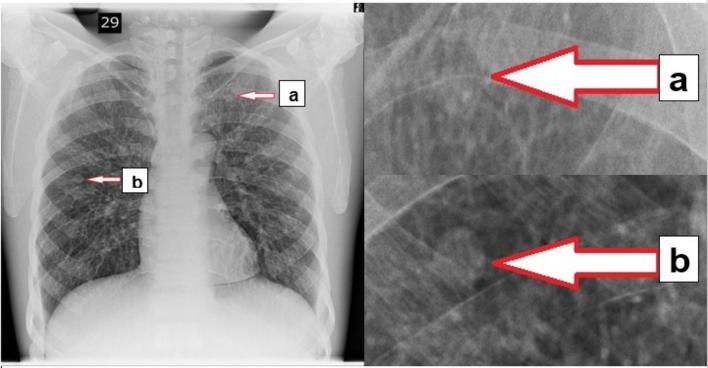
Methods: Using a range of acquisition parameters, a CDRAD 2.0 phantom was used to acquire a set of images with different levels of image quality. LCD detectability using the CDRAD 2.0 phantom, represented by an image quality figure inverse (IQFinv) metric, was determined using the phantom analyser software. A Lungman chest phantom was loaded with two simulated lesions, of different sizes/placed in different locations, and was imaged using the same acquisition factors as the CDRAD 2.0 phantom. A relative visual grading analysis (VGA) was used by seven observers for IQ and LV evaluation of the Lungman images. Correlations between IQFinv, IQ and LV were investigated.

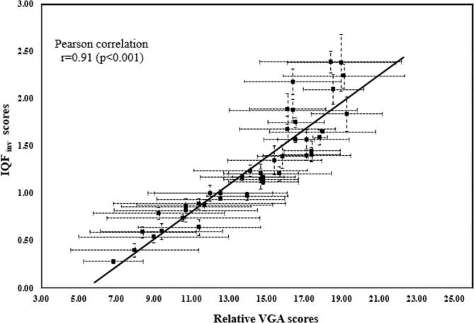
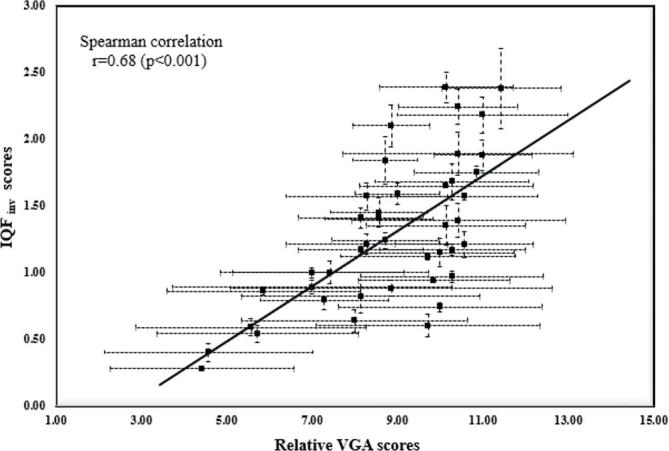
Results: Pearson's correlation demonstrated a strong positive correlation (r = 0.91; p < 0.001) between the IQ and the IQFinv. Spearman's correlation showed a good positive correlation (r = 0.79; p < 0.001) and (r = 0.68; p < 0.001) between the IQFinv and the LV for the first lesion (left upper lobe) and the second lesion (right middle lobe), respectively.

Conclusions: From results presented in this study, the automated evaluation of LCD detectability using CDRAD 2.0 phantom is likely to be a suitable option for IQ and LV evaluation in digital CXR optimisation studies. Advances in knowledge: This research establishes the potential of the CDRAD 2.0 phantom in digital CXR optimisation studies.


Figures

Figure 1.
Figure 1.
An example of a Lungman chest phantom image illustrating the two simulated lesions for the LV evaluation. (a) 10 mm + 100 HU lesion placed in left upper lobe. (b) The 12 mm + 100 HU lesion was placed in the right middle lobe. HU, Hounsfield unit; LV, lesion visibility.
Figure 2.
Figure 2.
Linear regression curve between mean IQFinv scores and mean IQ values. Error bars across the x axis represent the SD of the scores between the seven observers, while the error bars across the y axis represents the SD from the three automated CDRAD 2.0 image scores. IQFinv, image quality figure inverse; LV, lesion visibility; SD, standard deviation.
Figure 3.
Figure 3.
Linear regression curve between the mean IQFinv scores against the mean LV scores for the first lesion (left upper lobe). Error bars across the x axis represent the SD of the scores between the seven observers, while the error bars across the y axis represents the SD of the three CDRAD 2.0 image scores. IQFinv, image quality figure inverse; LV, lesion visibility; SD, standard deviation.
Figure 4.
Figure 4.
Linear regression curve between the mean IQFinv scores against the mean LV scores for the second lesion (right middle lobe). Error bars across the x axis represent the SD of the scores between the seven observers, while the error bars across the y axis represents the SD of the three CDRAD 2.0 image scores. IQFinv, image quality figure inverse; LV, lesion visibility; SD, standard deviation.

Similar articles

Cited by

References

    1. Busch HP, Faulkner K. Image quality and dose management in digital radiography: a new paradigm for optimisation. Radiat Prot Dosimetry 2005; 117(1–3): 143–7. doi: 10.1093/rpd/nci728 - DOI - PubMed
    1. Instrument S. The Ionising Radiation (Medical Exposure) Regulations 2000. 2000.
    1. Lanhede B, Båth M, Kheddache S, Sund P, Björneld L, Widell M, et al. . The influence of different technique factors on image quality of chest radiographs as evaluated by modified CEC image quality criteria. Br J Radiol 2002; 75: 38–49. doi: 10.1259/bjr.75.889.750038 - DOI - PubMed
    1. Redlich U, Hoeschen C, Doehring W. Assessment and optimisation of the image quality of chest-radiography systems. Radiat Prot Dosimetry 2005; 114: 264–8. doi: 10.1093/rpd/nch559 - DOI - PubMed
    1. Goo JM, Im JG, Lee HJ, Chung MJ, Seo JB, Kim HY, et al. . Detection of simulated chest lesions by using soft-copy reading: comparison of an amorphous silicon flat-panel-detector system and a storage-phosphor system. Radiology 2002; 224: 242–6. doi: 10.1148/radiol.2241011441 - DOI - PubMed

Publication types

MeSH terms