. 2018 Jun 14;173(7):1770-1782.e14.
doi: 10.1016/j.cell.2018.04.034.

Inactivation of CDK12 Delineates a Distinct Immunogenic Class of Advanced Prostate Cancer

Affiliations

Inactivation of CDK12 Delineates a Distinct Immunogenic Class of Advanced Prostate Cancer

Yi-Mi Wu et al. Cell. .

Abstract

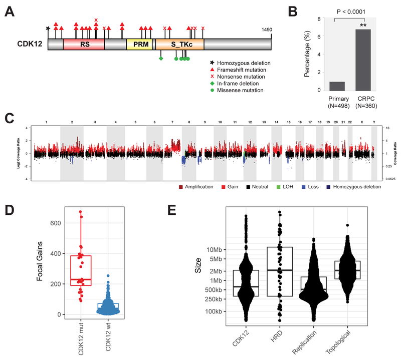
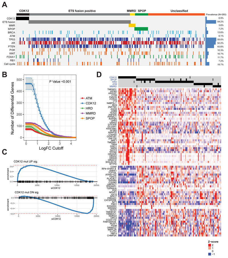
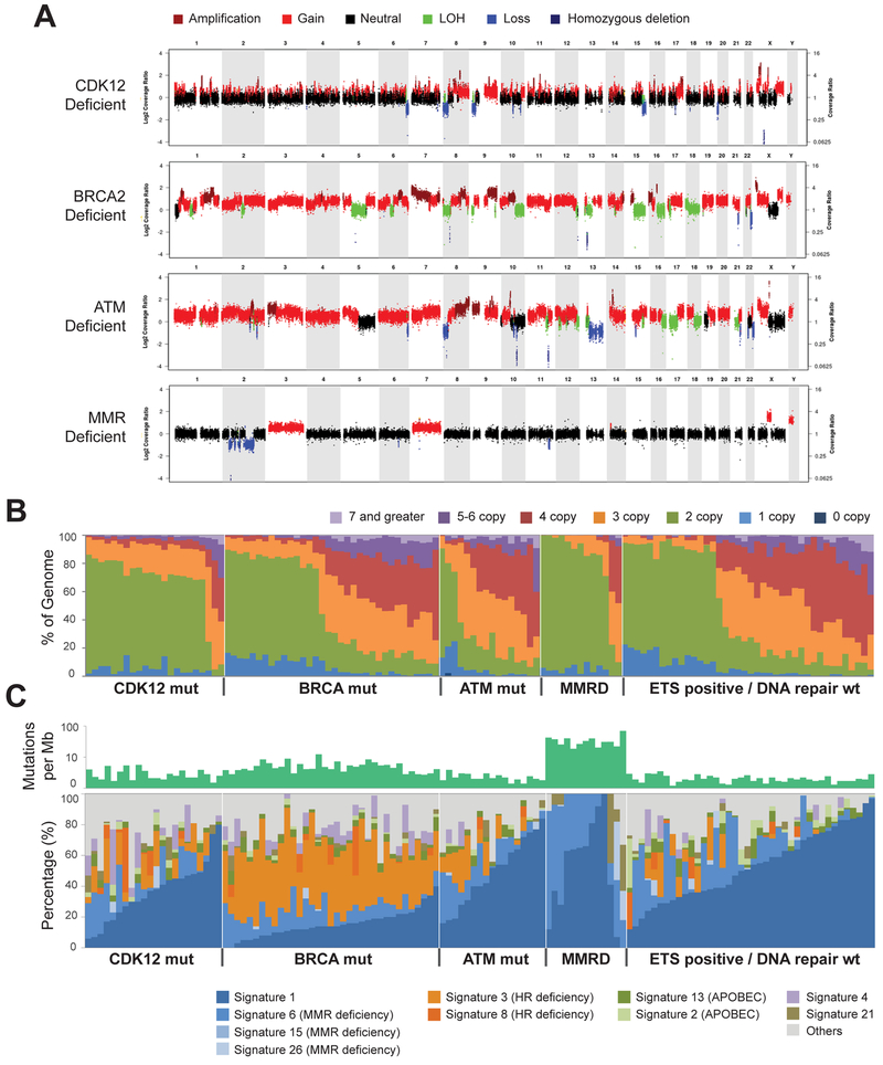
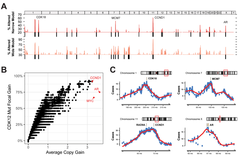
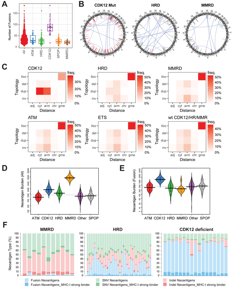
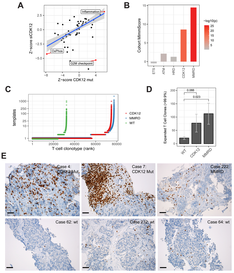
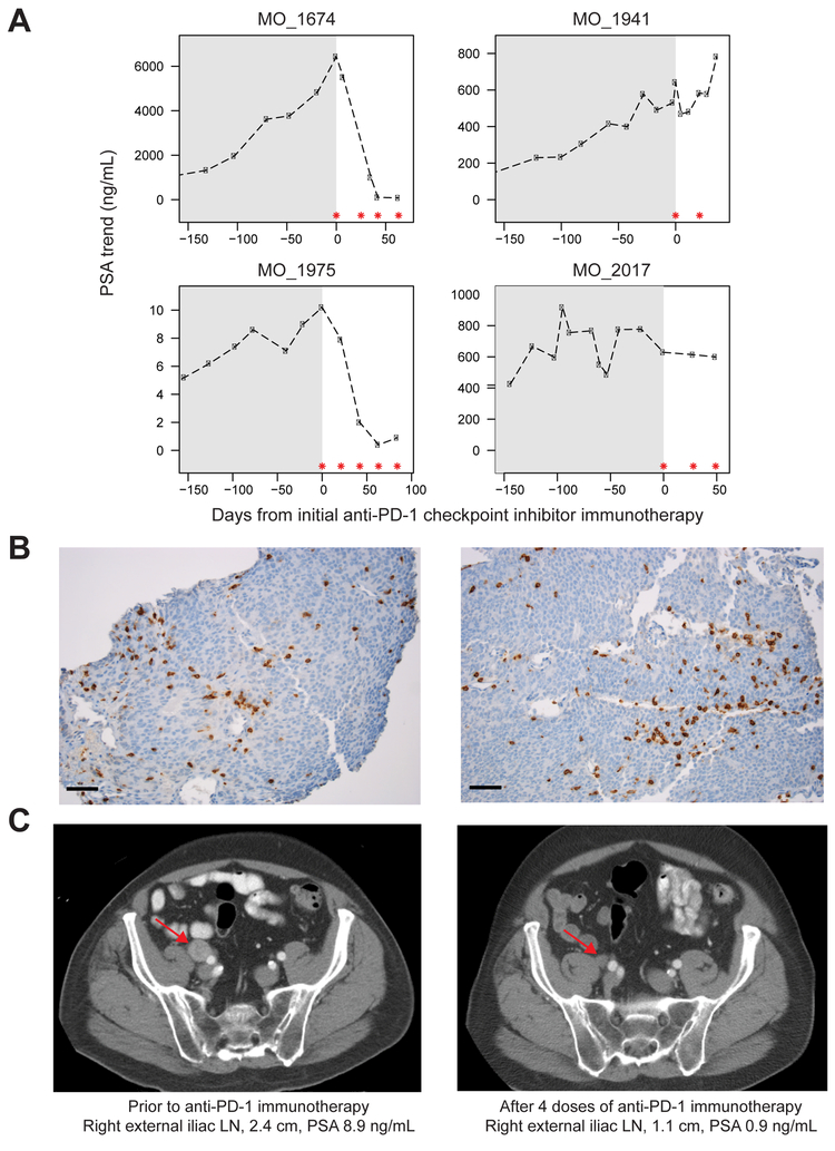
Using integrative genomic analysis of 360 metastatic castration-resistant prostate cancer (mCRPC) samples, we identified a novel subtype of prostate cancer typified by biallelic loss of CDK12 that is mutually exclusive with tumors driven by DNA repair deficiency, ETS fusions, and SPOP mutations. CDK12 loss is enriched in mCRPC relative to clinically localized disease and characterized by focal tandem duplications (FTDs) that lead to increased gene fusions and marked differential gene expression. FTDs associated with CDK12 loss result in highly recurrent gains at loci of genes involved in the cell cycle and DNA replication. CDK12 mutant cases are baseline diploid and do not exhibit DNA mutational signatures linked to defects in homologous recombination. CDK12 mutant cases are associated with elevated neoantigen burden ensuing from fusion-induced chimeric open reading frames and increased tumor T cell infiltration/clonal expansion. CDK12 inactivation thereby defines a distinct class of mCRPC that may benefit from immune checkpoint immunotherapy.

Keywords: CDK12; focal tandem duplications; gene fusions; immunotherapy; metastatic castration-resistant prostate cancer; neoantigens.


Figures

Figure 1.
Figure 1.. Biallelic loss of CDK12 is enriched in mCRPC and results in focal tandem duplications.
(A) Schematic of mutations in CDK12. (B) Increased frequency of CDK12 loss in metastatic castration-resistant prostate cancer (CRPC) compared to primary disease. (C) Characteristic pattern of genomic instability found in all cases with CDK12 loss. Copy gains are indicated in shades of red. LOH, loss of heterozygosity. (D) Number of focal copy gains (< 8Mb) by CDK12 mutational status, as determined by whole-exome analysis. (E) Size of copy gains (tandem duplications), as ascertained by whole-genome sequencing of index cases with CDK12 mutations (CDK12) and homologous recombination deficiency (HRD). Sizes of replication domains and topological domains in normal tissues are shown for comparison. See also Figures S1-S3 and Tables S1-S5.
Figure 2.
Figure 2.. CDK12-mutant prostate cancer is a novel molecular subtype of mCRPC.
(A) Mutual exclusivity of CDK12 loss, ETS fusions, mismatch repair deficiency (MMRD), SPOP mutations, and homologous recombination deficiency (HRD). (B) Number of significantly differentially expressed genes (DEGs) for the prostate tumors with different primary genetic drivers. (C) Enrichment plot for signatures of up- (top) and downregulated (bottom) genes in CDK12 mutant tumors. Genes are ranked by their fold change following siCDK12 knockdown in LNCaP cells, with CDK12-loss signature genes indicated as black dashes. The increased relative frequency (enrichment score) of genes at either end of this spectrum is shown as a blue line. (D) Heatmap of the top DEGs in CDK12-mutant prostate cancer. Differential expression for all samples (columns) in this heatmap is relative to tumors that are wild-type for primary genetic drivers of prostate cancer (as in B). See also Figure S4 and Tables S1, S6.
Figure 3.
Figure 3.. CDK12 loss results in a distinct pattern of genomic instability.
(A) Representative copy-number plots for prostate tumors with deficiencies in key DNA damage response or repair pathways. (B) Spectrum of copy-number aberrations in tumors with distinct genetic drivers. (C) Spectrum of inferred mutational signatures in tumors with distinct genetic drivers. See also Figure S4.
Figure 4.
Figure 4.. Recurrence of focal tandem duplications (FTDs) associated with CDK12 loss.
(A) Genome-wide frequency (percentage of CDK12-mutant patients) of FTDs based on a narrow (<2Mb) and wide (<8Mb) definition of focality. (B) FTD recurrence and average copy-number gain of FTDs at the individual gene level. Genes with the highest average copy-number are highlighted in red. (C) Delineation of minimal common regions (MCR) for loci with the most recurrent gains specific to CDK12-loss tumors. Genes related to the cell cycle are highlighted in each MCR. The AR locus is presented as a positive control. See also Figures S5-S6.
Figure 5.
Figure 5.. Signatures of structural variation and neoantigen presentation in CDK12-mutant tumors.
(A) Total number of detected gene fusions for prostate tumors with different genetic drivers. (B) Representative examples of circos plots showing the pattern structural variation in tumors with major types of genomic instability. Structural variants (SVs) detected from RNA-seq are classified into translocations, deletions, duplications, and inversions based on the topology of the breakpoints. Color code: blue - translocations, red - duplication, green - inversion, black - deletion. (C) Classification of SVs based on the topology and distance between the breakpoints. adj – breakpoints in adjacent loci, cyt – in same cytoband, arm – on same chromosome arm, gme – genomic translocation, inv – inversion, dup – duplication, del – deletion, tloc – translocation. Heatmap color indicates frequency of a SV class across all index cases. (numbers of patients: CDK12 = 24, HRD = 47, MMRD = 11, ATM = 21, ETS = 190, WT = 31). (D-E) Antigen burden in tumors with distinct types of genetic instability. Overall burden based on single nucleotide variants, insertions/deletions, and fusions is shown in D. Fusion-specific burden is shown in E. (F) Distribution of neoantigens based on genetic variant type and predicted MHC class-I (MHC-I) binding affinity. See also Figure S6.
Figure 6.
Figure 6.. Immunogenomic properties of CDK12-mutant tumors.
(A) Differential expression of MSigDB cancer hallmark gene-sets in CDK12-mutant patients and in LNCaP cells depleted with CDK12 by siRNA. Highlighted hallmarks are significant (FDR < 0.05, limma moderated t-test). (B) Levels of global immune infiltration across prostate tumors with distinct genetic drivers compared to genetically stable (PGD wild-type) tumors. The “Cohort MImmScore” is defined as the gene-set enrichment Z-score and p-value based on Random-Set test and moderated cohort DE log2 fold-changes. (C) Overview of T cell clonotypes across CDK12-mutant (n=10), MMRD (n=10), and WT (n=10) tumors. T cell clonotypes (i.e. identical CDR3 sequences) are ranked by their frequency (number of templates). CDK12-mutant and MMRD tumors show, overall, an increase in the total number of T cells (X-axis), and higher levels of clonal expansion (Y-axis). (D) Comparison of clonal expansion between immunogenic (MMRD, CDK12) and wild-type mCRPC tumors (t-test). Expanded clones are defined as those with the highest number of clonal expansion (estimated number of templates > 99.9 percentile across all cohorts; n > 12). (E) Immunohistochemistry (IHC) performed on formalin-fixed paraffin-embedded tumor sections using anti-CD3 antibody. Six representative cases are shown, including two CDK12-mutant tumors, one MMRD tumor, and three tumors which are wild type for CDK12, MMR genes, and HR genes. Black bar: 50 μm. See also Figure S7.
Figure 7.
Figure 7.. Response of CDK12-mutant patients to anti-PD1 checkpoint inhibitor immunotherapy.
(A) PSA levels of four CDK12-mutant prostate cancer patients treated with anti-PD1 monotherapy. Gray shading represents PSA levels prior to anti-PD-1 therapy. Asterisks indicate anti-PD1 doses of 200 mg IV. (B) Representative CD3 IHC images of metastatic lymph node biopsies of patient MO_1975 prior to anti-PD1 treatment. Cells exhibited membranous and cytoplasmic staining of CD3, highlighting the presence of T lymphocytes. Black bar: 50 μm. (C) CT imaging of patient MO_1975 pre- and post-immunotherapy treatment. Arrows indicate metastatic lymph node.

Comment in

  • New genomic drivers identified.
    Bradley CA. Bradley CA. Nat Rev Urol. 2018 Sep;15(9):525. doi: 10.1038/s41585-018-0057-2. Nat Rev Urol. 2018. PMID: 29977054 No abstract available.
  • CDK12 Changes Telling in Prostate Cancer.
    [No authors listed] [No authors listed] Cancer Discov. 2018 Sep;8(9):1055. doi: 10.1158/2159-8290.CD-NB2018-093. Epub 2018 Jul 13. Cancer Discov. 2018. PMID: 30006379

References

    1. Abida W, Armenia J, Gopalan A, Brennan R, Walsh M, Barron D, Danila D, Rathkopf D, Morris M, Slovin S, et al. (2017). Prospective genomic profiling of prostate cancer across disease states reveals germline and somatic alterations that may affect clinical decision making. JCO Precis Oncol 2017. - PMC - PubMed
    1. Alexandrov LB, Nik-Zainal S, Wedge DC, Aparicio SA, Behjati S, Biankin AV, Bignell GR, Bolli N, Borg A, Borresen-Dale AL, et al. (2013). Signatures of mutational processes in human cancer. Nature 500, 415–421. - PMC - PubMed
    1. Bajrami I, Frankum JR, Konde A, Miller RE, Rehman FL, Brough R, Campbell J, Sims D, Rafiq R, Hooper S, et al. (2014). Genome-wide profiling of genetic synthetic lethality identifies CDK12 as a novel determinant of PARP1/2 inhibitor sensitivity. Cancer Res 74, 287–297. - PMC - PubMed
    1. Barbieri CE, Baca SC, Lawrence MS, Demichelis F, Blattner M, Theurillat J-P, White TA, Stojanov P, Van Allen E, Stransky N, et al. (2012). Exome sequencing identifies recurrent SPOP, FOXA1 and MED12 mutations in prostate cancer. Nat Genet 44, 685–689. - PMC - PubMed
    1. Bartkowiak B, Liu P, Phatnani HP, Fuda NJ, Cooper JJ, Price DH, Adelman K, Lis JT, and Greenleaf AL (2010). CDK12 is a transcription elongation-associated CTD kinase, the metazoan ortholog of yeast Ctk1. Genes Dev 24, 2303–2316. - PMC - PubMed

Publication types

MeSH terms