. 2018 Oct;94(4):728-740.
doi: 10.1016/j.kint.2018.03.012. Epub 2018 Jun 12.

TACI deletion protects against progressive murine lupus nephritis induced by BAFF overexpression

Affiliations

TACI deletion protects against progressive murine lupus nephritis induced by BAFF overexpression

Tanvi Arkatkar et al. Kidney Int. 2018 Oct.

Abstract

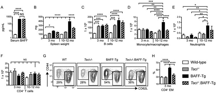
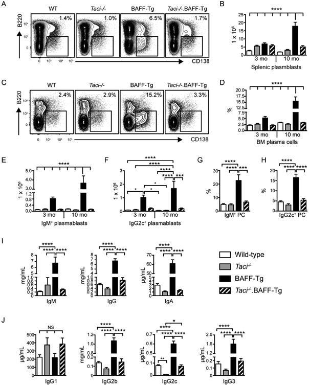
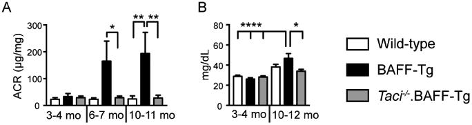
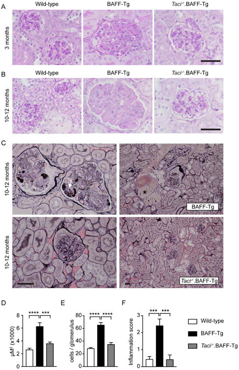
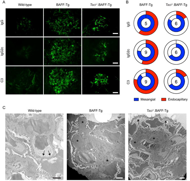
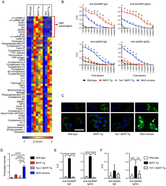
B cells are known to promote the pathogenesis of systemic lupus erythematosus (SLE) via the production of pathogenic anti-nuclear antibodies. However, the signals required for autoreactive B cell activation and the immune mechanisms whereby B cells impact lupus nephritis pathology remain poorly understood. The B cell survival cytokine B cell activating factor of the TNF Family (BAFF) has been implicated in the pathogenesis of SLE and lupus nephritis in both animal models and human clinical studies. Although the BAFF receptor has been predicted to be the primary BAFF family receptor responsible for BAFF-driven humoral autoimmunity, in the current study we identify a critical role for signals downstream of Transmembrane Activator and CAML Interactor (TACI) in BAFF-dependent lupus nephritis. Whereas transgenic mice overexpressing BAFF develop progressive membranoproliferative glomerulonephritis, albuminuria and renal dysfunction, TACI deletion in BAFF-transgenic mice provided long-term (about 1 year) protection from renal disease. Surprisingly, disease protection in this context was not explained by complete loss of glomerular immune complex deposits. Rather, TACI deletion specifically reduced endocapillary, but not mesangial, immune deposits. Notably, although excess BAFF promoted widespread breaks in B cell tolerance, BAFF-transgenic antibodies were enriched for RNA- relative to DNA-associated autoantigen reactivity. These RNA-associated autoantibody specificities were specifically reduced by TACI or Toll-like receptor 7 deletion. Thus, our study provides important insights into the autoantibody specificities driving proliferative lupus nephritis, and suggests that TACI inhibition may be novel and effective treatment strategy in lupus nephritis.

Keywords: B-cell activating factor of the TNF family (BAFF); autoantibodies; lupus nephritis; systemic lupus erythematosus; transmembrane activator and CAML interactor (TACI).


Figures

Figure 1
Figure 1. Excess BAFF promotes progressive, TACI-dependent immune activation
(A) Serum BAFF levels in 6-month-old WT (n=5), BAFF-Tg (n=8) and Taci-/-.BAFF-Tg (n=8) mice. (B-F) Spleen weight (B) and total number of splenic CD19+ B cells (C), CD11b+GR1lo monocyte/macrophages (D), CD11b+GR1+ neutrophils (E) and CD4+ T cells (F) in WT, Taci-/-, BAFF-Tg and Taci-/-.BAFF-Tg mice sacrificed at 3 and 10-12 months old. (G, H) Representative flow plots (G; gated on CD4+ T cells) and percentage of CD4+ T cells exhibiting CD44hiCD62Llo/hi effector/memory phenotype (H) from 3-month-old mice of indicated genotypes. Number equals percentage in CD44hiCD62Llo/hi EM gate. (B-H) n=11-16 mice analyzed per group. (A-H) Error bars indicate SEM; *, P<0.05; **, P<0.01; ***, P<0.001; ****, P<0.0001, NS, not significant; by one-way ANOVA, followed by Tukey's multiple comparison test.
Figure 2
Figure 2. BAFF signals via TACI to promote B cell activation
(A, B) Representative flow plots (A) and total number (B) of splenic B220loCD138+ plasma cells/plasmablasts in WT, Taci-/-, BAFF-Tg and Taci-/-.BAFF-Tg mice sacrificed at indicated ages. (C, D) Representative flow plots (C) and percentage (D) of BM B220negCD138+ plasma cells/plasmablasts in WT, Taci-/-, BAFF-Tg and Taci-/-.BAFF-Tg mice sacrificed at indicated ages. (A, C) Flow plots are representative of indicated genotypes sacrificed at 10-12 months, gated on total splenocytes (A) and total BM cells (C), respectively. Number equals percentage within gate. (E, F) Number of isotype-specific IgM+ (E) and IgG2c+ (F) splenic plasmablasts in WT, Taci-/-, BAFF-Tg and Taci-/-.BAFF-Tg mice. (G, H) Percentage of BM plasma cells expressing IgM (G) and IgG2c (H) by intracellular staining, from indicated genotypes sacrificed at 10-12 months. (I, J) Total serum IgM, IgG and IgA titers (I), and lgG1, lgG2b, lgG2c and lgG3 subclass titers (J) in 3-month-old mice of indicated genotype. (A-H) Error bars indicate SEM; *, P<0.05; ***, P<0.001; ****, P<0.0001; by one-way ANOVA, followed by Tukey's multiple comparison test (n=9-12 mice analyzed per group).
Figure 3
Figure 3. Excess BAFF promotes TACI-dependent albuminuria and renal dysfunction
(A) Urine albumin:creatinine (ACR) ratio (μg/mg) in aged cohorts of WT, BAFF-Tg and Taci-/-.BAFF-Tg mice, demonstrating that BAFF over-expression promotes progressive albuminuria in a TACI-dependent manner. Total number of animals analyzed at 3-4 months, 6-7 months and 10-11 months: WT (n=5, 7, 10); BAFF-Tg (n=5, 7, 6); and Taci-/-.BAFF-Tg (n=5, 10, 11). (B) Serum BUN in aged cohorts of indicated genotypes, showing that increased BAFF promotes TACI-dependent renal dysfunction. Total number of animals analyzed at 3-4 months and 10-12 months: WT (n=7, 6); BAFF-Tg (n=7, 7); and Taci-/-.BAFF-Tg (n=7, 7). (A, B) Error bars indicate SEM. *, P<0.05; **, P<0.01; ****, P<0.0001; by one-way ANOVA, followed by Tukey's multiple comparison test.
Figure 4
Figure 4. TACI deletion prevents progressive glomerulonephritis in BAFF-Tg mice
(A, B) Representative periodic acid-Schiff (PAS)-stained renal sections showing mild mesangial expansion and inflammatory cell infiltration in 3-month-old (A) BAFF-Tg mice, with progressive mesangial expansion, hypercellularity and endocapillary eosinophilic deposits in aged BAFF-Tg mice (B). These pathologic changes were inapparent in Taci-/-.BAFF-Tg mice. (C) Jones' methenamine silver stained kidney sections showing marked mesangiolysis, capillary basement membrane duplication and accumulation of silver-dense material (arrow) within the glomerular capillaries in representative aged BAFF-Tg mouse (upper left panel). Aged BAFF-Tg animals also exhibited prominent proteinaceous casts (*) within dilated tubules (upper right panel). Notably, TACI deletion prevented these pathologic changes for up to 1 year (lower panels). (A-C) Bars: 50μm. (D, E) Mean glomerular size (D) and mean cell count per glomerulus (E) in 10-12-month-old WT (white), BAFF-Tg (black) and Taci-/-.BAFF-Tg (gray) mice. (F) Severity of glomerular inflammation scored as: normal (0+); focal glomerular changes with mild mesangial expansion and hypercellularity (1+); moderate mesangial expansion, GBM thickening/reduplication and glomerular hypercellularity (2+); and diffuse glomerular changes with severe mesangial expansion, GBM thickening/reduplication and glomerular hypercellularity (3+). Pathology was scored by two independent observers blinded to genotype. (D-F) Error bars indicate SEM. ***, P<0.001; ****, P<0.0001; by one-way ANOVA, followed by Tukey's multiple comparison test. (A-F) Total mice analyzed: n=9-11 per genotype.
Figure 5
Figure 5. TACI deletion prevents BAFF-driven endocapillary, but not mesangial, immune complex deposits
(A) Representative IF staining for glomerular IgG, lgG2c, and C3 complement in indicated genotypes, showing both mesangial and capillary wall IC deposits in BAFF-Tg mice. In contrast, deposits were limited to the mesangium in Taci-/-.BAFF-Tg mice. Bars: 20μm. (B) Location of glomerular deposits, scored as mesangial (blue) and/or endocapillary (red), in BAFF-Tg vs. Taci-/-.BAFF-Tg mice. Unfilled portion of circle represents animals with absent or undefinable IF staining pattern, and number within circle indicates number of animals analyzed. Scoring was performed by observer blinded to genotype. (C) Representative electron micrographs showing prominent mesangial (arrowheads) and capillary wall immune complexes (stars) in BAFF-Tg mice, but no capillary wall deposits in wild type (WT) mice (arrows). In contrast to BAFF-Tg mice, immune deposits were limited to the mesangium in Taci-/-.BAFF-Tg mice (arrowheads), while capillary loops lacked deposits (arrows). Bars: 2μm.
Figure 6
Figure 6. TACI signals promote class-switched autoantibodies against RNA-associated autoantigens in BAFF-Tg mice
(A) Serum IgG autoantibodies in wild-type, BAFF-Tg and Taci-/-.BAFF-Tg mice, determined using an autoantigen microarray. Specific autoantigens are ordered from top to bottom based on intensity of BAFF-Tg IgG reactivity. Surprisingly, TACI deletion results in the loss of a limited subset of BAFF-Tg autoantibodies (depicted by red bracket), enriched for ribonucleoprotein (RNP) reactivity. Data are represented as a heat map of Z-scores ranging from -1 (blue) to 3 (red). Each column represents an independent animal. (B) Anti-Sm/RNP (upper panels) and anti-dsDNA (lower panels) IgG and lgG2c autoantibodies in sera from wild type (black, n=2), BAFF-Tg (red, n=5), Taci-/-.BAFF-Tg (gray, n=5), and the WAS chimera lupus model (blue, n=5). Whereas antibodies binding the RNA-associated autoantigen Sm/RNP were significantly increased in BAFF-Tg mice compared with the WAS chimera model, titers of dsDNA autoantibodies were markedly lower in BAFF-Tg vs. WAS chimera mice. Error bars indicate SEM; *, P<0.05, BAFF-Tg vs. WAS chimera reactivity by two-tailed Student's t test. (C) Representative images of C. luciliae stained with IgG (green) and DAPI (blue). Arrow highlights prominent IgG and DAPI counterstaining of the C. luciliae kinetoplast by WAS chimera sera. In contrast, BAFF-Tg and Taci-/-.BAFF-Tg sera exhibited variable diffuse staining of C. luciliae cytoplasm, without increased kinetoplast reactivity. Bars: 10μm. (D) Anti-dsDNA IgG Ab by intensity of C. luciliae kinetoplast staining in wild-type (black, n=12), BAFF-Tg (red, n=18), Taci-/-.BAFF-Tg (gray, n=5), and WAS chimera (blue, n=14) sera. Kinetoplast staining was scored from 0-3 by observers blinded to genotype. (E, F) Anti-Sm/RNP (E) and anti-dsDNA (F) IgG and lgG2c autoantibodies in 4-month-old wild-type (white, n=6), BAFF-Tg (black, n=7), and Tlr7-/-.BAFF-Tg (gray, n=4) sera. (D-F) Error bars indicate SEM. **, P<0.01, ***, P<0.001; ****, P<0.0001; by one-way ANOVA, followed by Tukey's multiple comparison test.

Similar articles

Cited by

References

    1. Mackay F, Schneider P. Cracking the BAFF code. Nature reviews Immunology. 2009;9:491–502. - PubMed
    1. Mackay F, Woodcock SA, Lawton P, et al. Mice transgenic for BAFF develop lymphocytic disorders along with autoimmune manifestations. The Journal of experimental medicine. 1999;190:1697–1710. - PMC - PubMed
    1. Gavin AL, Duong B, Skog P, et al. deltaBAFF, a splice isoform of BAFF, opposes full-length BAFF activity in vivo in transgenic mouse models. J Immunol. 2005;175:319–328. - PubMed
    1. Stohl W, Metyas S, Tan SM, et al. B lymphocyte stimulator overexpression in patients with systemic lupus erythematosus: longitudinal observations. Arthritis and rheumatism. 2003;48:3475–3486. - PubMed
    1. Steri M, Orru V, Idda ML, et al. Overexpression of the Cytokine BAFF and Autoimmunity Risk. The New England journal of medicine. 2017;376:1615–1626. - PMC - PubMed

Publication types

MeSH terms

LinkOut - more resources