Review
. 2018 Jul;22(7):569-582.
doi: 10.1016/j.tics.2018.04.004. Epub 2018 Jun 12.

Why Does the Cortex Reorganize after Sensory Loss?

Affiliations
Review

Why Does the Cortex Reorganize after Sensory Loss?

Amy Kalia Singh et al. Trends Cogn Sci. 2018 Jul.

Abstract

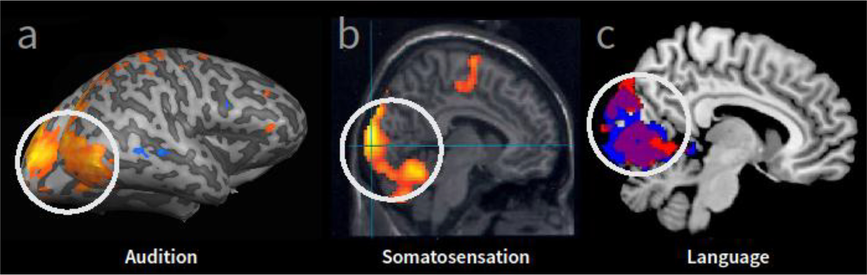
A growing body of evidence demonstrates that the brain can reorganize dramatically following sensory loss. Although the existence of such neuroplastic crossmodal changes is not in doubt, the functional significance of these changes remains unclear. The dominant belief is that reorganization is compensatory. However, results thus far do not unequivocally indicate that sensory deprivation results in markedly enhanced abilities in other senses. Here, we consider alternative reasons besides sensory compensation that might drive the brain to reorganize after sensory loss. One such possibility is that the cortex reorganizes not to confer functional benefits, but to avoid undesirable physiological consequences of sensory deafferentation. Empirical assessment of the validity of this and other possibilities defines a rich program for future research.

Keywords: cortical reorganization; multimodal activations; plasticity; sensory compensation; sensory loss.


Figures

Figure 1.
Figure 1.. Examples of cortical reorganization (i.e. crossmodal occipital activation) observed in early blind individuals using fMRI.
Occipital cortical areas (white circle, shown in sagittal plane) typically associated with processing visual information are found to respond to stimulation from non-visual modalities. (A) Occipital cortical regions that were activated more in early blind compared to sighted controls in response to auditory processing (sound localization task [22], modified from Renier et al., 2010). (B) Occipital cortical responses within a group of blind subjects reading Braille (compared to rest) [23] (modified from Gizewski et al., 2003). (C) Greater activity within occipital cortex in early blind compared to sighted controls in response to auditory linguistic stimuli [24] (modified from Bedny et al., 2011).
Figure 2.
Figure 2.. A performance comparison of blind and sighted individuals across a variety of tasks.
(a – c) Examples of tasks on which blind participants show an advantage relative to their sighted counterparts. (d – f) Examples of tasks on which there are either no differences between the two groups or a disadvantage for the blind. (Sources: a: [28]; b: [42]; c: [37]; d: [40]; e: [58]; f: [73]
Figure 3.
Figure 3.. Schematic depictions of three accounts of cortical reorganization after partial sensory loss.
In all figures, V and A refer to ‘vision’ and ‘audition’ as example modalities. (a) Possibility 1: The cortical area devoted to the ‘lost’ sense (here, vision) is co-opted by a different modality (here, audition). This leads to changes in functional capabilities, although the dimensions along which these changes happen may be different from the conventionally assumed ones of low-level detection and discrimination thresholds. (b) Possibility 2: Loss of one modality leads to the unmasking of already present connections from a different sense. (c) Possibility 3: Structured sensory inputs (here depicted as square waves) lead to structured cortical activation. With the loss of this input, cortical activity can become paroxysmal. Structured inputs from a different modality help re-impose structured activity in the cortical area.

References

    1. Gougoux F et al. (2005) A functional neuroimaging study of sound localization: visual cortex activity predicts performance in early-blind individuals. PLoS Biol. 3, e27. - PMC - PubMed
    1. Voss P et al. (2011) Relevance of spectral cues for auditory spatial processing in the occipital cortex of the blind. Front. Psychol 2, 48. - PMC - PubMed
    1. Voss P and Zatorre RJ (2015) Early visual deprivation changes cortical anatomical covariance in dorsal-stream structures. NeuroImage 108, 194–202 - PubMed
    1. Burton H et al. (2004) Cortical activity to vibrotactile stimulation: an fMRI study in blind and sighted individuals. Hum. Brain Mapp 23, 210–228 - PMC - PubMed
    1. Gagnon L et al. (2010) Tactile maze solving in congenitally blind individuals. Neuroreport 21, 989–992 - PubMed

Publication types

LinkOut - more resources