. 2018 Aug 10;293(32):12350-12359.
doi: 10.1074/jbc.RA118.003586. Epub 2018 Jun 15.

Structure of human ADP-ribosyl-acceptor hydrolase 3 bound to ADP-ribose reveals a conformational switch that enables specific substrate recognition

Affiliations

Structure of human ADP-ribosyl-acceptor hydrolase 3 bound to ADP-ribose reveals a conformational switch that enables specific substrate recognition

Yasin Pourfarjam et al. J Biol Chem. .

Abstract

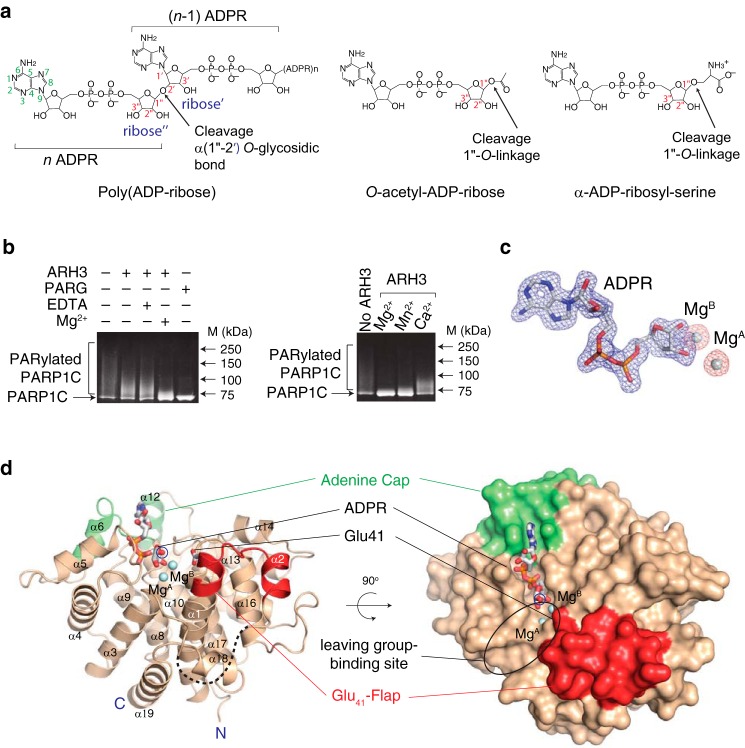
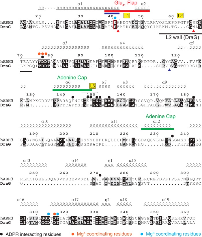
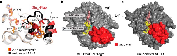
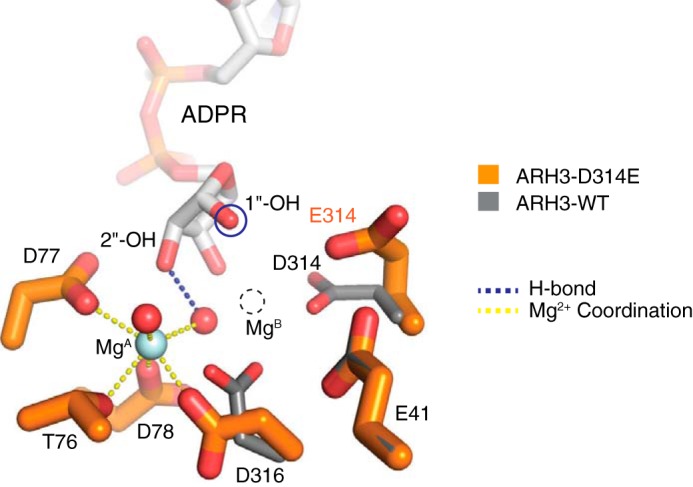
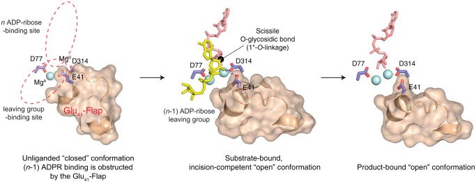
ADP-ribosyl-acceptor hydrolase 3 (ARH3) plays important roles in regulation of poly(ADP-ribosyl)ation, a reversible post-translational modification, and in maintenance of genomic integrity. ARH3 degrades poly(ADP-ribose) to protect cells from poly(ADP-ribose)-dependent cell death, reverses serine mono(ADP-ribosyl)ation, and hydrolyzes O-acetyl-ADP-ribose, a product of Sirtuin-catalyzed histone deacetylation. ARH3 preferentially hydrolyzes O-linkages attached to the anomeric C1″ of ADP-ribose; however, how ARH3 specifically recognizes and cleaves structurally diverse substrates remains unknown. Here, structures of full-length human ARH3 bound to ADP-ribose and Mg2+, coupled with computational modeling, reveal a dramatic conformational switch from closed to open states that enables specific substrate recognition. The glutamate flap, which blocks substrate entrance to Mg2+ in the unliganded closed state, is ejected from the active site when substrate is bound. This closed-to-open transition significantly widens the substrate-binding channel and precisely positions the scissile 1″-O-linkage for cleavage while securing tightly 2″- and 3″-hydroxyls of ADP-ribose. Our collective data uncover an unprecedented structural plasticity of ARH3 that supports its specificity for the 1″-O-linkage in substrates and Mg2+-dependent catalysis.

Keywords: ADP-ribosylation; ARH3; PARP1; conformational change; hydrolase; structural biology; substrate specificity.


Conflict of interest statement

The authors declare that they have no conflicts of interest with the contents of this article. The content is solely the responsibility of the authors and does not necessarily represent the official views of the National Institutes of Health.

Figures

Figure 1.
Figure 1.
Structure of full-length human ARH3 bound to ADP-ribose and Mg2+. a, structures of ARH3 substrates: a linear unbranched poly(ADP-ribose) (left), O-acetyl-ADP-ribose (middle), and α-ADP-ribosylserine (right). ARH3 cleaves the 1″-O-linkage in substrates. The exoglycohydrolase activity of ARH3 cleaves the α(1″-2′) O-glycosidic bond between n and n − 1 ADP-ribose, releasing ADP-ribose as a product. b, left, Mg2+ enhances the ADP-ribosyl-acceptor hydrolase activity of ARH3. The ARH3-mediated hydrolysis of PAR on PARylated PARP1C was monitored in the presence and absence of Mg2+ using a gel-based assay. PARG has stronger PAR turnover activity than ARH3 and was used as a positive control. Right, metal preference of ARH3. ARH3 prefers Mg2+ for catalysis followed by Mn2+ and Ca2+. c, difference electron density maps (FoFc) for ADPR and Mg2+ ions contoured at 3.0 σ (blue, ADPR; orange, Mg2+). d, structure of the ARH3–ADPR–Mg2+ complex revealing two unique and flexible structural elements, adenine cap (green) and glutamate flap (Glu41-flap) (red), that undergo conformational changes and strongly contribute to specific substrate recognition. The 1″-OH of ADPR (blue circled), corresponding to the scissile 1″-O-linkage in substrates, is exposed to solvent, consistent with ARH3 specificity for 1″-O-linkage for cleavage. A putative binding site for the leaving group is highlighted with a black ellipse. The N and C termini of ARH3 are indicated by N and C, respectively.
Figure 2.
Figure 2.
Structure-based alignment of ARH3 and DraG with structural elements and residues that are important for function. The Glu41 residue of the Glu41-flap of ARH3 that undergoes a large conformational change upon ADPR binding is indicated by a red box. A part of L2 of DraG (L2 wall) completely blocks the conformational change of α1 and restricts its activity to cleavage of mono(ADP-ribosyl)ated substrates. The end of α1 in ARH3, which exists as 310-helix in the unliganded form and undergoes 310-to-α transition upon ADPR binding (Fig. 4a), is indicated by a blue bar. Two aromatic residues in DraG that stabilize the L2 wall are indicated by a red triangle. Asp97 in DraG that is essential for the cleavage of MARylated arginine is indicated by a blue triangle.
Figure 3.
Figure 3.
ARH3 specifically exposes the scissile 1″-O-linkage in substrates for cleavage. a, structural superposition of ADPR-bound forms of ARH3 (wheat) and DraG (gray) reveals a distinctive ADPR-binding mode in ARH3. The adenine cap of ARH3 (green) grasps the adenine ring and is essential for ARH3 activities. Hydrogen bonds contributed by the main-chain atoms of the adenine cap to N6 and N7 of the adenine ring impart specificity. b, diagram showing interactions between ADPR and ARH3. c, close-up of the binuclear Mg2+ catalytic center and ADPR binding in ARH3. A matrix of Mg2+-mediated coordination and hydrogen-bonding interactions secure ADPR in the active site, which is consistent with Mg2+-dependent catalysis by ARH3. The 1″-OH of ADPR (blue circled), corresponding to the scissile 1″-O-linkage, is specifically exposed to solvent, strongly supporting ARH3 specificity for the 1″-O-linkage as a site for cleavage. d, close-up of the ARH3–di-ADPR–Mg2+ complex model. The energy-minimized computational model for the ARH3–di-ADPR–Mg2+ complex was generated using the ARH3–ADPR–Mg2+ complex as a starting model (see “Experimental procedures”). In this model, 3′-OH of the n − 1 ADPR leaving group is directly coordinated by MgB, replacing the W1 water ligand, and MgB tightly secures both n and n − 1 ADPR units for efficient cleavage to occur.
Figure 4.
Figure 4.
A conformational change of ARH3 enables specific substrate recognition and cleavage. a, structural comparison of the unliganded (gray/black) and ADPR-bound forms (orange/red) of ARH3 reveals a conformational switch in the Glu41-flap. The end of α1 undergoes a 310-to-α transition upon ADPR binding, which results in displacement of Glu41 away from MgB and significantly widens the leaving group–binding site of ARH3. b and c, ARH3 switches conformations to enable specific poly(ADP-ribose) recognition. The structure of the ARH3–di-ADPR–Mg2+ complex model is superimposed onto those of the ARH3–ADPR–Mg2+ complex and the apo-ARH3ΔN16, and di-ADPR is shown on the surfaces of ARH3. Comparison of solvent-accessible surfaces reveals an open platform that can accommodate n − 1 ADPR in the ARH3–ADPR–Mg2+ complex (b), whereas n − 1 ADPR binding is completely obstructed by the Glu41-flap in the unliganded ARH3 (c).
Figure 5.
Figure 5.
Asp314 is essential for the formation of the binuclear metal center. Structural comparison of the WT and catalytically inactive ARH3-D314E mutant reveals a side-chain flipping of Glu314, which leads to the loss of MgB. This suggests that Asp314 is essential for the formation of the binuclear metal center in ARH3 and that MgB is required for catalysis.
Figure 6.
Figure 6.
ARH3 switches between closed and open conformations to specifically recognize and cleave substrates. ARH3 switches between a self-inhibited and an incision-competent conformation for specific substrate recognition and cleavage. In the absence of PAR substrates, the Glu41-flap caps MgB, constituting a closed, self-inhibited enzyme state. Substrate binding induces a conformational switch in the Glu41-flap, allowing substrate entrance to the active site. This active-site rearrangement in ARH3 specifically exposes the scissile 1″-O-linkage to the catalytic Asp314 for cleavage, explaining ARH3 specificity for the 1″-O-linkage in structurally diverse substrates.

References

    1. Hakmé A., Wong H. K., Dantzer F., and Schreiber V. (2008) The expanding field of poly(ADP-ribosyl)ation reactions. 'Protein modifications: beyond the usual suspects' review series. EMBO Rep. 9, 1094–1100 10.1038/embor.2008.191 - DOI - PMC - PubMed
    1. Gibson B. A., and Kraus W. L. (2012) New insights into the molecular and cellular functions of poly(ADP-ribose) and PARPs. Nat. Rev. Mol. Cell Biol. 13, 411–424 10.1038/nrm3376 - DOI - PubMed
    1. Mégnin-Chanet F., Bollet M. A., and Hall J. (2010) Targeting poly(ADP-ribose) polymerase activity for cancer therapy. Cell. Mol. Life Sci. 67, 3649–3662 10.1007/s00018-010-0490-8 - DOI - PMC - PubMed
    1. El-Khamisy S. F., Masutani M., Suzuki H., and Caldecott K. W. (2003) A requirement for PARP-1 for the assembly or stability of XRCC1 nuclear foci at sites of oxidative DNA damage. Nucleic Acids Res. 31, 5526–5533 10.1093/nar/gkg761 - DOI - PMC - PubMed
    1. Beck C., Robert I., Reina-San-Martin B., Schreiber V., and Dantzer F. (2014) Poly(ADP-ribose) polymerases in double-strand break repair: focus on PARP1, PARP2 and PARP3. Exp. Cell Res. 329, 18–25 10.1016/j.yexcr.2014.07.003 - DOI - PubMed

Publication types

Substances

Associated data

LinkOut - more resources