

NLRP3 in somatic non-immune cells of rodent and primate testes
- PMID: 29907661
- PMCID: PMC6098733
- DOI: 10.1530/REP-18-0111
NLRP3 in somatic non-immune cells of rodent and primate testes
Abstract
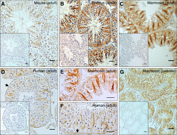
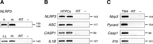
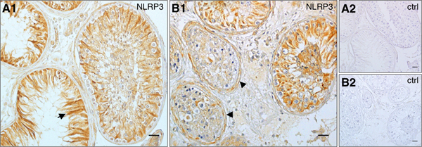
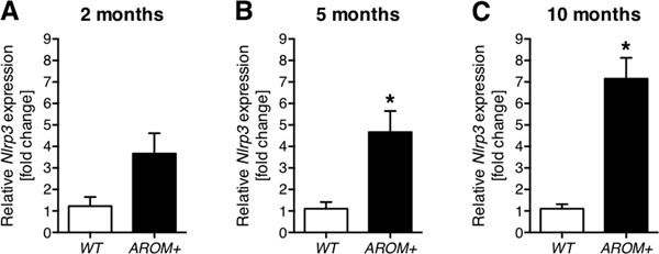
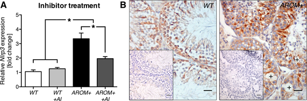
NLRP3 is part of the NLRP3 inflammasome and a global sensor of cellular damage. It was recently discovered in rodent Sertoli cells. We investigated NLRP3 in mouse, human and non-human primate (marmoset and rhesus macaque) testes, employing immunohistochemistry. Sertoli cells of all species expressed NLRP3, and the expression preceded puberty. In addition, peritubular cells of the adult human testes expressed NLRP3. NLRP3 and associated genes (PYCARD, CASP1, IL1B) were also found in isolated human testicular peritubular cells and the mouse Sertoli cell line TM4. Male infertility due to impairments of spermatogenesis may be related to sterile inflammatory events. We observed that the expression of NLRP3 was altered in the testes of patients suffering from mixed atrophy syndrome, in which tubules with impairments of spermatogenesis showed prominent NLRP3 staining. In order to explore a possible role of NLRP3 in male infertility, associated with sterile testicular inflammation, we studied a mouse model of male infertility. These human aromatase-expressing transgenic mice (AROM+) develop testicular inflammation and impaired spermatogenesis during aging, and the present data show that this is associated with strikingly elevated Nlrp3 expression in the testes compared to WT controls. Interference by aromatase inhibitor treatment significantly reduced increased Nlrp3 levels. Thus, throughout species NLRP3 is expressed by somatic cells of the testis, which are involved in testicular immune surveillance. We conclude that NLRP3 may be a novel player in testicular immune regulation.
© 2018 Society for Reproduction and Fertility.
Figures
Similar articles
-
Exploring the Ion Channel TRPV2 and Testicular Macrophages in Mouse Testis.Int J Mol Sci. 2021 Apr 29;22(9):4727. doi: 10.3390/ijms22094727. Int J Mol Sci. 2021. PMID: 33946947 Free PMC article.
-
Irradiation affects germ and somatic cells in prepubertal monkey testis xenografts.Mol Hum Reprod. 2017 Mar 1;23(3):141-154. doi: 10.1093/molehr/gax003. Mol Hum Reprod. 2017. PMID: 28130393
-
Expression of the oestrogen receptor GPER by testicular peritubular cells is linked to sexual maturation and male fertility.Andrology. 2014 Sep;2(5):695-701. doi: 10.1111/j.2047-2927.2014.00243.x. Epub 2014 Jul 23. Andrology. 2014. PMID: 25052196 Free PMC article.
-
Human testicular peritubular cells, mast cells and testicular inflammation.Andrologia. 2018 Dec;50(11):e13055. doi: 10.1111/and.13055. Andrologia. 2018. PMID: 30569646 Review.
-
Testicular inflammation and infertility: Could chlamydial infections be contributing?Am J Reprod Immunol. 2020 Sep;84(3):e13286. doi: 10.1111/aji.13286. Epub 2020 Jun 24. Am J Reprod Immunol. 2020. PMID: 32533905 Review.
Cited by
-
Genes underlying the evolution of tetrapod testes size.BMC Biol. 2021 Aug 18;19(1):162. doi: 10.1186/s12915-021-01107-z. BMC Biol. 2021. PMID: 34407824 Free PMC article.
-
Prokineticin 2 via Calcium-Sensing Receptor Activated NLRP3 Inflammasome Pathway in the Testicular Macrophages of Uropathogenic Escherichia coli-Induced Orchitis.Front Immunol. 2020 Oct 23;11:570872. doi: 10.3389/fimmu.2020.570872. eCollection 2020. Front Immunol. 2020. PMID: 33193351 Free PMC article.
-
The NLRP3 inflammasome: molecular activation and regulation in spermatogenesis and male infertility; a systematic review.Basic Clin Androl. 2022 May 30;32(1):8. doi: 10.1186/s12610-022-00157-9. Basic Clin Androl. 2022. PMID: 35637440 Free PMC article. Review.
-
The Inflammasome in Reproductive Biology: A Promising Target for Novel Therapies.Front Endocrinol (Lausanne). 2020 Jan 28;11:8. doi: 10.3389/fendo.2020.00008. eCollection 2020. Front Endocrinol (Lausanne). 2020. PMID: 32047476 Free PMC article. Review.
-
The role of inflammasome dysregulation in obstructive and non-obstructive azoospermia: a comparative molecular analysis of blood, tissue, and seminal plasma.Front Immunol. 2024 Dec 6;15:1507885. doi: 10.3389/fimmu.2024.1507885. eCollection 2024. Front Immunol. 2024. PMID: 39712014 Free PMC article.
References
-
- Albrecht M, Ramsch R, Kohn FM, Schwarzer JU & Mayerhofer A 2006. Isolation and cultivation of human testicular peritubular cells: a new model for the investigation of fibrotic processes in the human testis and male infertility. J Clin Endocrinol Metab 91 1956–1960. - PubMed
-
- Bazrafkan M, Nikmehr B, Shahverdi A, Hosseini SR, Hassani F, Poorhassan M, Mokhtari T, Abolhassani F, Choobineh H, Beyer C, et al. 2018. Lipid Peroxidation and Its Role in the Expression of NLRP1a and NLRP3 Genes in Testicular Tissue of Male Rats: a Model of Spinal Cord Injury. Iran Biomed J 22 151–159. - PMC - PubMed
-
- Broz P & Dixit VM 2016. Inflammasomes: mechanism of assembly, regulation and signalling. Nat Rev Immunol 16 407–420. - PubMed
Publication types
MeSH terms
Substances
Grants and funding
LinkOut - more resources
Full Text Sources
Other Literature Sources
Molecular Biology Databases
Miscellaneous
