

Obesity/Type II Diabetes Promotes Function-limiting Changes in Murine Tendons that are not reversed by Restoring Normal Metabolic Function
- PMID: 29907811
- PMCID: PMC6003963
- DOI: 10.1038/s41598-018-27634-4
Obesity/Type II Diabetes Promotes Function-limiting Changes in Murine Tendons that are not reversed by Restoring Normal Metabolic Function
Abstract
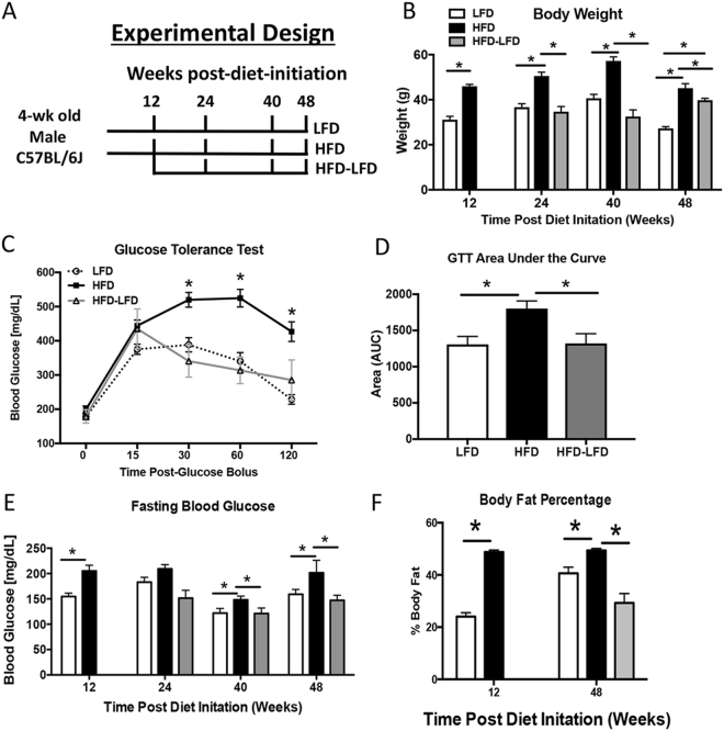
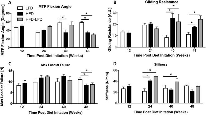
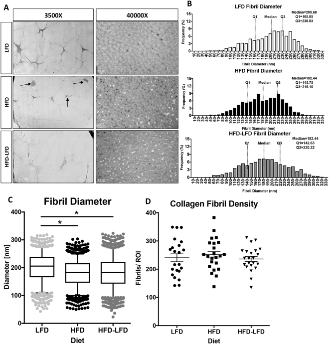
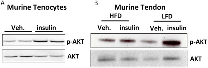
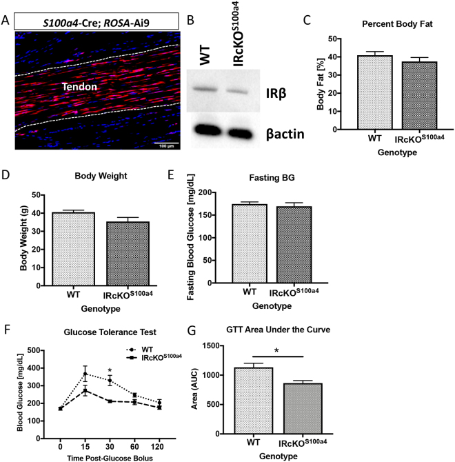
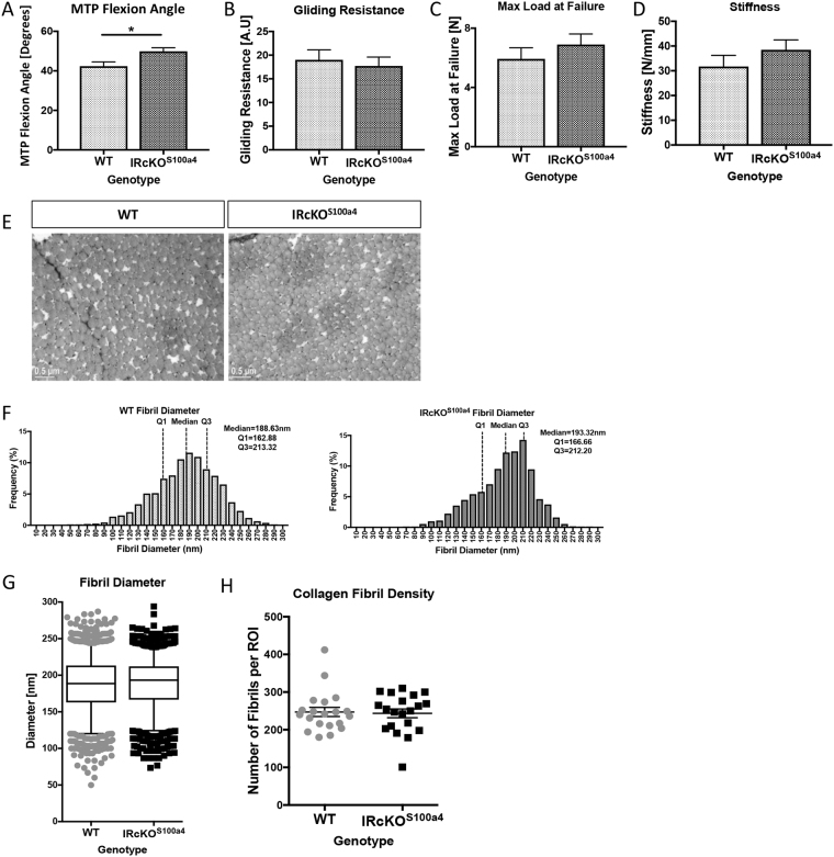
Type II Diabetes (T2DM) negatively alters baseline tendon function, including decreased range of motion and mechanical properties; however, the biological mechanisms that promote diabetic tendinopathy are unknown. To facilitate identification of therapeutic targets we developed a novel murine model of diabetic tendinopathy. Mice fed a High Fat Diet (HFD) developed diet induced obesity and T2DM and demonstrated progressive impairments in tendon gliding function and mechanical properties, relative to mice fed a Low Fat Diet (LFD). We then determined if restoration of normal metabolic function, by switching mice from HFD to LFD, was sufficient to halt the pathological changes in tendon due to obesity/T2DM. However, switching from a HFD to LFD resulted in greater impairments in tendon gliding function than mice maintained on a HFD. Mechanistically, IRβ signaling is decreased in obese/T2DM murine tendons, suggesting altered IRβ signaling as a driver of diabetic tendinopathy. However, knock-down of IRβ expression in S100a4-lineage cells (IRcKOS100a4) was not sufficient to induce diabetic tendinopathy as no impairments in tendon gliding function or mechanical properties were observed in IRcKOS100a4, relative to WT. Collectively, these data define a murine model of diabetic tendinopathy, and demonstrate that restoring normal metabolism does not slow the progression of diabetic tendinopathy.
Conflict of interest statement
The authors declare no competing interests.
Figures
Similar articles
-
Deletion of EP4 in S100a4-lineage cells reduces scar tissue formation during early but not later stages of tendon healing.Sci Rep. 2017 Aug 17;7(1):8658. doi: 10.1038/s41598-017-09407-7. Sci Rep. 2017. PMID: 28819185 Free PMC article.
-
Obesity/Type II diabetes alters macrophage polarization resulting in a fibrotic tendon healing response.PLoS One. 2017 Jul 7;12(7):e0181127. doi: 10.1371/journal.pone.0181127. eCollection 2017. PLoS One. 2017. PMID: 28686669 Free PMC article.
-
Effects of Type II Diabetes Mellitus on Tendon Homeostasis and Healing.J Orthop Res. 2020 Jan;38(1):13-22. doi: 10.1002/jor.24388. Epub 2019 Jun 24. J Orthop Res. 2020. PMID: 31166037 Free PMC article. Review.
-
High fat diet attenuates hyperglycemia, body composition changes, and bone loss in male streptozotocin-induced type 1 diabetic mice.J Cell Physiol. 2018 Feb;233(2):1585-1600. doi: 10.1002/jcp.26062. Epub 2017 Aug 4. J Cell Physiol. 2018. PMID: 28631813 Free PMC article.
-
Rehabilitation of Tendon Problems in Patients with Diabetes Mellitus.Adv Exp Med Biol. 2016;920:199-208. doi: 10.1007/978-3-319-33943-6_19. Adv Exp Med Biol. 2016. PMID: 27535262 Review.
Cited by
-
A Maternal High Fat Diet Leads to Sex-Specific Programming of Mechanical Properties in Supraspinatus Tendons of Adult Rat Offspring.Front Nutr. 2021 Sep 13;8:729427. doi: 10.3389/fnut.2021.729427. eCollection 2021. Front Nutr. 2021. PMID: 34589513 Free PMC article.
-
Diabetes Mellitus as a Risk Factor for Trigger Finger -a Longitudinal Cohort Study Over More Than 20 Years.Front Clin Diabetes Healthc. 2021 Nov 2;2:708721. doi: 10.3389/fcdhc.2021.708721. eCollection 2021. Front Clin Diabetes Healthc. 2021. PMID: 36994346 Free PMC article.
-
Obesity Increases the Risk of Tendinopathy, Tendon Tear and Rupture, and Postoperative Complications: A Systematic Review of Clinical Studies.Clin Orthop Relat Res. 2020 Aug;478(8):1839-1847. doi: 10.1097/CORR.0000000000001261. Clin Orthop Relat Res. 2020. PMID: 32732565 Free PMC article.
-
Effect of high-fat diet on the lipid profile of ovarian granulosa cells and female reproduction in mice.PLoS One. 2023 Jun 27;18(6):e0287534. doi: 10.1371/journal.pone.0287534. eCollection 2023. PLoS One. 2023. PMID: 37368884 Free PMC article.
-
High fat diet induced obesity model using four strainsof mice: Kunming, C57BL/6, BALB/c and ICR.Exp Anim. 2020 Aug 5;69(3):326-335. doi: 10.1538/expanim.19-0148. Epub 2020 Mar 19. Exp Anim. 2020. PMID: 32188837 Free PMC article.
References
-
- Williamson RT. Causes of diabetes. 1909. Practitioner. 2009;253:37. - PubMed
-
- Rodriguez A, Delgado-Cohen H, Reviriego J, Serrano-Rios M. Risk factors associated with metabolic syndrome in type 2 diabetes mellitus patients according to World Health Organization, Third Report National Cholesterol Education Program, and International Diabetes Federation definitions. Diabetes Metab Syndr Obes. 2011;4:1–4. - PMC - PubMed
Publication types
MeSH terms
Substances
Grants and funding
LinkOut - more resources
Full Text Sources
Other Literature Sources
Medical
Molecular Biology Databases
Research Materials
