. 2018 Jun 15;8(1):9194.
doi: 10.1038/s41598-018-27568-x.

Ex vivo biomechanical characterization of syringe-needle ejections for intracerebral cell delivery

Affiliations

Ex vivo biomechanical characterization of syringe-needle ejections for intracerebral cell delivery

Brendon Wahlberg et al. Sci Rep. .

Abstract

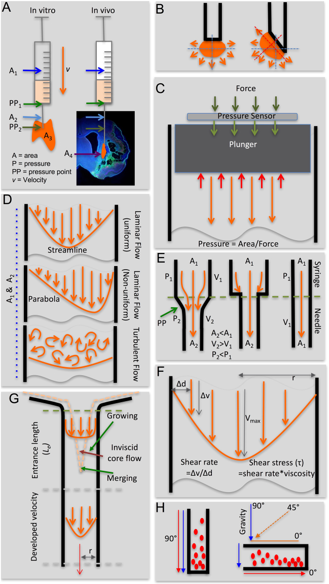
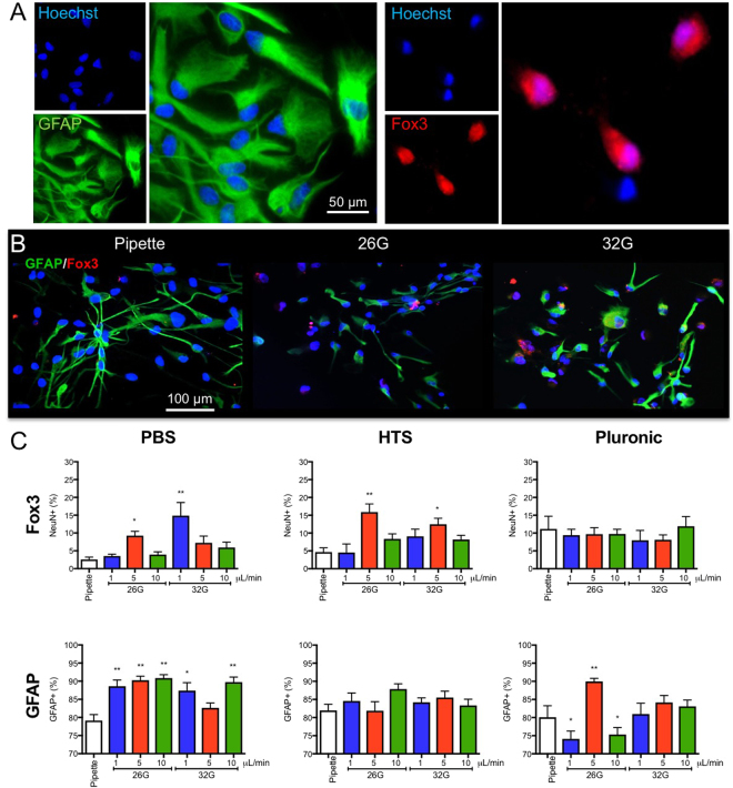
Intracerebral implantation of cell suspensions is finding its clinical translation with encouraging results in patients with stroke. However, the survival of cells in the brain remains poor. Although the biological potential of neural stem cells (NSCs) is widely documented, the biomechanical effects of delivering cells through a syringe-needle remain poorly understood. We here detailed the biomechanical forces (pressure, shear stress) that cells are exposed to during ejection through different sized needles (20G, 26G, 32G) and syringes (10, 50, 250 µL) at relevant flow rates (1, 5, 10 µL/min). A comparison of 3 vehicles, Phosphate Buffered Saline (PBS), Hypothermosol (HTS), and Pluronic, indicated that less viscous vehicles are favorable for suspension with a high cell volume fraction to minimize sedimentation. Higher suspension viscosity was associated with greater shear stress. Higher flow rates with viscous vehicle, such as HTS reduced viability by ~10% and also produced more apoptotic cells (28%). At 5 µL/min ejection using a 26G needle increased neuronal differentiation for PBS and HTS suspensions. These results reveal the biological impact of biomechanical forces in the cell delivery process. Appropriate engineering strategies can be considered to mitigate these effects to ensure the efficacious translation of this promising therapy.


Conflict of interest statement

The authors declare no competing interests.

Figures

Figure 1
Figure 1
Biophysical and biomechanical considerations for syringe-needle ejections. (A) The ejection pressure from needle-syringe is defined by the area of the barrel and the velocity (v) to move the plunger. The area of the barrel inside the syringe (A1) can be of a different size than inside the needle (A2). Ejection in vitro into an empty space (A3) versus in tissue in vivo (A4) further influences the force required to push the plunger at a given velocity. As the area of the ejectate changes between the syringe and the needle, as well as the needle and the environment, pressure points (PP) are formed. (B) The point of ejection is defined by the shape of the needle tip being flat or beveled, which will influence the dispersion of the ejectate. (C) Pressure within the syringe and needle barrel was calculated based on the measurement of the applied force using a pressure sensor placed onto of the plunger. (D) Based on Reynold’s numbers, the flow characteristics within the barrel were defined to be uniform or non-uniform laminar flow or turbulent flow. (E) The interface between syringe and needle defined pressure points and potentially affects flow characteristics. A straight barrel between syringe and needle is the most optimal arrangement to avoid pressure points as well as to minimize the formation of a plug that could block the ejection. (F) Within the syringe and needle, shear rate and stress can be calculated based on the radius of the barrel, the viscosity of the material, and the flow rate. (G) During the transition between the syringe and the needle, the suspension will comply with an entrance length (Le) which will allow the streamline to move along the barrel and develop velocity. (H) Cells in the suspension along the path of ejection will sediment if their density is higher than the vehicle. The sedimentation rate is dependent on the angle of the barrel with an orientation along the path of gravity (90°) exerting maximal sedimentation and least sediment along the barrel being observed with a horizontal orientation (0°).
Figure 2
Figure 2
Syringe and needle pressures of suspension vehicle ejections. (A) Ejection pressure was calculated based on the measured force applied to eject phosphate buffered saline (PBS), hypothermosol (HTS) or pluronic at defined flow rates using different syringe-needle combinations. (B) A direction comparison of ejection pressures for the 3 suspension vehicles for syringe and needle combinations illustrates that faster ejection using large bore needle and less viscous vehicles reduces ejection pressure.
Figure 3
Figure 3
Cell suspension viscosity, sedimentation and Reynold’s numbers. (A) The density of cell suspensions is dependent on the volume fractions of cells and suspension vehicle. However, practically a ~60% volume fraction (150,000 cells/μL) is the highest achievable concentration due to the maximum packing density of spheres being 0.636. As the density of cells is higher than vehicles, increased volume fraction defined suspension density. (B) The viscosity of the cell suspension is also related to volume fraction. An exponential increase in viscosity is seen with increased cell volume fraction. (C) A contour plot further highlights the interaction between volume fraction and fluid density to define the viscosity of the cell suspension. (D) Sedimentation of cells at a horizontal orientation is minimal, whereas a significant sedimentation is seen at a vertical orientation with a speed of sedimentation >160 μm/s, if a 10% cell volume fraction (25,000 cells/μL) is used. Higher cell volume fractions reduce sedimentation rate. (E) Reynolds numbers for cell suspensions inside the needle remain well below the Re < 0.1 threshold to indicate that these would still flow in a uniform laminar streamline.
Figure 4
Figure 4
Contour plots for Stoke’s drag force and shear stress influencing cells in suspension. (A) The drag force (μN) in a 20G and 26G needle is neglible for all practical cell suspension (<0.6 volume fraction). The smaller dimater of the 32G needle, as well as the higher viscosity of HTS, exert greated drag force on cells than other conditions, but these are still very low and unlikely to influence cells or their sedimentation. (B) Shear stress (N/m2) is increased with smaller diameter needles, higher viscosity and flow rate. A 32G needle with a high cell volume fraction in HTS therefore will be exposed to the highest shear rate. However, the use of a 26G needle significantly reduces this shear stress.
Figure 5
Figure 5
Cell viability, cell membrane damage and apoptosis after syringe-needle ejection. (A) Viability measurements using trypan blue were used to determine the percentage of dead cells upon preparation of cell suspensions (20% volume fraction, 50,000 cells/μL) with different suspension vehicles, as well as after ejection. (B) To determine if the ejection procedure induced cell membrane damage, lactate dehydrogenase (LDH) was measured 24 hours after ejection and compared to pipetting of cells. (C) To model in vitro the injection procedure, cell suspension were placed as a deposit on a cover slip with gentile agitation to spread cells. (D) This in vitro injection model produce a greater cell density at the centre of the coverlsip with a lower cell density at the edge of the deposit. Caspase-3 (CSP-3) immunocytochemistry was used to define cells undergoing apoptosis. A greater degree of apoptosis was evident after 24 hours at the core of the deposit compared to the corona. (E) The number of CSP-3+ cells were counted for each ejection. Very few apoptotic cells were evident in the PBS condition compared to HTS and Pluronic. It was further evident that a higher flow rate and thinner needle increased the number of apoptotic cells. (F) Ejection of cells using pluronic as suspension vehicle through a 32G needle at a high flow rate produced the highest proportion of apoptotic cells, whereas a slow ejection using a larger needle and PBS had minimal impact.
Figure 6
Figure 6
Ejection conditions affect neuronal and astrocytic differentiation of NSCs. (A) Neural stem cells differentiate into both astrocytic (GFAP+) and neuronal cells (Fox3) with distinct morphologies. Typically astrocytes are found in dense cell clusters, whereas neurons were more common in less densely populated areas. (B) Astrocytic and neuronal differentiation of NSCs after ejection from a pipette, 26G or 32G needle, at a 20% volume fraction (50,000 cells/μL) revealed both morphological, as well as phenotypic differences after 7 days of differentiation. (C) Ejection parameters influenced neuronal and astrocytic differentiation of NSCs compared to pipette only ejection. Especially PBS resulted in an overall increased astrocytic differentiation, while at 5 and 1 μL/min ejection using a 26G and 32G needles respectively produced more neuronal differentiation. HTS reduced the impact of ejection on cell differentiation, although a 5 μl/min flow rate using a 26G needle increased neuronal differentiation. Neuronal differentiation was also maintained constant with pluronic as suspension vehicle, but significant shift in astrocytic differentiation were evident.

Similar articles

Cited by

References

    1. Amer MH, Rose FRA, Shakesheff KM, Modo M, White LJ. Translational considerations in injectable cell-based therapeutics for neurological applications: concepts, progress and challenges. npj Regenerative Medicine. 2017;2:23. doi: 10.1038/s41536-017-0028-x. - DOI - PMC - PubMed
    1. Kondziolka D, et al. Transplantation of cultured human neuronal cells for patients with stroke. Neurology. 2000;55:565–569. doi: 10.1212/WNL.55.4.565. - DOI - PubMed
    1. Kalladka D, et al. Human neural stem cells in patients with chronic ischaemic stroke (PISCES): a phase 1, first-in-man study. Lancet. 2016;388:787–796. doi: 10.1016/S0140-6736(16)30513-X. - DOI - PubMed
    1. Steinberg GK, et al. Clinical Outcomes of Transplanted Modified Bone Marrow-Derived Mesenchymal Stem Cells in Stroke: A Phase 1/2a Study. Stroke. 2016;47:1817–1824. doi: 10.1161/STROKEAHA.116.012995. - DOI - PMC - PubMed
    1. Smith EJ, et al. Implantation site and lesion topology determine efficacy of a human neural stem cell line in a rat model of chronic stroke. Stem Cells. 2012;30:785–796. doi: 10.1002/stem.1024. - DOI - PubMed

Publication types