Clinical Trial
. 2018 Jun 15;8(1):9228.
doi: 10.1038/s41598-018-27347-8.

Diagnostic yield and accuracy of coronary CT angiography after abnormal nuclear myocardial perfusion imaging

Affiliations
Clinical Trial

Diagnostic yield and accuracy of coronary CT angiography after abnormal nuclear myocardial perfusion imaging

Felix G Meinel et al. Sci Rep. .

Abstract

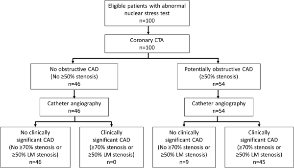
We aimed to determine the diagnostic yield and accuracy of coronary CT angiography (CCTA) in patients referred for invasive coronary angiography (ICA) based on clinical concern for coronary artery disease (CAD) and an abnormal nuclear stress myocardial perfusion imaging (MPI) study. We enrolled 100 patients (84 male, mean age 59.6 ± 8.9 years) with an abnormal MPI study and subsequent referral for ICA. Each patient underwent CCTA prior to ICA. We analyzed the prevalence of potentially obstructive CAD (≥50% stenosis) on CCTA and calculated the diagnostic accuracy of ≥50% stenosis on CCTA for the detection of clinically significant CAD on ICA (defined as any ≥70% stenosis or ≥50% left main stenosis). On CCTA, 54 patients had at least one ≥50% stenosis. With ICA, 45 patients demonstrated clinically significant CAD. A positive CCTA had 100% sensitivity and 84% specificity with a 100% negative predictive value and 83% positive predictive value for clinically significant CAD on a per patient basis in MPI positive symptomatic patients. In conclusion, almost half (48%) of patients with suspected CAD and an abnormal MPI study demonstrate no obstructive CAD on CCTA.


Conflict of interest statement

This study was supported by a research grant provided by GE Healthcare (Wauwatosa/WI/United States). UJS is a consultant for and/or receives institutional research support from Astellas, Bayer, Bracco, GE, Guerbet, HeartFlow, and Siemens Healthineers. FGM has received institutional research support from Siemens, unrelated to this investigation. The other authors declare that they have no competing interests.

Figures

Figure 1
Figure 1
Flowchart demonstrating the results of CCTA and ICA. The presence of ≥50% stenosis was used to define a positive CCTA study as commonly used in clinical routine. We assessed the diagnostic accuracy of CCTA for the detection of clinically significant coronary artery disease, which we defined as any ≥70% stenosis or ≥50% left main stenosis on catheter angiograms. CAD = coronary artery disease, CCTA = coronary computed tomography angiography, LM = left main coronary artery.
Figure 2
Figure 2
Example of a true positive CCTA examination. This 60 year old male patient underwent stress nuclear myocardial perfusion imaging because of recurrent chest pain. Stress images in the vertical short axis (B) and horizontal short axis views (D) are suggestive of decreased perfusion to the anterior, septal and apical portions of the left ventricular myocardium (arrowheads) and are largely reversible at rest (A,C). The examination was thus interpreted as concerning for ischemia involving the anteroseptal wall and the cardiac apex. At CCTA, transverse section (E) and curved multiplanar reformat (F) of the left anterior descending artery demonstrate a severe proximal stenosis caused by a predominantly non-calcified plaque (arrow). Another severe stenosis with mixed plaque is noted further distally (dotted arrow). The right coronary artery (G) and the circumflex artery show diffuse calcified and non-calcified plaque but no ≥50% stenosis. Absence of critical stenosis in the right coronary artery was confirmed on the catheter angiogram (H). The angiogram also confirmed the two critical stenoses in the left anterior descending artery (I). Both lesions were successfully treated with angioplasty and stent placement. CCTA = coronary computed tomography angiography.
Figure 3
Figure 3
Example of a true negative CCTA examination. This 58 year old male patient underwent stress nuclear myocardial perfusion imaging because of chest pain and dyspnea on exertion. Stress images in the vertical long axis (B) and horizontal short axis views (D) are suggestive of decreased perfusion to the inferior wall of the left ventricle (arrow heads), which are at significantly reversible at rest without extracardiac activity (A,C). The examination was thus interpreted as concerning for inferior wall ischemia. At CCTA, curved multiplanar reformats of the right coronary artery (E), left anterior descending artery (F,G) and circumflex artery (H) demonstrate no evidence of stenosis. Myocardial bridging of the left anterior descending artery with a relatively long intra-myocardial course as a potential cause of the patient’s symptoms is noted (arrows in F,G). On catheter angiograms (I,K), there is no evidence of coronary artery disease. CCTA = coronary computed tomography angiography.
Figure 4
Figure 4
Example of a false positive CCTA examination. This 63 years old male patient underwent stress nuclear myocardial perfusion imaging because of exertional dyspnea. Stress images in the vertical short axis (B) and horizontal short axis views (D) are suggestive of decreased perfusion to the lateral wall of the left ventricle (arrow heads), which is reversible at rest (A,C). The examination was thus interpreted as concerning for lateral wall ischemia. At CCTA, transverse section (E) and curved multiplanar reformat (F) of the right coronary artery were interpreted as showing 50% stenosis with focal soft plaque (arrow). No luminal narrowing is noted in the left anterior descending (G) and circumflex (H) arteries. On catheter angiograms (I,K), there is no evidence of luminal narrowing in any vessel. The false positive finding on CCTA likely represents an artifact. CCTA = coronary computed tomography angiography.

Similar articles

Cited by

References

    1. Patel MR, et al. Low diagnostic yield of elective coronary angiography. N Engl J Med. 2010;362:886–895. doi: 10.1056/NEJMoa0907272. - DOI - PMC - PubMed
    1. Knuuti, J. et al. Risks and benefits of cardiac imaging: an analysis of risks related to imaging for coronary artery disease. Eur Heart J (2013). - PubMed
    1. Taylor AJ, et al. ACCF/SCCT/ACR/AHA/ASE/ASNC/NASCI/SCAI/SCMR 2010 Appropriate Use Criteria for Cardiac Computed Tomography. Circulation. 2010;122:e525–555. doi: 10.1161/CIR.0b013e3181fcae66. - DOI - PubMed
    1. Meijboom WB, et al. Comprehensive assessment of coronary artery stenoses: computed tomography coronary angiography versus conventional coronary angiography and correlation with fractional flow reserve in patients with stable angina. J Am Coll Cardiol. 2008;52:636–643. doi: 10.1016/j.jacc.2008.05.024. - DOI - PubMed
    1. Schuijf JD, Bax JJ. CT angiography: an alternative to nuclear perfusion imaging? Heart. 2008;94:255–257. doi: 10.1136/hrt.2006.105833. - DOI - PubMed

Publication types