. 2018 Jun 15;8(1):9202.
doi: 10.1038/s41598-018-27379-0.

Deletion of DDB1- and CUL4- associated factor-17 (Dcaf17) gene causes spermatogenesis defects and male infertility in mice

Affiliations

Deletion of DDB1- and CUL4- associated factor-17 (Dcaf17) gene causes spermatogenesis defects and male infertility in mice

Asmaa Ali et al. Sci Rep. .

Erratum in

Abstract

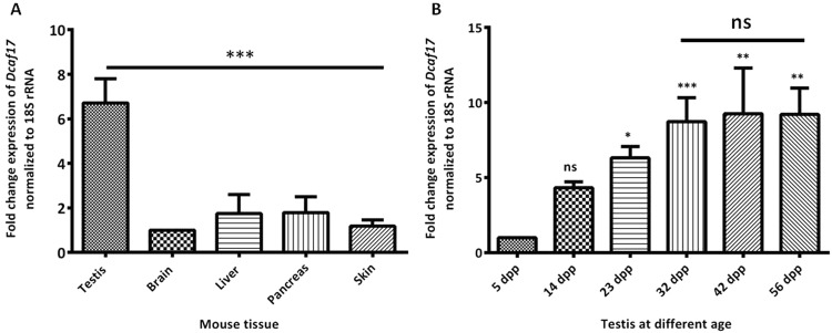
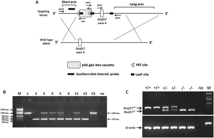
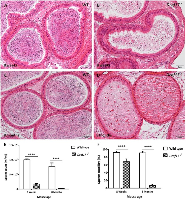
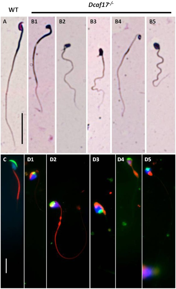
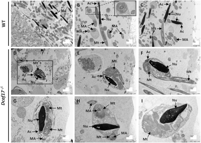
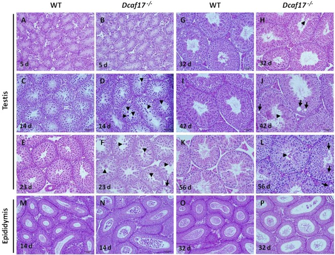
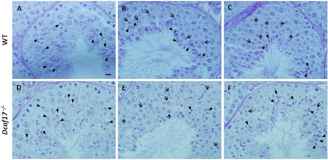
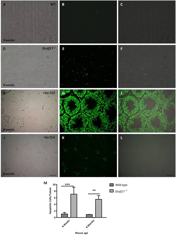
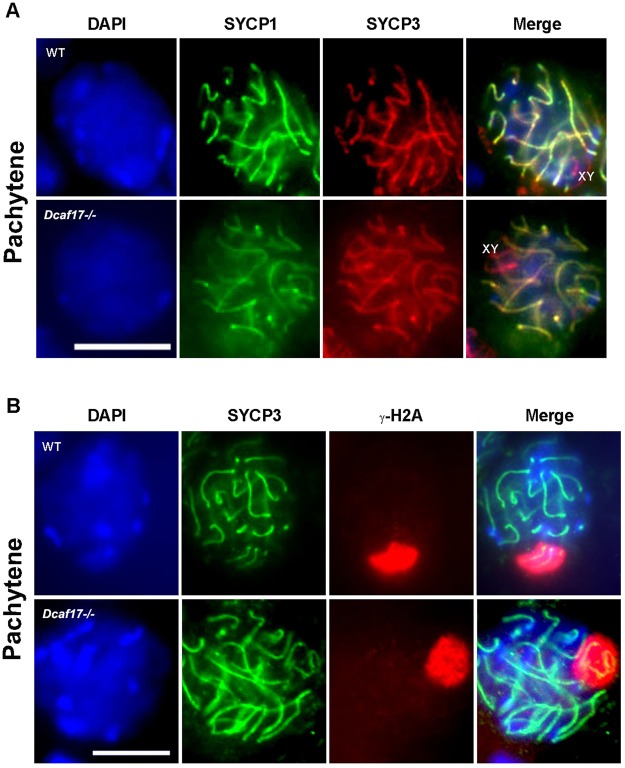
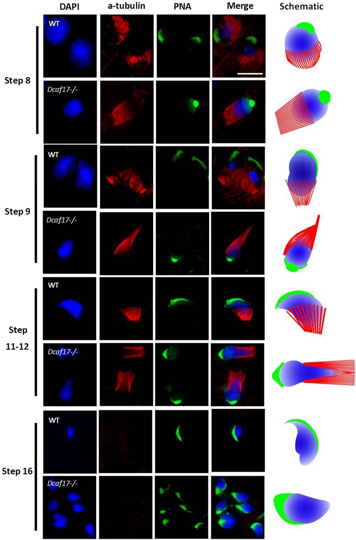
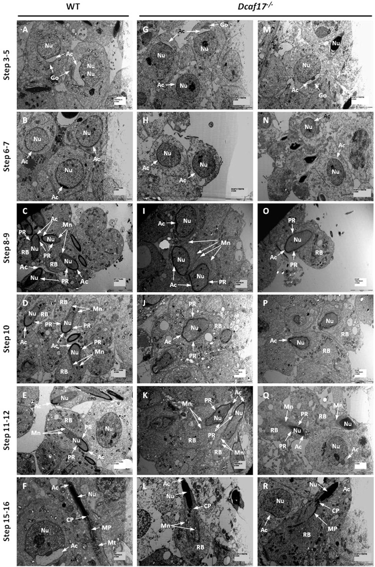
DDB1- and CUL4-associated factor 17 (Dcaf17) is a member of DCAF family genes that encode substrate receptor proteins for Cullin-RING E3 ubiquitin ligases, which play critical roles in many cellular processes. To unravel the function of DCAF17, we performed expression profiling of Dcaf17 in different tissues of wild type mouse by qRT-PCR and generated Dcaf17 knockout mice by gene targeting. Expression profiling of Dcaf17 showed highest expression in testis. Analyses of Dcaf17 transcripts during post-natal development of testis at different ages displayed gradual increase in Dcaf17 mRNA levels with the age. Although Dcaf17 disruption did not have any effect on female fertility, Dcaf17 deletion led to male infertility due to abnormal sperm development. The Dcaf17 -/- mice produced low number of sperm with abnormal shape and significantly low motility. Histological examination of the Dcaf17 -/- testis revealed impaired spermatogenesis with presence of vacuoles and sloughed cells in the seminiferous tubules. Disruption of Dcaf17 caused asymmetric acrosome capping, impaired nuclear compaction and abnormal round spermatid to elongated spermatid transition. For the first time, these data indicate that DCAF17 is essential for spermiogenesis.


Conflict of interest statement

The authors declare no competing interests.

Figures

Figure 1
Figure 1
Investigating Dcaf17 mRNA levels by real time PCR. (A) Relative expression of Dcaf17 in different tissues of adult mouse. Relative expression values of Dcaf17 mRNA were normalized to 18S rRNA expression and the level of Dcaf17 mRNA in the brain was arbitrarily set at 1. The Dcaf17 is highly expressed in testis, while other tissues show low level of expression. (B) Relative expression of Dcaf17 in testis at different ages. Relative expression values of Dcaf17 mRNA were normalized to 18S rRNA expression and the level of Dcaf17 mRNA in the 5 days postpartum (dpp) testis was arbitrarily set at 1. Expression of Dcaf17 in the testis increased with the age till the age of 32 dpp and then after it remained constant. Asterisks indicate statistical significance (p < 0.0001 for A and p < 0.0036 for B). Error bars represent SEM. Three to five animals were used to investigate the transcript levels of Dcaf17. Experiments were performed in triplicates. Data were analyzed on Graphpad prism 5 software using one-way ANOVA technique followed by post hoc Bonferroni’s (A) or Tukey’s (B) multiple comparison test. ns - not significant.
Figure 2
Figure 2
Diagrammatic representation (A) of Dcaf17 gene targeting approach in mouse by homologous recombination and genotyping (B,C) of different alleles of Dcaf17 in mice. (A) Homologous recombination strategy in mouse ES cells. The Dcaf17 targeting vector (top) was constructed to replace wild type exon 4 and introduce neomycin drug selection marker, LoxP and FRT sites. (B) Agarose gel image of PCR genotyping of representative Dcaf17 mutant mice. PCR amplification of wild type genotype gives 1 kbps amplicon (1, C3), heterozygous genotype for Dcaf17 mutation gives 1 kbps and 193 bps amplicons (3–5, C1) and homozygous genotype for Dcaf17 mutation gives 193 bps amplicon (2, 6 and C2). 1–6 – genomic DNA samples of different Dcaf17 genotypes; C1-C3 – Different Dcaf17 genotype controls; −Ve – no template control. (C) Agarose gel image of RT-PCR of different Dcaf17 genotypes. PCR products of various Dcaf17 alleles and β-actin were run on the same agarose gel and single image was taken. +/+ - Dcaf17+/+ (WT); +/− - Dcaf17+/− (heterozygous Dcaf17 mutant); −/− - Dcaf17−/− (homozygous Dcaf17 mutant); −Ve – no template control; M – DNA ladder. PCR fragment size for β-actin is 190 bps; for Dcaf17+/+ is 284 bps and for Dcaf17−/− is 148 bps. Gel images were taken using ImageQuant LAS 4000 imaging system.
Figure 3
Figure 3
Histology of cauda epididymides and cauda sperm analyses from WT and Dcaf17−/− adult mice. Hematoxylin and eosin (H,E) staining of cauda epididymides sections (5 µm thick) from 8 weeks (A,B) and 8 months (C,D) old WT (A,C) and Dcaf17−/− (B,D) mice. The lumen of WT cauda epididymis (A,C) is full of mature sperm, whereas the lumen of Dcaf17−/− mutant cauda epididymidis (B,D) contains oval shaped sloughed cells of various size. Sperm count (E) and motility (F) analyses of WT and Dcaf17−/− mutant mice at the age of 8 weeks and 8 months old reveal severe and progressive reduction in sperm count (E) and motility (F) in Dcaf17−/− mice compared to WT mice. Around 300 sperm in a total of five fields in each replicate were analyze for sperm motility analysis. Number of animals used for each genotype to analyze sperm count (E) and motility (F) was 5. Data were analyzed on Graphpad prism 5 using two-way ANOVA technique followed by post hoc Bonferroni’s multiple comparison test. For images A to D the magnification is 200X and the scale bars are 5 µm. Error bars in panels E and F represent SEM. Asterisks in E and F indicate statistical significance (p < 0.0001 for E and F).
Figure 4
Figure 4
Bright field and fluorescence microscopy of cauda epididymal sperm from WT and Dcaf17−/− adult mice. Representative images of Diff-Quick staining (bright field images, top panels) and MitoTracker staining (fluorescence images, bottom panels) of epididymal sperm spreads from WT (A,C) and Dcaf17−/− (B,D) adult mice. The WT sperm (A,C) show typical hook-shaped head, patent midpiece and tail morphology. Whereas, the Dcaf17−/− sperm (B,D) show variety of morphological defects in head shape, midpiece and tail. To categorize different sperm defects in Dcaf17 KO mice we analyzed total 150 sperms from 3 different mice. Major categories of sperm head defects were triangular (cupcake) (B1,D1,D3), oval (B5,D2,D4,D5) or amorphous (B2,B3) shaped with many times either bent head or coiled midpiece surrounding the head. Fluorescently labeled WT sperm (C) shows normal crescent shaped acrosome (green), uniformly distributed mitochondria (red) along the midpiece and highly condensed nucleus (blue). Dcaf17−/− sperm (D1–5) show spatially dysmorphic sperm structure with abnormal acrosome (green), ectopic localization of mitochondria (red) and diffused chromatin structure (blue). Images C and D1–5 are merged fluorescence images of sperm stained for acrosome (green), midpiece (red) and nucleus (blue). Separate fluorescence images for each sperm acrosome, midpiece and nucleus staining are shown in supplementary figure S5. Magnification of bright images (A,B1–5) is 400X and magnification of fluorescence images (C,D1–5) is 1000X. Scale bar in the image A is 1 mm and in the image C is 10 µm.
Figure 5
Figure 5
Transmission electron microscopy (TEM) of cauda epididymides from 8 weeks old WT (A–C) and Dcaf17 −/− (D–I) adult mice. Representative TEM images of WT cauda epididymis (A–C) show spermatozoa with typical elongated nucleus (Nu) with homogeneously condensed chromatin. The acrosome (Ac) in WT sperm is covering the anterior portion of the head and is tightly attached to the nucleus through the acrosome-acroplaxome complex. The mid-piece of WT sperm show spirally arranged mitochondria (Mt) that are enclosed by well-defined mitochondrial sheath. WT sperm tail sections show the typical “9 + 2” pattern of the microtubular axoneme (MA). TEM images of Dcaf17−/− cauda epididymis (D–I) show abnormal sperm with misshaped head containing defective nuclear (Nu) chromatin condensation, malformed and detached acrosome (Ac), disorganized mitochondria (Mt) trapped inside large cytoplasmic droplets and ectopic localization of the microtubular axoneme (MA). Inset image in panel B is enlarged image of squared region of image B. Image E is a higher magnification image of square region shown in the image D. Scale bar are shown at the bottom right corner to each image. Scale bar: A, D 5 µm; B, C, E, F, and G 2 µm; H and I 1 µm.
Figure 6
Figure 6
Histology of testes and cauda epididymides from WT and Dcaf17 −/− mice at different ages. Sections (5 µm thick) of testes and cauda epididymes from WT and Dcaf17−/− mice at different ages were stained with Hematoxylin and Eosin. Testes of WT (A) and Dcaf17−/− (B) mice at 5 days postpartum (dpp) show normal Sertoli cells with no apparent morphological or histological differences. At 14 dpp, normal spermatocytes are seen in the WT (C) and Dcaf17−/− (D) testes sections. Unlike WT testis at 14 dpp, the lumen of Dcaf17−/− seminiferous tubules (ST) (D) show large shaded cells (arrow heads). Spermatocytes at most advanced stage and round spermatids are observed in the ST of the WT (E) and Dcaf17−/− (F) testes at 23 dpp. The lumen of ST in Dcaf17−/− testis at 23 dpp (F) shows giant, multinucleated and prematurely sloughed cells (arrow heads). Epithelial vacuoles (arrow) are also seen in the Dcaf17−/− testis at 23 dpp. At 32 dpp, the WT testis (G) shows numerous elongating and elongated spermatids with normal morphology and chromatin condensation. Dcaf17−/− testis at 32 dpp (H) shows fewer elongating and elongated spermatids with abnormal shape and chromatin condensation. Testes from 42 dpp (I) and 56 dpp (K) old WT mice show a complete cycle of spermatogenesis with seminiferous tubules depicting various stages of highly organized spermatogenic cells. Testes of Dcaf17−/− mice at 42 dpp (J) and 56 dpp (L) show abnormal spermatogenesis with sever defects in post-meiotic stages of spermatogenesis. Vacuoles (arrows) are observed in the epithelium of some ST of the Dcaf17−/− testis (J,L). The lumen of cauda epididymis (M) from sexually immature (14 dpp) WT mouse is devoid of any degenerated cells. In Dcaf17−/− mouse at 14 dpp, the lumen of cauda epididymis shows numerous prematurely degenerated oval shaped cells of various size. Sexually mature cauda epididymis of WT mouse (32 dpp) has lumen (O) full of normal mature sperm. Prematurely degenerated spermatogenic cells of various size and few abnormal sperm are observed in the lumen of Dcaf17−/− mouse cauda epididymis (P) at 32 dpp. Magnification 400X. Scale bar 2 mm.
Figure 7
Figure 7
Periodic acid-Schiff (PAS) staining of testes sections from 8 weeks old WT and Dcaf17−/− mice. To visualize the glycoproteins/acrosomes (violet) and nuclei (blue), the testis sections from WT (A–C) and Dcaf17−/− (D–F) mice were stained with PAS-stain and hematoxylin counter stain. WT testis sections (A–C) show normal spermatogenesis with well-organized stages of germ cell development, round spermatids with PAS-positive normal acrosomal caps (arrows), elongating and elongated spermatids (arrow heads) and chromatin condensation. Dcaf17−/− testis sections (D–F) show defective spermatogenesis with abnormal acrosomal caps (arrows), distorted elongated spermatids (arrows heads) and chromatin condensation. Magnification – 1000X. Scale bar: 10 µm.
Figure 8
Figure 8
TUNEL staining of testes sections from 8 weeks old WT and Dcaf17−/− mice to assess germ cell apoptosis. Testes sections of 8 weeks old WT (A–C) and Dcaf17−/− (D–F) mice were subjected to TUNEL assay, which detects fragmented DNA in the apoptotic germ cells (green). WT testis section (A–C) shows fewer TUNEL-positive cells (green) (B,C) compare to Dcaf17−/− testis section (E,F). Image H is positive control for TUNEL assay where the testis section was treated with DNaseI enzyme to generate fragmented genomic DNA. Image K is a negative control where testis section was treated only with labelling solution without terminal transferase. Images A,D,G and J are bright field images of respective TUNEL stained fluorescence images B, E, H, and K. Images C,F,I,L are merged images of respective bright field and fluorescence images. Magnification 200X. Scale Bar (J): 50 µm. The number of TUNEL-positive cells (green) per tubule in the testes sections of WT and Dcaf17−/− mice aged 8 weeks and 8 months were plotted in the graph (M). The Histogram (M) represents mean ± SEM of 3 animals analyzed for TUNEL assay from each strain. Asterisks indicate statistical significance (p < 0.001 at 8 weeks and P < 0.01 at 8 month). Data were analyzed on Graphpad prism 5 using two-way ANOVA technique followed by post hoc Bonferroni’s multiple comparison test.
Figure 9
Figure 9
Immunofluorescence analysis of meiotic spermatocytes of 8 weeks old WT and Dcaf17−/− testes. Immunostaining of the synaptonemal complex with SYCP1 (green) and SYCP3 (red) antibodies in pachytene spermatocytes of WT and Dcaf17 mutant mice (Panel A). Immunofluorescnece staining of γ-H2AX (red) and the lateral element of the synaptonemal complex, SYCP3 (Green) in pachytene spermatocytes of WT and Dcaf17−/− mice (Panel B). Spermatocytes with synaptic X and Y chromosomes (green) around which γ-H2AX (red) was normally distributed in WT and Dcaf17−/− strains. Nucleus is counter stained with DAPI (blue). Magnification 1000X. Scale bar: 10 µm.
Figure 10
Figure 10
Manchette and acrosome formation based on α-tubulin and PNA immunofluorescence staining of testes squash preparations from WT and Dcaf17−/− mice. Testes squash preparations from WT and Dcaf17−/− testes from adult mice were stained using antibodies against α-tubulin (red) and lectin PNA (green) to analyze manchette and acrosome formation, respectively. Nucleus was stained with DAPI (blue). Representative fluorescence images of microtubule (red), acrosome (green) and nucleus (blue) staining and corresponding merge images of post-meiotic male germ cells at different steps of spermiogenesis are shown for both the genotypes (WT and Dcaf17−/−) at similar stages. Different steps of sperimioenesis are depicted on the basis of nuclear and acrosome staining. WT post-meiotic germ cells show normal microtubule bundles (manchette) assembly (red), acrosome morphogenesis (green) and nuclear (blue) elongation during spermiogenesis. In the Dcaf17 mutant spermiogenic cells, the organization of microtubule bundles (red) around the nucleus (blue) is disrupted resulting in abnormal manchette formation, defective nuclear (blue) condensation and elongation, and abnormal acrosome (green) morphogenesis. Ectopic microtubules, defective acrosome and abnormal nuclear head shaping are noticeable in Dcaf17−/− post-meiotic germ cells. Magnification 1000X. Scale bar: 10 µm. Schematic representations of microtubule bundles (red) assembly, acrosome (green) formation in relation to the nucleus (blue) shaping during spermiogenesis in wild-type and Dcaf17 KO spermatids are shown.
Figure 11
Figure 11
Testicular ultrastructure of WT and Dcaf17−/− adult mice by transmission electron microscopy (TEM). Representative TEM images of 8 weeks old WT (A–F) and Dcaf17−/− (G–R) mice testes ultrathin sections displaying different steps of spermiogenesis. Spermatids in step 3–5 (A,G,M), step 6–7 (B,H,N), step 8–9 (C,I,O), step 10 (D,J,P), step 11–12 (E,K,Q) and step 15–16 (F,L,R) are represented. In WT (A) and Dcaf17−/− (G,M) testes, round spermatids during steps 3–5(A,G,M) show acrosome (Ac) attached with spherical nucleus (Nu) at one end. Some round spermatids show Golgi (Go) apparatus located close to acrosome (Ac). During steps 6 and 7 of spermiogenesis in WT (B) and Dcaf17−/− (H,N) testes, the acrosome (Ac) flattens and grows to form a cap covering approximately half of the nuclear (Nu) surface. During steps 8–12 of WT spermiogenesis, the spermatid nucleus (Nu) starts to elongate and the acrosome (Ac) extends along the nuclear envelope (C,D,E). The manchette (Mn) in WT elongating spermatids (C,D,E) extends from perinuclear ring (PR) into residual body (RB) and attached to posterior region of nuclear membrane. The Dcaf17−/− elongating spermatids in steps 8–12 (I–K and O–Q) show abnormal nuclear elongation and asymmetric extension of the acrosome (Ac) that is associated with distorted nucleus (Nu). The perinuclear ring (PR) in the elongated spermatids of Dcaf17−/− mutant (I–K and O–Q) is abnormally positioned and microtubule bundles of manchette (Mn) are severely disorganized along the posterior surface of the deformed nucleus (Nu). WT elongated spermatids in steps 15–16 show elongated nucleus (Nu), a midpiece (MP) region containing uniformly distributed mitochondria (Mt) and connecting piece (CP) joining the midpiece region of the tail to the head of maturing spermatozoa. The manchette (Mn) and residual body (RB) gradually disappear during the maturation steps 15–16 in WT. Elongated spermatids at steps 15–16 in Dcaf17−/− testis (L,R) show oval shaped nucleus (Nu), abnormal manchette (Mn) (L) and residual body (RB). Scale bars: (A–E), (G–K) and (M–R) 5 µm; F and L 2 µm.

References

    1. Neto FT, Bach PV, Najari BB, Li PS, Goldstein M. Genetics of Male Infertility. Current urology reports. 2016;17:70. doi: 10.1007/s11934-016-0627-x. - DOI - PubMed
    1. Agarwal A, Mulgund A, Hamada A, Chyatte MR. A unique view on male infertility around the globe. Reproductive biology and endocrinology: RB&E. 2015;13:37. doi: 10.1186/s12958-015-0032-1. - DOI - PMC - PubMed
    1. Singh K, Jaiswal D. Human male infertility: a complex multifactorial phenotype. Reproductive sciences. 2011;18:418–425. doi: 10.1177/1933719111398148. - DOI - PubMed
    1. O’Flynn O’Brien KL, Varghese AC, Agarwal A. The genetic causes of male factor infertility: a review. Fertility and sterility. 2010;93:1–12. doi: 10.1016/j.fertnstert.2009.10.045. - DOI - PubMed
    1. Krausz C. Male infertility: pathogenesis and clinical diagnosis. Best practice & research. Clinical endocrinology & metabolism. 2011;25:271–285. doi: 10.1016/j.beem.2010.08.006. - DOI - PubMed

Publication types

Substances