

The Role of MicroRNA-181a in Myocardial Fibrosis Following Myocardial Infarction in a Rat Model
- PMID: 29908129
- PMCID: PMC6036961
- DOI: 10.12659/MSM.908056
The Role of MicroRNA-181a in Myocardial Fibrosis Following Myocardial Infarction in a Rat Model
Abstract
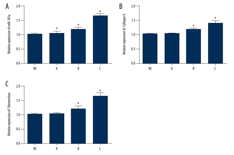
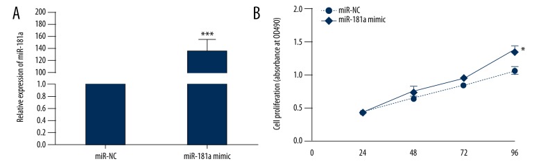
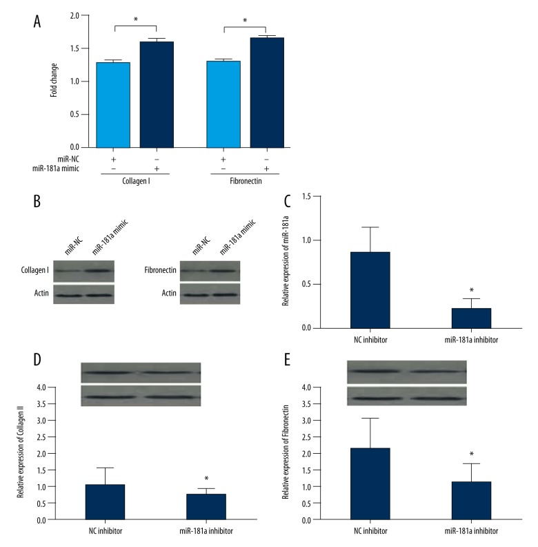
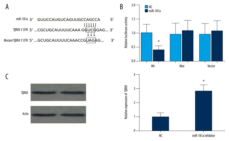
BACKGROUND The role of miR-181a in the development of cardiac disease and in particular, myocardial fibrosis following myocardial infarction (MI) remains unknown. The aim of this study was to explore the role of miR-181a in myocardial fibrosis in a rat model of MI and the expression of TGF-β receptor III (TβRIII). MATERIAL AND METHODS Forty adult male Wistar rats were randomly divided into an MI model group (n=30) and a control group with (n=10). The rat MI model involved ligating the left anterior descending (LAD) coronary artery in the model group; the control group was treated with a sham operation. Cardiac function was assessed using cardiac ultrasound. Myocardial fibroblasts were extracted from the rat hearts and transfected with a miR-mimic or miR-inhibitor, and cell growth was measured using an MTT assay. The level of miR-181a expression was detected using quantitative reverse transcription polymerase chain reaction (qRT-PCR) and Western blots. RESULTS miR-181a expression was significantly increased during the progression of MI (P<0.05). Over-expression of miR-181a was associated with increased deposition of extracellular matrix (ECM) components, collagen I and fibronectin. This effect was reversed with the use of a miR-181a inhibitor (P<0.05). Upregulation of miR-181a suppressed the expression of TGF-β receptor III (TβRIII) by binding with 3'-UTR. CONCLUSIONS In this rat model of MI, the findings were that miR-181a had a role in the progression of myocardial fibrosis. The findings require further studies to determine whether miR-181a might provide a novel therapeutic target to limit myocardial fibrosis following MI.
Conflict of interest statement
All authors declare no conflicts of interest.
Figures
References
-
- Mozaffarian D, Benjamin EJ, Go AS, et al. Heart disease and stroke statistics – 2015 update: a report from the American Heart Association. Circulation. 2015;131(4):e29–322. - PubMed
-
- Crackower MA, Oudit GY, Kozieradzki I, et al. Regulation of myocardial contractility and cell size by distinct PI3K-PTEN signaling pathways. Cell. 2002;110(6):737–49. - PubMed
-
- Filipowicz W, Jaskiewicz L, Kolb FA, Pillai RS. Post-transcriptional gene silencing by siRNAs and miRNAs. Curr Opin Struct Biol. 2005;15(3):331–41. - PubMed
-
- Jing Q, Huang S, Guth S, et al. Involvement of microRNA in AU-rich element-mediated mRNA instability. Cell. 2005;120(5):623–34. - PubMed
MeSH terms
Substances
LinkOut - more resources
Full Text Sources
Other Literature Sources
Medical
