

Platelet-derived growth factor-coated decellularized meniscus scaffold for integrative healing of meniscus tears
- PMID: 29908335
- PMCID: PMC6090559
- DOI: 10.1016/j.actbio.2018.06.021
Platelet-derived growth factor-coated decellularized meniscus scaffold for integrative healing of meniscus tears
Abstract
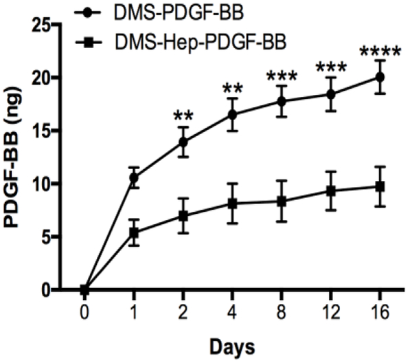
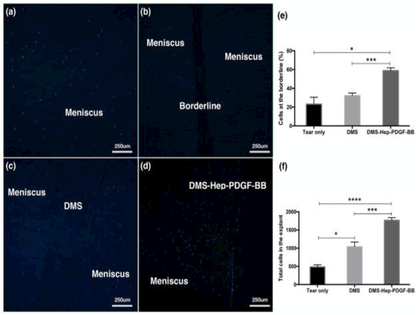
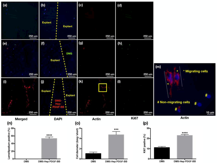
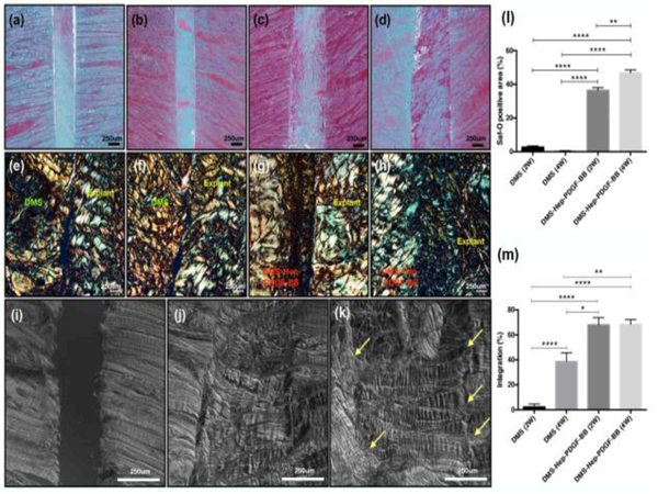
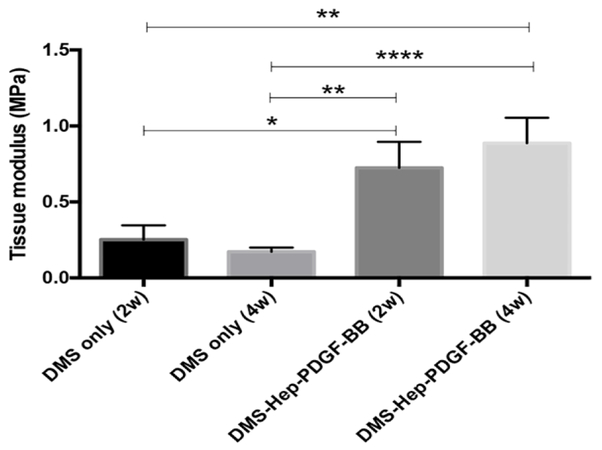
The aim of this study was to examine the potential of platelet-derived growth factor (PDGF)-coated decellularized meniscus scaffold in mediating integrative healing of meniscus tears by inducing endogenous cell migration. Fresh bovine meniscus was chemically decellularized and covalently conjugated with heparin and PDGF-BB. In vitro PDGF release kinetics was measured. The scaffold was transplanted into experimental tears in avascular bovine meniscus explants and cultured for 2 and 4 weeks. The number migrating and proliferating cells at the borderline between the scaffold and injured explant and PDGF receptor-β (PDGFRβ) expressing cells were counted. The alignment of the newly produced ECM and collagen was analyzed by Safranin-O, picrosirius red staining, and differential interference contrast (DIC). Tensile testing of the explants was performed after culture for 2 and 4 weeks. Heparin conjugated scaffold showed immobilization of high levels of PDGF-BB, with sustained release over 2 weeks. Insertion of the PDGF-BB treated scaffold in defects in avascular meniscus led to increased PDGFRβ expression, cell migration and proliferation into the defect zone. Safranin-O, picrosirius red staining and DIC showed tissue integration between the scaffold and injured explants. Tensile properties of injured explants treated with PDGF-BB coated scaffold were significantly higher than in the scaffold without PDGF. In conclusion, PDGF-BB-coated scaffold increased PDGFRβ expression and promoted migration of endogenous meniscus cells to the defect area. New matrix was formed that bridged the space between the native meniscus and the scaffold and this was associated with improved biomechanical properties. The PDGF-BB-coated scaffold will be promising for clinical translation to healing of meniscus tears.
Statement of significance: Meniscus tears are the most common injury of the knee joint. The most prevalent forms that occur in the inner third typically do not spontaneously heal and represent a major risk factor for the development of knee osteoarthritis. The goal of this project was to develop an approach that is readily applicable for clinical use. We selected a natural and readily available decellularized meniscus scaffold and conjugated it with PDGF, which we had previously found to have strong chemotactic activity for chondrocytes and progenitor cells. The present results show that insertion of the PDGF-conjugated scaffold in defects in avascular meniscus led to endogenous cell migration and proliferation into the defect zone with tissue integration between the scaffold and injured explants and improved tensile properties. This PDGF-conjugated scaffold will be promising for a translational approach to healing of meniscus tears.
Keywords: Cell migration; Decellularization; Heparin; Meniscus; PDGF.
Copyright © 2018 Acta Materialia Inc. Published by Elsevier Ltd. All rights reserved.
Conflict of interest statement
Disclosures
Authors have no conflicts of interest to disclose.
Figures

References
-
- Sanders TG, Medynski MA, Feller JF, Lawhorn KW, Bone contusion patterns of the knee at MR imaging: footprint of the mechanism of injury, Radiographics 20 Spec No (2000) S135–51. - PubMed
-
- Hagino T, Ochiai S, Watanabe Y, Senga S, Wako M, Ando T, Sato E, Haro H, Complications after arthroscopic knee surgery, Arch Orthop Trauma Surg 134(11) (2014) 1561–4. - PubMed
-
- Tuman J, Haro MS, Foley S, Diduch D, All-inside meniscal repair devices and techniques, Expert Rev Med Devices 9(2) (2012) 147–57. - PubMed
-
- Arnoczky SP, Warren RF, The microvasculature of the meniscus and its response to injury. An experimental study in the dog, Am J Sports Med 11(3) (1983) 131–41. - PubMed
Publication types
MeSH terms
Substances
Grants and funding
LinkOut - more resources
Full Text Sources
Other Literature Sources
Medical
Research Materials
