. 2018 Sep:18:25-32.
doi: 10.1016/j.redox.2018.06.003. Epub 2018 Jun 9.

Adropin regulates pyruvate dehydrogenase in cardiac cells via a novel GPCR-MAPK-PDK4 signaling pathway

Affiliations

Adropin regulates pyruvate dehydrogenase in cardiac cells via a novel GPCR-MAPK-PDK4 signaling pathway

Dharendra Thapa et al. Redox Biol. 2018 Sep.

Abstract

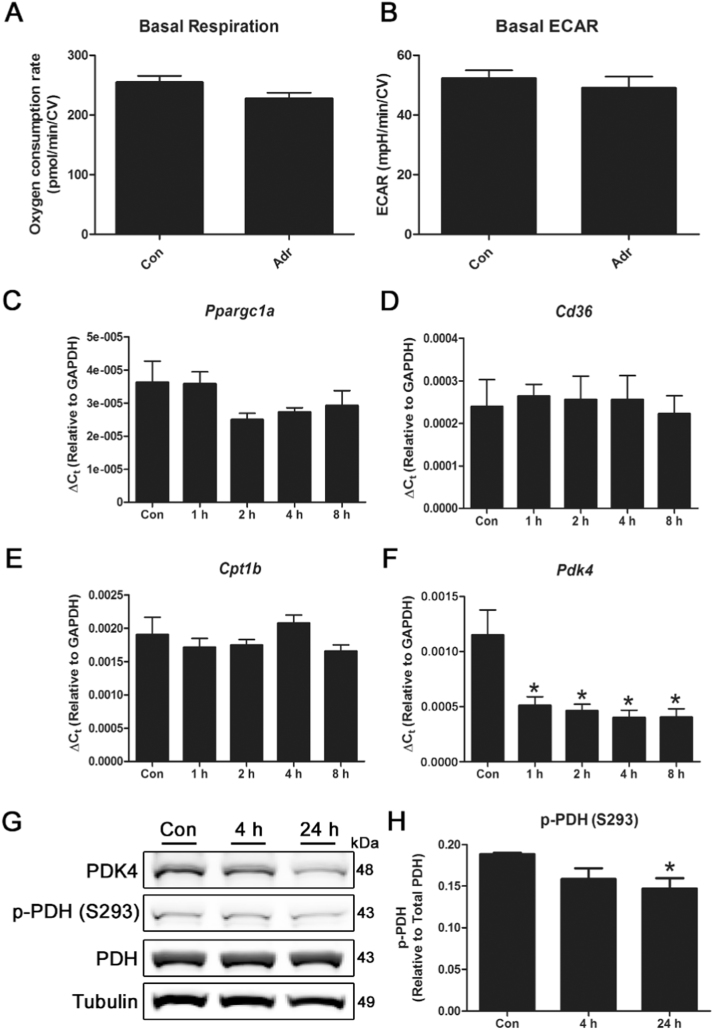
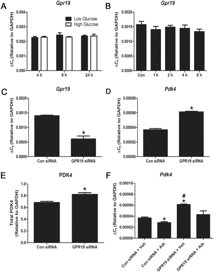
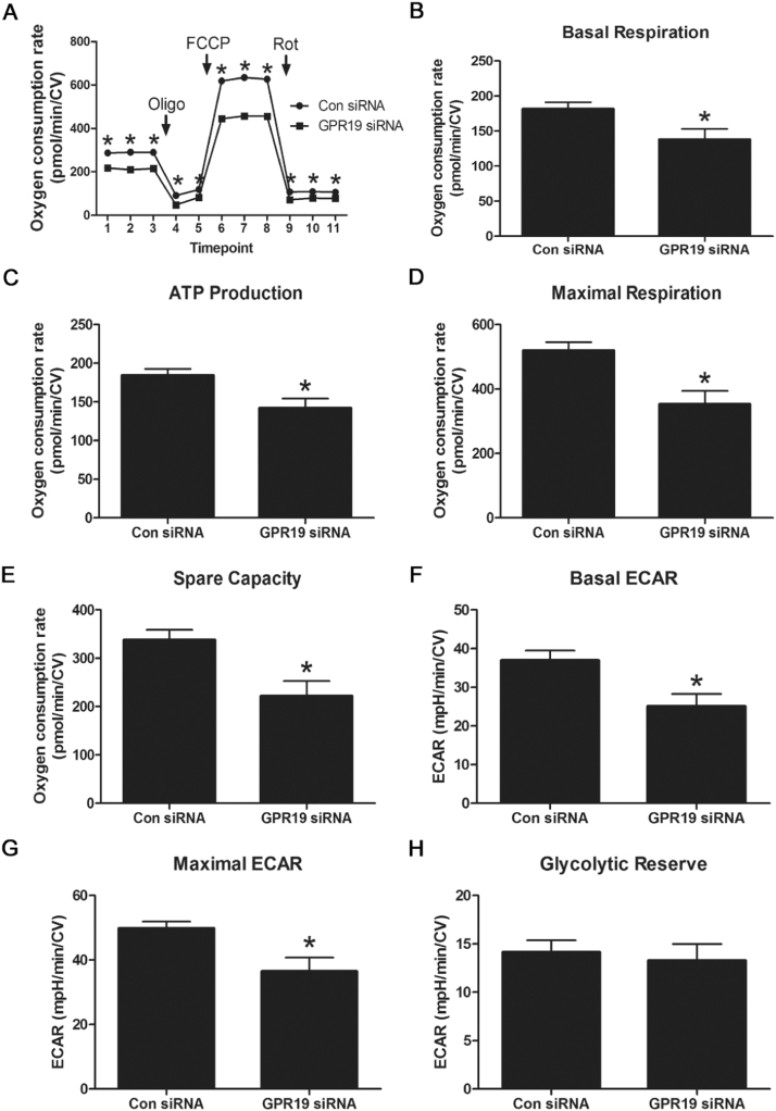
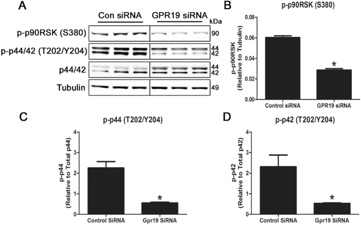
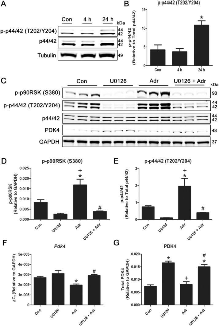
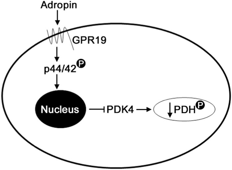
Mitochondria supply ~90% of the ATP required for contractile function in cardiac cells. While adult cardiomyocytes preferentially utilize fatty acids as a fuel source for oxidative phosphorylation, cardiac mitochondria can switch to other substrates when required. This change is driven in part by a combination of extracellular and intracellular signal transduction pathways that alter mitochondrial gene expression and enzymatic activity. The mechanisms by which extracellular metabolic information is conveyed to cardiac mitochondria are not currently well defined. Recent work has shown that adropin - a liver-secreted peptide hormone - can induce changes in mitochondrial fuel substrate utilization in skeletal muscle, leading to increased glucose use. In this study, we examined whether adropin could regulate mitochondrial glucose utilization pathways in cardiac cells. We show that stimulation of cultured cardiac cells with adropin leads to decreased expression of the pyruvate dehydrogenase (PDH) negative regulator PDK4, which reduces inhibitory PDH phosphorylation. The downregulation of PDK4 expression by adropin is lost when GPR19 - a putative adropin receptor - is genetically depleted in H9c2 cells. Loss of GRP19 expression alone increased PDK4 expression, leading to a reduction in mitochondrial respiration. Finally, we show that adropin-mediated GPR19 signaling relies on the p44/42 MAPK pathway, and that pharmacological disruption of this pathway blocks the effects of adropin on PDK4 in cardiac cells. These findings suggest that adropin may be a key regulator of fuel substrate utilization in the heart, and implicates an orphan G-protein coupled receptor in a novel signaling pathway controlling mitochondrial fuel metabolism.

Keywords: Adropin; GPCR; GRP19; Metabolism; Mitochondria; PDK4; Pyruvate dehydrogenase.


Figures

fx1
Graphical abstract
Fig. 1
Fig. 1
Treatment of cardiac cells with adropin reduces PDK4 expression and PDH phosphorylation. Treatment of H9c2 cardiac cells with 0.5 μg/mL adropin for 4 h does not have a significant effect on basal respiration or glycolysis in Seahorse XF assays (A-B). N = 12. Exposure of cells to adropin for 0–8 h leads to a reduction in Pdk4 expression, while levels of Ppargc1a, Cd36 and Cpt1b remain unchanged (C-F). The reduction in PDK4 protein abundance results in a decrease in PDH phosphorylation at serine 293 after 24 h (G-H). N = 3–4. * = P < 0.05.
Fig. 2
Fig. 2
Genetic depletion of the putative adropin receptor GPR19 promotes PDK4 expression and blocks adropin-mediated PDK4 downregulation. Expression of the putative adropin receptor, Gpr19, is not regulated by exposure to changing nutrient levels (low glucose = 5.5 mM, high glucose = 25 mM) or 0.5 μg/mL adropin over 0–24 h (A-B) in H9c2 cells. Knockdown of Gpr19 using siRNA led to a concomitant increase in Pdk4 gene and protein expression (C-E). Knockdown of Gpr19 prevented the reduction of Pdk4 expression seen in control cells treated with adropin for 4 h (F). N = 3–4, * = P < 0.05 all vs. Con siRNA+Veh; # = P < 0.05 Con siRNA+Adr vs. GPR19 siRNA+Veh.
Fig. 3
Fig. 3
Genetic depletion of GPR19 blunts mitochondrial respiration. Knockdown of Gpr19 using siRNA in H9c2 cells led to a significant decrease in both basal and maximal mitochondrial oxygen consumption during Seahorse XF respirometry analyses (A-E). Loss of Gpr19 expression limited basal and maximal glycolysis, but had no effect on glycolytic capacity (F-H). N = 16–18, * = P < 0.05.
Fig. 4
Fig. 4
Genetic depletion of GPR19 blocks the p44/42 MAPK signaling cascade in cardiac cells. Knockdown of Gpr19 using siRNA in H9c2 cells led to a significant reduction in p44/42 phosphorylation at threonine 202/tyrosine 204, leading to a decrease in downstream p90RSK phosphorylation at serine 380 (A-D). N = 3–4, * = P < 0.05.
Fig. 5
Fig. 5
Pharmacological inhibition of the p44/42 MAPK signaling cascade abrogates the response of cardiac cells to adropin. Treatment of H9c2 cells with 0.5 μg/mL adropin for 24 h leads to significantly increased p44/42 phosphorylation at threonine 202/tyrosine 204 (A-B). Treatment with the p44/42 inhibitor U0126 blocked phosphorylation of p44/42 at threonine 202/tyrosine 204 and p90RSK phosphorylation at serine 380 (D-E). The interruption of p44/42 signaling by U0126 prevented the inhibitory action of adropin on PDK4 gene expression and protein abundance (F-G). N = 3–4. * = P < 0.05 all vs. Con; # = P < 0.05 U0126 +Adr vs. Adr; + = P < 0.05 Adr vs. U0126.
Fig. 6
Fig. 6
Proposed model of the adropin-GPR19-p44/42-PDK4 signaling cascade in cardiac cells. Exposure of cardiac cells to circulating adropin leads to the activation of the membrane-bound GPCR protein GPR19. This activation stimulates p44/42 phosphorylation, which results in decreased Pdk4 gene expression and inhibitory phosphorylation of PDH at serine 293.

References

    1. Cornall L.M., Mathai M.L., Hryciw D.H., Simcocks A.C., O'Brien P.E., Wentworth J.M., McAinch A.J. GPR119 regulates genetic markers of fatty acid oxidation in cultured skeletal muscle myotubes. Mol. Cell. Endocrinol. 2013;365:108–118. - PubMed
    1. Fillmore N., Mori J., Lopaschuk G.D. Mitochondrial fatty acid oxidation alterations in heart failure, ischaemic heart disease and diabetic cardiomyopathy. Br. J. Pharmacol. 2014;171:2080–2090. - PMC - PubMed
    1. Fujie S., Hasegawa N., Sato K., Fujita S., Sanada K., Hamaoka T., Iemitsu M. Aerobic exercise training-induced changes in serum adropin level are associated with reduced arterial stiffness in middle-aged and older adults. Am. J. Physiol. - Heart Circ. Physiol. 2015;309:H1642–H1647. - PubMed
    1. Gao S., McMillan R.P., Zhu Q., Lopaschuk G.D., Hulver M.W., Butler A.A. Therapeutic effects of adropin on glucose tolerance and substrate utilization in diet-induced obese mice with insulin resistance. Mol. Metab. 2015;4:310–324. - PMC - PubMed
    1. Gao S. Regulation of substrate oxidation preferences in muscle by the peptide hormone adropin. Diabetes. 2014;63:3242–3252. - PMC - PubMed

Publication types

MeSH terms

LinkOut - more resources