Comparative Study
. 2018 Oct;155(4):1177-1191.e16.
doi: 10.1053/j.gastro.2018.06.034. Epub 2018 Jun 15.

Diet Modifies Colonic Microbiota and CD4+ T-Cell Repertoire to Induce Flares of Colitis in Mice With Myeloid-Cell Expression of Interleukin 23

Affiliations
Comparative Study

Diet Modifies Colonic Microbiota and CD4+ T-Cell Repertoire to Induce Flares of Colitis in Mice With Myeloid-Cell Expression of Interleukin 23

Lili Chen et al. Gastroenterology. 2018 Oct.

Abstract

Background & aims: Several studies have shown that signaling via the interleukin 23 (IL23) receptor is required for development of colitis. We studied the roles of IL23, dietary factors, alterations to the microbiota, and T cells in the development and progression of colitis in mice.

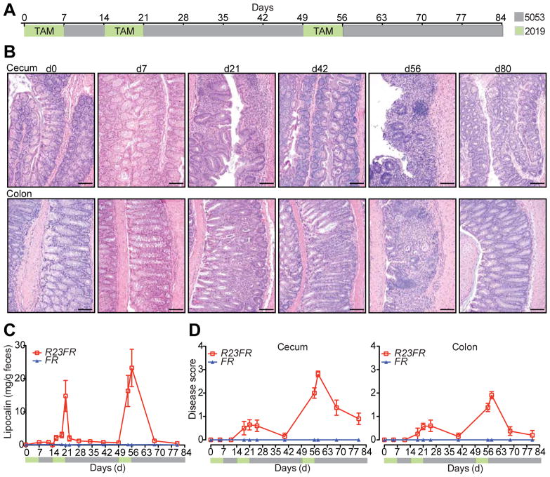
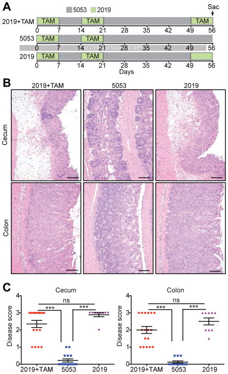
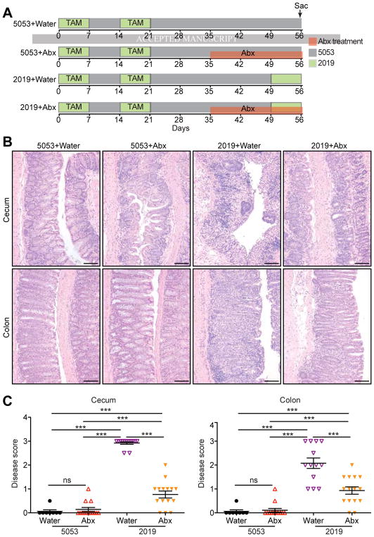
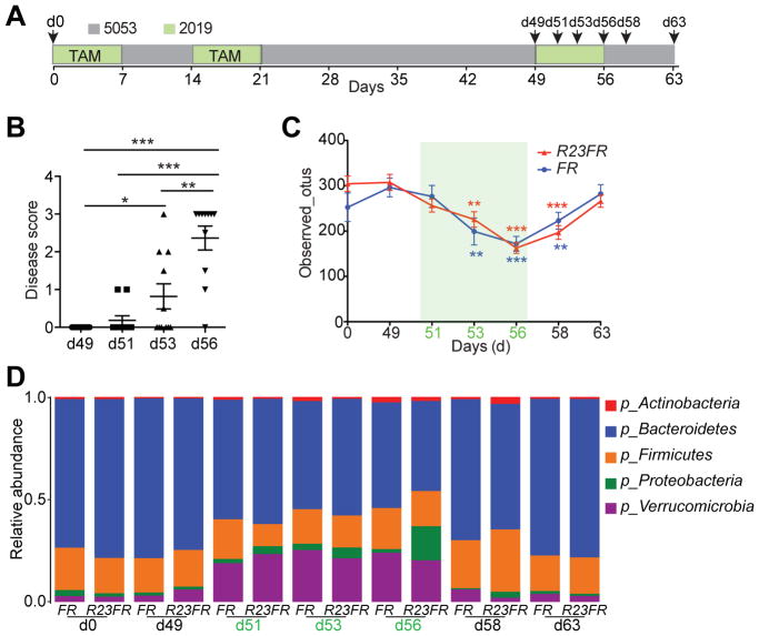
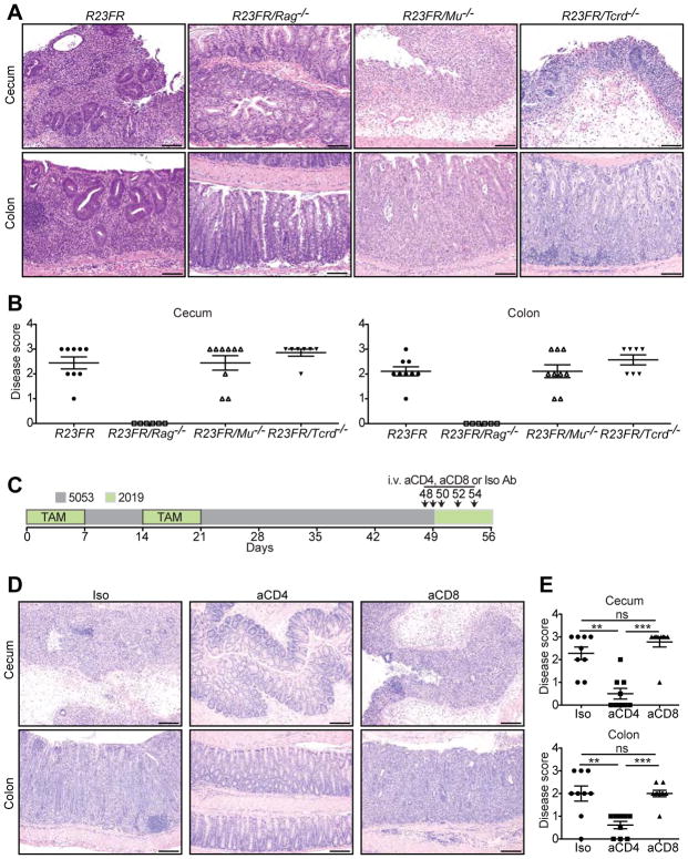
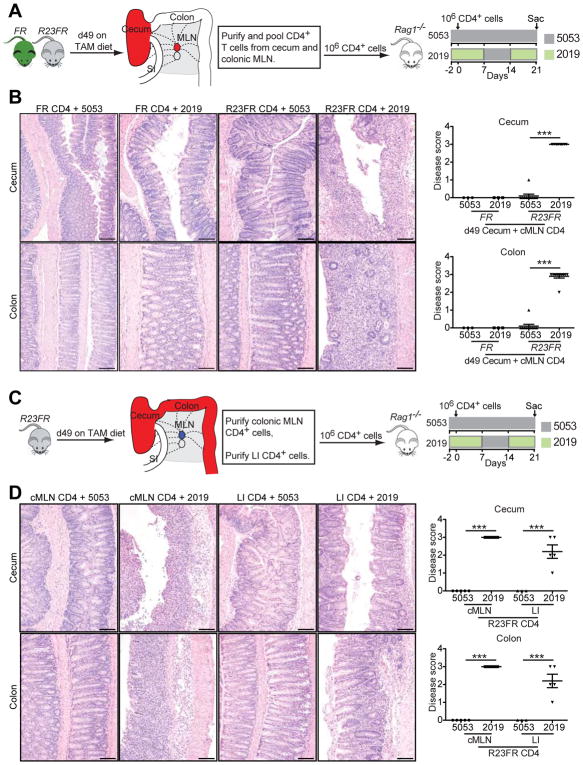
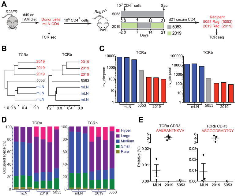
Methods: All mice were maintained on laboratory diet 5053, unless otherwise noted. We generated mice that express IL23 in CX3CR1-positive myeloid cells (R23FR mice) upon cyclic administration of tamoxifen dissolved in diet 2019. Diets 2019 and 5053 have minor differences in the overall composition of protein, fat, fiber, minerals, and vitamins. CX3CR1CreER mice (FR mice) were used as controls. Some mice were given antibiotics, and others were raised in a germ-free environment. Intestinal tissues were collected and analyzed by histology and flow cytometry. Feces were collected and analyzed by 16S rDNA sequencing. Feces from C57/Bl6, R23FR, or FR mice were fed to FR and R23FR germ-free mice in microbiota transplant experiments. We also performed studies with R23FR/Rag-/-, R23FR/Mu-/-, and R23FR/Tcrd-/- mice. R23FR mice were given injections of antibodies against CD4 or CD8 to deplete T cells. Mesenteric lymph nodes and large intestine CD4+ cells from R23FR or FR mice in remission from colitis were transferred into Rag-/- mice. CD4+ cells were isolated from donor R23FR mice and recipient Rag-/- mice, and T-cell receptor sequences were determined.

Results: Expression of IL23 led to development of a relapsing-remitting colitis that was dependent on the microbiota and CD4+ T cells. The relapses were caused by switching from the conventional diet used in our facility (diet 5053) to the diet 2019 and were not dependent on tamoxifen after the first cycle. The switch in the diet modified the microbiota but did not alter levels of IL23 in intestinal tissues compared with mice that remained on the conventional diet. Mesenteric lymph nodes and large intestine CD4+ cells from R23FR mice in remission, but not from FR mice, induced colitis after transfer into Rag-/- mice, but only when these mice were placed on the diet 2019. The CD4+ T-cell receptor repertoire of Rag-/- mice with colitis (fed the 2019 diet) was less diverse than that from donor mice and Rag-/- mice without colitis (fed the 5053 diet) because of expansion of dominant T-cell clones.

Conclusions: We developed mice that express IL23 in CX3CR1-positive myeloid cells (R23FR mice) and found that they are more susceptible to diet-induced colitis than mice that do not express IL23. The R23FR mice have a population of CD4+ T cells that becomes activated in response to dietary changes and alterations to the intestinal microbiota. The results indicate that alterations in the diet, intestinal microbiota, and IL23 signaling can contribute to pathogenesis of inflammatory bowel disease.

Keywords: Cytokine; Immune Response; Inflammatory Bowel Disease Model; Microbiome.


Conflict of interest statement

Conflicts of interest

Both R.S. and A.J. are co-founders of Girihlet Inc., which has licensed the TCR sequencing technology from Mount Sinai, with the goal of developing it as a commercial product. JF.C. has served as consultant, advisory board member or speaker for AbbVie, Amgen, Boehringer-Ingelheim, Celgene Corporation, Celltrion, Enterome, Ferring, Genentech, Janssen and Janssen, Lilly, Medimmune, Merck & Co., Pfizer, PPM Services, Protagonist, Second Genome, Seres, Shire, Takeda, Theradiag, Theravance Biopharma. Stock options: Intestinal Biotech Development, Genfit. JF.C. has research Grants from AbbVie, Takeda, Janssen. The remaining authors disclose no conflicts.

Figures

Figure 1
Figure 1
IL23 Expression by CX3CR1+ Cells Induces Colonic and Cecal Inflammation. (A) Tamoxifen (TAM) in diet 2019 (green) was fed to R23FR and FR mice during the indicated times. After each cycle of TAM treatment, animals were switched to our mouse facility diet 5053 (gray). (B) Representative H&E-stained cecum and colon sections of R23FR mice at different time points. Scale bars, 100 μm. (C) Fecal lipocalin-2 levels in the stools of R23FR and FR mice were measured by ELISA (n= 5–15 per group per time point). (D) Histological scores of the colon and cecum of R23FR and FR mice at different time points (n= 5–15 per group per time point). Error bars represent mean ± SEM.
Figure 2
Figure 2
Change in Diet Causes Colitis Relapse in R23FR Mice. (A) Schematic representation of the diet switch experiments. Animals were treated with 2 cycles of TAM and subsequently treated with diet 2019 with TAM, diet 5053 without TAM, or diet 2019 without TAM. (B) Representative H&E stained cecum and colon sections of R23FR mice at d56 from each group. Scale bars, 100 μm. (C) Histological scores of the colon and cecum of R23FR mice at d56. *** p<0.001, ns, not significant; by nonparametric Mann-Whitney test.
Figure 3
Figure 3
Microbiota is Required for Colitis Relapse. (A) Schematic representation of the experimental design. R23FR mice after treatment with 2 cycles of TAM were treated with an antibiotic cocktail (ampicillin metronidazole, neomycin, vancomycin) or water from d35 to d56 with different diets. (B) Representative H&E stained sections of cecum and colon of R23FR mice at d56 from each group. Scale bars, 100 μm. (C) Histological scores of the colon and cecum of R23FR mice at d56. *** p<0.001, ns, not significant; by nonparametric Mann-Whitney test.
Figure 4
Figure 4
Diet Switch Changes the Composition of the Intestinal Microbiota. (A) Schematic representation of the experimental design. The fecal samples were collected for microbiota analysis at different time points (d0, d49, d51, d53 and d56, d58 and d63) from R23FR and FR mice. (B) Histological scores of the colon and cecum of R23FR mice at d49, d51, d53 and d56. *p<0.05, **p<0.01, *** p<0.001; by nonparametric Mann-Whitney test. (C) Alpha diversity (estimated as number of observed OTUs) at a sequencing depth of 12,000 seqs/sample. Asterisks (colored according to the different genotyping group) indicate a statistically significant difference in the number of observed OTUs compared to OTUs at day 49 within each group. N=8–15 per time point for each group. **p<0.01, *** p<0.001; by nonparametric Mann-Whitney test. (D) Bacterial phylum-level community composition in the stool of R23FR and FR mice at each time point.
Figure 5
Figure 5
T Cells Are Required for Intestinal Inflammation in R23FR Mice. (A) Representative H&E stained sections from R23FR, R23FR/Rag−/−, R23FR/Mu−/− and R23FR/Tcrd−/− mice at d56. Scale bars, 100 μm. (B) Histological scores of the colon and cecum of R23FR, R23FR/Rag−/−, R23FR/Mu−/− and R23FR/Tcrd−/− mice at d56. Notice that disease was completely absent in R23FR/Rag−/− mice but not in R23FR/Mu−/− and R23FR/Tcrd−/− mice. (C) Schematic representation depicts experimental design for cell depletion experiments. (D) Representative H&E-stained colon and cecum sections (left) and histological scores (right) of R23FR mice after treatment with anti-CD4 and anti-CD8 antibodies. Depletion of CD4+, but not CD8+ T cells significantly reduced disease severity. Scale bars, 100 μm. **p<0.01, *** p<0.001, ns, not significant; by nonparametric Mann-Whitney test.
Figure 6
Figure 6
IL23-induced CD4+ T Cells Drive Inflammation in Adoptively Transferred Mice. (A) Experimental setup for adoptive transfer of pooled CD4+ T cells from cecum and mLN of R23FR or FR mice into Rag−/− mice. (B) Representative H&E staining (left) and histological scores (right) of the colon and cecum of Rag−/− mice that received R23FR and FR mice CD4+ T cells fed with different diets. Rag−/− mice that received R23FR CD4+ T cells, but not FR CD4+ T cells, and were fed with 2 cycles of 2019 diet developed severe colitis. Scale bars, 100 μm. *** p<0.001; by nonparametric Mann-Whitney test. (C) Experimental setup for adoptive transfer of CD4+ T cells from large intestine (LI) or colonic mLN (cMLN) of R23FR mice into Rag−/− mice fed with different diets. (D) Representative H&E stained sections (left) and histological scores (right) of the colon and cecum of Rag−/− mice that received mLN or intestinal CD4+ T cells fed with different diets. Scale bars, 100 μm. Both mLN and colonic CD4+ T cells from R23FR mice caused severe colitis in reconstituted Rag−/− mice after a diet switch. *p<0.05, *** p<0.001; by nonparametric Mann-Whitney test.
Figure 7
Figure 7
T Cell Receptor Clonality Analysis. (A) Experimental setup for TCR sequencing analysis. Note that donor cells from mLN of R23FR mice were obtained at remission stage (d49). (B) Unsupervised hierarchical clustering of TCR repertoire from donor mLN cells, cecum CD4+ T cells recovered from recipient Rag−/− mice treated with diets 5013 and 2019. N=3 individual mice in both donor group and 2019 group; n=1 of pooled 10 mice for group 5053. (C) Inverse Simpson’s diversity index was used to assess the diversity of TCR repertoires for each sample. (D) The percent of clonal space occupied by clones of a given type (classified by size, Supplementary Table 1). (E) Frequencies of selected TCRα and TCRβ clonotypes in different samples.

Similar articles

Cited by

References

    1. Tontini GE, Vecchi M, Pastorelli L, et al. Differential diagnosis in inflammatory bowel disease colitis: state of the art and future perspectives. World J Gastroenterol. 2015;21:21–46. - PMC - PubMed
    1. Ng SC, Shi HY, Hamidi N, et al. Worldwide incidence and prevalence of inflammatory bowel disease in the 21st century: a systematic review of population-based studies. Lancet. 2017;390:2769–2778. - PubMed
    1. Gkouskou KK, Deligianni C, Tsatsanis C, et al. The gut microbiota in mouse models of inflammatory bowel disease. Frontiers in Cellular and Infection Microbiology. 2014:4. - PMC - PubMed
    1. Kamada N, Seo SU, Chen GY, et al. Role of the gut microbiota in immunity and inflammatory disease. Nature Reviews Immunology. 2013;13:321–335. - PubMed
    1. Nishikawa J, Kudo T, Sakata S, et al. Diversity of mucosa-associated microbiota in active and inactive ulcerative colitis. Scandinavian Journal of Gastroenterology. 2009;44:180–186. - PubMed

Publication types

MeSH terms