Review
. 2018 Nov:191:1-22.
doi: 10.1016/j.pharmthera.2018.06.004. Epub 2018 Jun 22.

Autophagy as an emerging target in cardiorenal metabolic disease: From pathophysiology to management

Affiliations
Review

Autophagy as an emerging target in cardiorenal metabolic disease: From pathophysiology to management

Yingmei Zhang et al. Pharmacol Ther. 2018 Nov.

Abstract

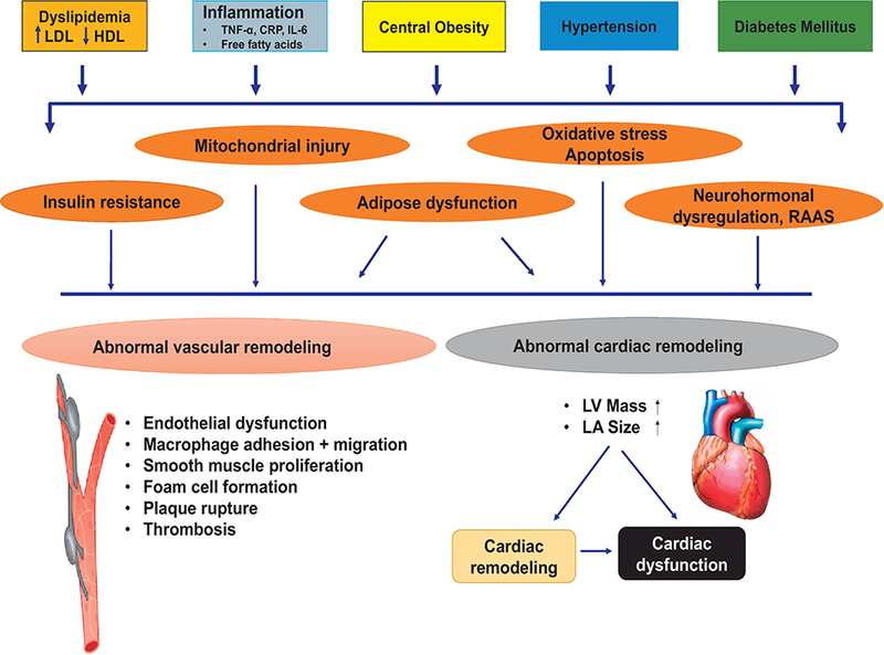
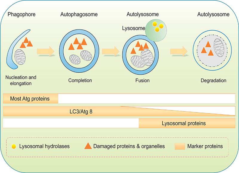
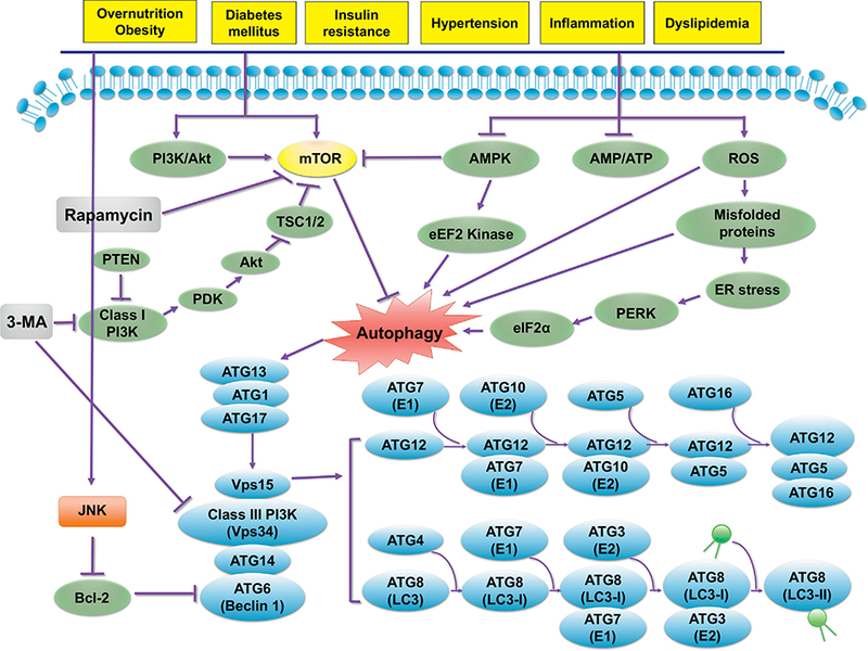
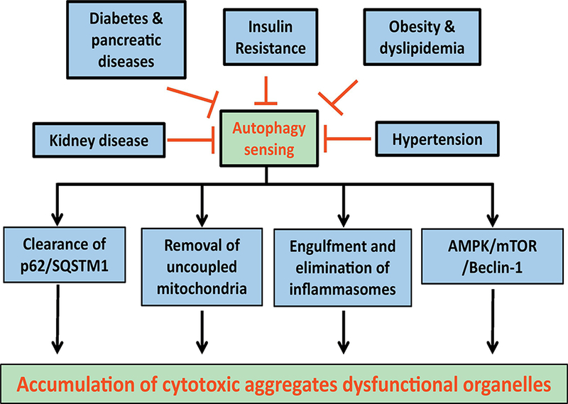
Although advances in medical technology and health care have improved the early diagnosis and management for cardiorenal metabolic disorders, the prevalence of obesity, insulin resistance, diabetes, hypertension, dyslipidemia, and kidney disease remains high. Findings from numerous population-based studies, clinical trials, and experimental evidence have consolidated a number of theories for the pathogenesis of cardiorenal metabolic anomalies including resistance to the metabolic action of insulin, abnormal glucose and lipid metabolism, oxidative and nitrosative stress, endoplasmic reticulum (ER) stress, apoptosis, mitochondrial damage, and inflammation. Accumulating evidence has recently suggested a pivotal role for proteotoxicity, the unfavorable effects of poor protein quality control, in the pathophysiology of metabolic dysregulation and related cardiovascular complications. The ubiquitin-proteasome system (UPS) and autophagy-lysosomal pathways, two major although distinct cellular clearance machineries, govern protein quality control by degradation and clearance of long-lived or damaged proteins and organelles. Ample evidence has depicted an important role for protein quality control, particularly autophagy, in the maintenance of metabolic homeostasis. To this end, autophagy offers promising targets for novel strategies to prevent and treat cardiorenal metabolic diseases. Targeting autophagy using pharmacological or natural agents exhibits exciting new strategies for the growing problem of cardiorenal metabolic disorders.

Keywords: Adipose tissue; Autophagy; Cardiorenal metabolic syndrome; Cardiovascular; Liver.


Figures

Figure 1:
Figure 1:
Schematic diagram displaying various contributing factors for cardiorenal metabolic syndrome in the complex pathophysiology of cardiovascular complications. RAAS: renin angiotensin aldosterone system; CRP: C reactive peptide; TNF-α: tumor necrosis factor α; IL-6: interleukin 6 [adapted from (Y. Zhang & Ren, 2016) with modifications].
Figure 2:
Figure 2:
Schematic diagram of sequential autophagy events in a step-wise manner. Phagophore forms with initial sequestration of aged or damaged proteins and organelles; phagophores then undergo a series of further membrane expansion and elongation events to yie ld a completed double-membrane sequestering vesicle named autophagosome. During formation of autophagosome, various substrates (cytosolic proteins, lipids, nucleic acids, glycogen, damaged organelles, and invasive microbes, also known as cargo) of autophagy are encapsulated within the autophagosomal vesicle. Autophagosomes fuse with lysosomes to form autophagolysosmes, where cargos are digested by lysosomal hydrolases. Protein markers are identified throughout each individual steps. Most ATG proteins are visible during early stages of autophagy initiation while the elevated levels of lipidated LC3/Atg8 can sustain for a much longer period of time.
Figure 3:
Figure 3:
Autophagy regulatory mechanisms in response to various cardiorenal metabolic stimuli (e.g., overnutrition/obesity, hyperlipidemia, insulin resistance, diabetes mellitus, inflammation and hypertension). The PI3K-Akt and AMPK signaling cascades represent the two major cell signaling pathways in response to cardiorenal metabolic stress. Typically, mTOR (with PI3K-Akt being the upstream activator) suppresses autophagy through inhibition of the ULK1 complex (ULK1-ATG13-FIP200) that is required for autophagy induction. AMPK activates the ULK1 complex or indirectly suppress mTOR to initiate autophagy. ROS directly or indirectly (through ER stress) turns on autophagy. The mTOR- independent autophagy regulating autophagy are less clear. One example is c-Jun N-terminal kinase (JNK)-regulated phosphphorylation (or inhibition) of Bcl-2, leading to relieve of Bcl-2-Beclin 1 coupling and its inhibition on autophagy induction. The ULK1 complex consisting of ATG1 (ULK1)-ATG13 and ATG17 recruits Vps34, Beclin-1, and Vpsl5 for autophagosome synthesis, possibly through mTOR-independent pathway(s). Two ubiquitin-like conjugation systems involving ATGs govern the elongation of phagophores. The ATG5-ATG12 conjugation involves ATG7 (El-like) and ATG10 (E2-like), whereas the light chain 3 (LC3, also commonly known as ATG8)-LC3-I- phosphatidylethanolamine (PE) conjugation involves ATG4 (a cysteine protease), ATG7 (El-like), and ATG3 (E2-like). The ATG5-ATG12 conjugate generates a complex with ATG16 to control LC3-I-PE conjugation (resulting in LC3-II). Arrowheads and “T” ended line lines represent activation and inhibition, respectively. AMPK, AMP-activated protein kinase; Akt: also known as Protein Kinase B (PKB); mTOR, mammalian target of rapamycin; Rheb, Ras homology enriched in brain; TSC, tuberous sclerosis complex. Part of this cartoon is modified from (Y. Zhang, et al., 2018).
Figure 4:
Figure 4:
Various components of the cardiorenal metabolic syndrome may compromise autophagy sensing mechanisms leading to compromised autophagy regulatory machineries such as lysosomal degradation, selective removal of mitochondria and other organelles, engulfment of inflammasome and a number of cell signalling molecules governing autophagy, leading to accumulation of cytotoxic aggregates and dysfunctional organelles.

Similar articles

Cited by

References

    1. Alberti KG, Eckel RH, Grundy SM, Zimmet PZ, Cleeman JI, Donato KA, Fruchart JC, James WP, Loria CM, & Smith SC Jr. (2009). Harmonizing the metabolic syndrome: a joint interim statement of the International Diabetes Federation Task Force on Epidemiology and Prevention; National Heart, Lung, and Blood Institute; American Heart Association; World Heart Federation; International Atherosclerosis Society; and International Association for the Study of Obesity. Circulation, 120, 1640–1645. - PubMed
    1. Alberti KG, Zimmet P, & Shaw J (2005). The metabolic syndrome--a new worldwide definition. Lancet, 366, 1059–1062. - PubMed
    1. Altman BJ, & Rathmell JC (2012). Metabolic stress in autophagy and cell death pathways. Cold Spring Harb Perspect Biol, 4, a008763. - PMC - PubMed
    1. Andres AM, Kooren JA, Parker SJ, Tucker KC, Ravindran N, Ito BR, Huang C, Venkatraman V, Van Eyk JE, Gottlieb RA, & Mentzer RM Jr. (2016). Discordant signaling and autophagy response to fasting in hearts of obese mice: Implications for ischemia tolerance. Am J Physiol Heart Circ Physiol, 311, H219–228. - PMC - PubMed
    1. Aon MA, Cortassa S, Juhaszova M, & Sollott SJ (2016). Mitochondrial health, the epigenome and healthspan. Clin Sci (Lond), 130, 1285–1305.. - PMC - PubMed

Publication types

MeSH terms