. 2018 Jun 27;6(6):722-733.e6.
doi: 10.1016/j.cels.2018.05.016. Epub 2018 Jun 13.

Systems Analyses Reveal Physiological Roles and Genetic Regulators of Liver Lipid Species

Affiliations

Systems Analyses Reveal Physiological Roles and Genetic Regulators of Liver Lipid Species

Pooja Jha et al. Cell Syst. .

Abstract

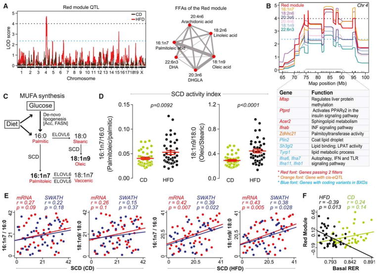
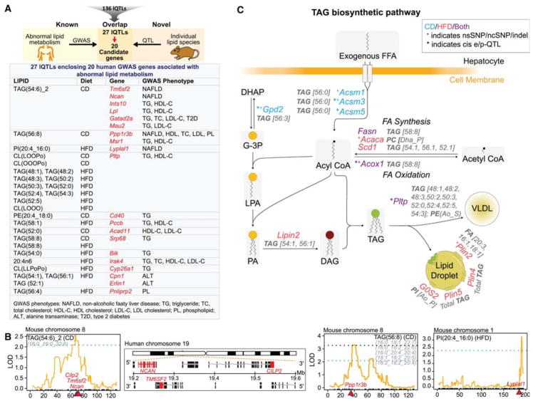
The genetics of individual lipid species and their relevance in disease is largely unresolved. We profiled a subset of storage, signaling, membrane, and mitochondrial liver lipids across 385 mice from 47 strains of the BXD mouse population fed chow or high-fat diet and integrated these data with complementary multi-omics datasets. We identified several lipid species and lipid clusters with specific phenotypic and molecular signatures and, in particular, cardiolipin species with signatures of healthy and fatty liver. Genetic analyses revealed quantitative trait loci for 68% of the lipids (lQTL). By multi-layered omics analyses, we show the reliability of lQTLs to uncover candidate genes that can regulate the levels of lipid species. Additionally, we identified lQTLs that mapped to genes associated with abnormal lipid metabolism in human GWASs. This work provides a foundation and resource for understanding the genetic regulation and physiological significance of lipid species.

Keywords: BXD; cardiolipin; fatty liver; genetic reference population, GRP; genetic variation; genome-wide association study, GWAS; lipid species; lipidomics; non-alcoholic fatty liver disease, NAFLD; quantitative trait locus, QTL.


Conflict of interest statement

Competing interest statement

The authors declare no competing interests.

Figures

Figure 1
Figure 1. Liver lipid species profile and their association with physiological and molecular traits
(A) Heatmap analysis with unsupervised hierarchical clustering of 96 lipid species for each BXD cohort shows mixed dietary and genetic impact. (B) Spearman correlation network (p<1e-04) of all lipid species measured in CD and HFD. Lipid species are color coded as 7 major lipid classes. The side chain FA composition of lipids has been abbreviated (O, oleic; P, palmitic; Po, palmitoleic; S, stearic; L, linoleic and Dha, docosahexaenoic acid. Refer to table S1 for abbreviation and composition). (C) Legend of the module composition, indicating the range of total number of carbons and degree of unsaturation. (D) Lipid module-clinical trait correlation. Each cell is color-coded by the Pearson’s correlation coefficient according to the legend color on the right. The stars in the cells represent the p-value of the correlation (p < 0.05 *, p < 0.01 **, p < 0.001 ***). (E) Module and its corresponding KEGG enriched pathway correlation. Red and blue cells represent the enriched pathways with the positively (scale bar: log10 p-value) and negatively (scale bar: -log10 p-value) correlated proteins respectively. Lipid classes hereafter are abbreviated as follows: triacylglycerol, TAG/TG; diacylglycerol, DAG; free-fatty acid, FFA; phospholipid, PL (phosphatidic acid, PA; phosphatidylcholine, PC; phosphatidylethanolamine, PE; phosphatidylglycerol, PG; phosphatidylinositol, PI; phosphatidylserine, PS); cardiolipin, CL; monolysocardiolipin; MLCL. See also Figures S1, S2 and Tables S1 and S2.
Figure 2
Figure 2. Identification of cardiolipin signatures of healthy and fatty liver
(A) Correlation diagram (corrgram) showing diet-independent association of lipid species with liver mass. Lipid species with Spearman’s correlation p-value <0.05 with liver mass (both normalized to body weight (%) and unnormalized (weight in g) in both CD and HFD were selected. (B) Spearman correlation network of diet specific significant correlation of lipid species with liver mass in CD (left) and HFD (right). (C) Corrgram of CLs that significantly correlate with liver mass in HFD. (D–F) C57BL6/J mice were fed with CD or high-fat high-sucrose (HFHS) diet for 18 weeks or nicotinamide riboside (NR) supplemented HFHS diet, 9 weeks after the start of the HFHS diet (HFHS+NR). Levels of healthy (D) and unhealthy (E) CL species in livers of the three cohorts. Note that the CLs—CL(LOOPo), CL(LLPoP) and CL(OOOP)—are shown in the figure with an additional CL species because the two are isobaric and were inseparable chromatographically. (F) Corrgram showing negative correlation of obesity and NAFLD traits with healthy CL species and positive correlation with unhealthy CL species.
Figure 3
Figure 3. Genetics of lipid species
(A) Circos plot of all lipids measured. Blue bars in the outermost ring represent the log2 fold change (HFD vs. CD) of the lipids. Lipids increased in CD or HFD are shown in green and blue font respectively. Orange bars represent the correlation of lipids between CD and HFD. Significant correlations (adj. p-value<0.05) are represented by “*”. Red bars represent lipid h2 in CD (light red) and HFD (dark red). The inner ring of yellow bars represents the strength of lQTLs in CD (light yellow) and HFD (dark yellow). Number of bars per lipid is equivalent to the number of lQTLs. The lines between the two innermost rings stem from the peak lQTL bar (with LOD > 3) and terminates on their approximate chromosomal position of the innermost ring. Lipid pairs marked with “_1” and “_2” (TAGs 54:5, 54:6, 56:7; PI(Dha_S) and CL(LLOPo)) indicate two isobaric peaks. (B) Schematic representation of the lQTLs. 136 lQTLs (55 CD and 81 HFD) were mapped from 37/46 lipids in CD/HFD. The number of QTLs (red font) per lipid species (blue font) is indicated. See also Figure S3 and Tables S3–S5.
Figure 4
Figure 4. Genetic assessment of lQTLs
(A) Manhattan plot of lipid species in CD and HFD. Names of the lipid species with genome-wide p-value < 0.05 are indicated. (B) All genes under the lQTLs (±5 Mb from the peak) were filtered through four independent pipelines as indicated. Genes fulfilling 2 or more of the filtering criteria were analyzed for enrichment of GO biological process (BP). (C) Enriched GO BP from 566 filtered lQTL genes. (D) lQTL position of the top 14 QTLs (p-value < 0.05). The candidate genes fulfilling 4 (red font), 3 (orange font) and 2 (grey font) of the 4 filtering criteria are indicated below each lQTL peak. Genes indicated with “*” are associated with metabolic phenotypes. (E) KEGG enrichment analysis of genes under the top 14 lQTLs passing 2 or more of the filtering criteria. For A and D, blue and black dotted lines represent suggestive and significant QTL threshold respectively. See also Table S5 and S6
Figure 5
Figure 5. FFA module identifies a genetic hotspot locus associated with FA metabolism and signaling
(A) Red module QTL showing significant peak on chromosome 4 (left) in HFD and the weighted correlation network of the FFAs (right) in the red module. (B) Hotspot region on chromosome 4 showing the overlapping QTL of the red module and the individual FFAs of the module. modQTL genes involved in FA metabolic processes and signaling are indicated along with their biological function. For A and B, blue and black dotted lines represent suggestive and significant QTL threshold respectively. (C) Schematic representation of monounsaturated FA (MUFA) synthesis. (D) Stearoyl-CoA Desaturase (SCD) activity index represented as ratios of MUFA to saturated FA (SFA). (E) Spearman correlation between hepatic expression of SCD transcript and protein (SWATH) with SCD activity index. (F) Pearson’s correlation between the basal RER and the red module.
Figure 6
Figure 6. lQTL genes associated with abnormal lipid metabolism in human GWAS and with TAG biosynthetic pathway
(A) lQTL genes fulfilling 2 or more filtering criteria as shown in Figure 4B were screened for any known association with abnormal lipid metabolism in human GWAS. The screening identified 20 GWAS genes from 27 lQTLs (Top). Each box of the table represents the lQTL(s) for the indicated gene(s) and the GWAS phenotype associated with the genes. (B) QTL position of the three lipid species (TAG(54:6)_2, TAG(56:8) and PI(20:4_16:0)) which map to the indicated genes (in red) implicated in hepatic steatosis in human GWAS studies. The loci of Cilp2, Tm6sf2 and Ncan is syntenic in mice (left) as well as in humans (middle). The genomic location of the genes is shown in red in the positive strand for NCAN and CILP2 and in the negative strand for TM6SF2. Blue and black dotted lines represent suggestive and significant QTL threshold respectively (C) Schematic representation of TAG biosynthetic pathways showing only those candidate genes that are under the lQTLs and fulfill 1 or more of our filtering criteria. lQTLs are represented in grey. Genes under CD lQTLs are in blue, those under HFD lQTLs in red, and those under both CD and HFD lQTLs in purple. See also Figures S3C, S4 and Table S7.

Similar articles

Cited by

References

    1. Andreux PA, Williams EG, Koutnikova H, Houtkooper RH, Champy MF, Henry H, Schoonjans K, Williams RW, Auwerx J. Systems genetics of metabolism: the use of the BXD murine reference panel for multiscalar integration of traits. Cell. 2012;150:1287–1299. - PMC - PubMed
    1. Anstee QM, Day CP. The genetics of NAFLD. Nature reviews Gastroenterology & hepatology. 2013;10:645–655. - PubMed
    1. Arends D, Prins P, Jansen RC, Broman KW. R/qtl: high-throughput multiple QTL mapping. Bioinformatics. 2010;26:2990–2992. - PMC - PubMed
    1. Belknap JK. Effect of within-strain sample size on QTL detection and mapping using recombinant inbred mouse strains. Behavior genetics. 1998;28:29–38. - PubMed
    1. Bigaud E, Corrales FJ. Methylthioadenosine (MTA) Regulates Liver Cells Proteome and Methylproteome: Implications in Liver Biology and Disease. Molecular & cellular proteomics: MCP. 2016;15:1498–1510. - PMC - PubMed

Publication types

LinkOut - more resources