. 2019 Mar;56(3):1607-1617.
doi: 10.1007/s12035-018-1184-8. Epub 2018 Jun 16.

Inhibition of Toll-Like Receptor-4 (TLR-4) Improves Neurobehavioral Outcomes After Acute Ischemic Stroke in Diabetic Rats: Possible Role of Vascular Endothelial TLR-4

Affiliations

Inhibition of Toll-Like Receptor-4 (TLR-4) Improves Neurobehavioral Outcomes After Acute Ischemic Stroke in Diabetic Rats: Possible Role of Vascular Endothelial TLR-4

Yasir Abdul et al. Mol Neurobiol. 2019 Mar.

Abstract

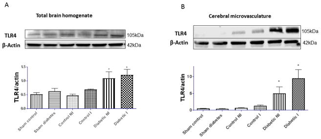
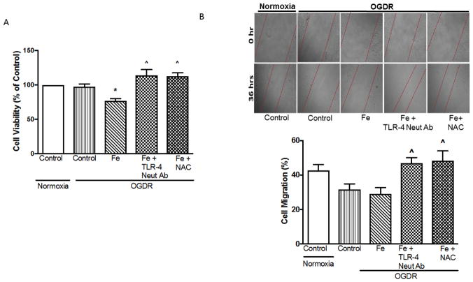
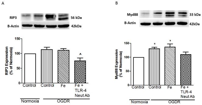
Diabetes increases the risk of occurrence and poor functional recovery after ischemic stroke injury. Previously, we have demonstrated greater hemorrhagic transformation (HT), edema, and more severe functional deficits after stroke in diabetic animals that also presented with cerebral vasoregression and endothelial cell death in the recovery period. Given that Toll-like receptor 4 (TLR-4) activation in microvascular endothelial cells triggers a robust inflammatory response, we hypothesized that inhibition of TLR-4 signaling prevents endothelial cell death and improves outcomes after stroke. Animals were treated with vehicle or TLR-4 inhibitor TAK242 (3 mg/kg; i.p.) following middle cerebral artery occlusion (MCAO). Neurobehavioral deficits were measured at baseline and day 3 after ischemic stroke. Primary brain microvascular endothelial cells (BMVECs) from diabetic animals were subjected to oxygen glucose deprivation re-oxygenation (OGDR) and treated with 0.1 mM iron(III)sulfate hydrate (iron) (to mimic the post-stroke bleeding) and TLR-4 inhibitors. Ischemic stroke increased the expression of TLR-4 in both hemispheres and in the microvasculature of diabetic animals. Cerebral infarct, edema, HT, and functional deficits were greater in diabetic compared to control animals. Inhibition of TLR-4 significantly reduced the neurovascular injury and improved functional outcomes. OGDR and iron reduced the cell viability and increased the expression of TLR-4 associated proteins (RIP3, MyD88, phospho-NF-kB, and release of IL-6) in BMVECs from diabetic animals. In conclusion, TLR-4 is highly upregulated in the microvasculature and that beneficial effects of TLR-4 inhibition are more profound in diabetes. This suggests that inhibition of vascular TLR-4 may provide therapeutic benefits for stroke recovery in diabetes.

Keywords: Brain vascular endothelial cells; Diabetes; Hemorrhagic transformation; Inflammation; Neurovascular injury; Stroke; TLR-4.


Figures

Figure 1
Figure 1. Diabetes increases expression of TLR-4 in total brain homogenate and cerebral microvasculature after ischemic stroke
Control and diabetic rats were subjected to 60 min of MCAO or sham surgery and sacrificed after 72 hours of reperfusion. (A) & (B) Representative Western blot images and measurement of TLR-4 protein expression in total brain homogenate and brain microvessels, respectively. Level of TLR-4 protein was significantly higher in both ischemic (I) and non-ischemic (NI) hemisphere of diabetic animals; *p< 0.05, compared with sham or control (n=4).
Figure 2
Figure 2. Inhibition of TLR-4 reduces the neurovascular injury in diabetic animals subjected to ischemic stroke
Control and diabetic rats were subjected to 60 min of MCAO and treated with vehicle or TLR-4 inhibitor TAK-242 (3mg/kg; i.p.) at reperfusion, 24 and 48 hours after reperfusion (n=5). After 72 hours of reperfusion animals were sacrificed to measure the neurovascular injury. (A) Diabetic animals had larger infarct size and treatment with TAK242 reduced infarct size only in the diabetic animals as indicated by the interaction. (B) Edema was greater in diabetic animals and treatment reduced edema in both control and diabetic animals. (C) Hemorrhagic transformation (HT) index was significantly higher compared to control animals. There was a treatment effect in both groups. (D) Diabetic animals showed excess Hb in ischemic hemisphere and inhibition of TLR-4 significantly reduced it only in diabetic animals.
Figure 3
Figure 3. Inhibition of TLR-4 improves the functional outcomes after ischemic stroke
(A) Composite score measured at day 1 after ischemic stroke injury was significantly lower in diabetic animals (*p<0.05 vs TAK242-treated control or diabetic animals; n=5 Control+TAK242, n=10 Control and Diabetic, n=9 Diabetic +TAK242). On Day 3, diabetic animals did not show any recovery (^p<0.05 vs all other groups). (B) Adhesive removal time was significantly reduced only in diabetic TAK242-treated animals (*p<0.05 vs diabetic).
Figure 4
Figure 4. OGDR increased the expression of TLR-4 in BMVECs of diabetic animals
(A) Representative immunofluorescent images of TLR-4 (green), nuclei stained with DAPI (blue) and merged images in BMVECs exposed to 6 hours OGD and 12 hours of reoxygenation. There was no positive staining when primary antibody was omitted (not shown). Scale bar is 50μm. (B) OGDR increased the membrane permeability and reduced the expression of tight junction proteins in BMVECs. (C) Representative images of immunoblots for occludin and claudin-5 after 6 hours OGD and 12 hours of reoxygenation in BMVECs isolated from diabetic GK rats. Treatment with TLR-4 inhibitor (TAK242; 30μM) reduced tight junction proteins under normoxic conditions but increased expression of both occludin and claudin5 under OGDR conditions indicated by the interaction (n=4–5).
Figure 5
Figure 5. Inhibition of TLR-4 improved cell viability and cell migration of BMVECs of diabetic rats upon OGDR and iron treatment
(A) Cell viability was measured after 6 hours OGD and 12 hours of reoxygenation and iron treatment in BMVECs isolated from diabetic GK rats. Cells were treated with iron(III)sulfate (Fe; 0.1mM) and TLR-4 neutralizing antibody (TLR4 Neu Ab;10μg/ml) or N-Acetyl Cysteine (NAC; 1mM). (B) Representative image and measurement of cell migration after 6 hours OGD and 30 hours of reoxygenation and iron treatment in BMVECs. Inhibition of TLR-4 or oxidative stress with NAC increased the viability and cell migration on OGDR and addition of iron. (*p<0.05 vs control, ^p<0.05 vs OGRD + Fe, n=4).
Figure 6
Figure 6. Inhibition of TLR-4 reduces expression of RIP3 and Myd88 proteins of upon OGDR and iron treatment in BMVECs of diabetic rats
Representative image and measurement of RIP3 (A) and Myd88 (B) expression after 6 hours of OGD followed by 12 hours of reoxygenation in BMVECs. Cells were treated with iron(III)sulfate (Fe; 0.1mM) and TLR-4 neutralizing antibody (TLR4 Neu Ab;10μg/ml). Addition of TLR-4 neutralizing antibody significantly reduced the expression of downstream proteins RIP3 and Myd88 after OGDR and addition of iron (^p<0.05 vs OGRD + Fe; *p< 0.05 vs control; n=4)
Figure 7
Figure 7. Inhibition of TLR-4 reduced the expression of NF-κB in cell lysate and secretion of IL-6 in media upon OGDR and iron treatment in BMVECs of diabetic rats
(A) Representative image and measurement of NF-κB and (B) measurement of IL-6 in collected media after 6 hours of OGD and 12 hours of reoxygenation and iron treatment in BMVECs isolated from diabetic GK rats. Cells were treated with iron(III)sulfate (Fe; 0.1mM) and TLR-4 neutralizing antibody (TLR4 Neu Ab;10μg/ml) or N-Acetyl Cysteine (NAC; 1mM). Inhibition of TLR-4 (TLR-4 neut Ab) and oxidative stress (NAC treatment) both reduced the NF-κB expression (*p< 0.05 vs control, (^p<0.05 vs OGDR + Fe; n=4).

References

    1. Krishnamurthi RV, Moran AE, Feigin VL, Barker-Collo S, Norrving B, Mensah GA, Taylor S, Naghavi M, Forouzanfar MH, Nguyen G, Johnson CO, Vos T, Murray CJ, Roth GA Group GBDSPE. Stroke Prevalence, Mortality and Disability-Adjusted Life Years in Adults Aged 20–64 Years in 1990–2013: Data from the Global Burden of Disease 2013 Study. Neuroepidemiol. 2015;45(3):190–202. doi: 10.1159/000441098. - DOI - PubMed
    1. Ergul A, Kelly-Cobbs A, Abdalla M, Fagan SC. Cerebrovascular complications of diabetes: focus on stroke. Endocr Metab Immune Disord Drug Targets. 2012;12(2):148–158. - PMC - PubMed
    1. Navaratna D, Guo S, Arai K, Lo EH. Mechanisms and targets for angiogenic therapy after stroke. Cell Adh Migr. 2009;3(2):216–223. - PMC - PubMed
    1. Xiong Y, Mahmood A, Chopp M. Angiogenesis, neurogenesis and brain recovery of function following injury. Current Opin Investig Drugs. 2010;11(3):298–308. - PMC - PubMed
    1. Ergul A, Alhusban A, Fagan SC. Angiogeneis: a harmonized target for recovery after stroke. Stroke. 2012;43(8):2270–2274. doi: 10.1161/STROKEAHA.111.642710. - DOI - PMC - PubMed

MeSH terms

Substances