

Single-Factor SOX2 Mediates Direct Neural Reprogramming of Human Mesenchymal Stem Cells via Transfection of In Vitro Transcribed mRNA
- PMID: 29909688
- PMCID: PMC6158546
- DOI: 10.1177/0963689718771885
Single-Factor SOX2 Mediates Direct Neural Reprogramming of Human Mesenchymal Stem Cells via Transfection of In Vitro Transcribed mRNA
Abstract
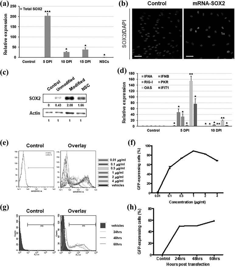
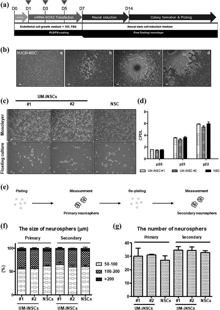
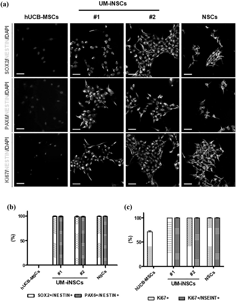
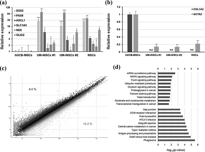
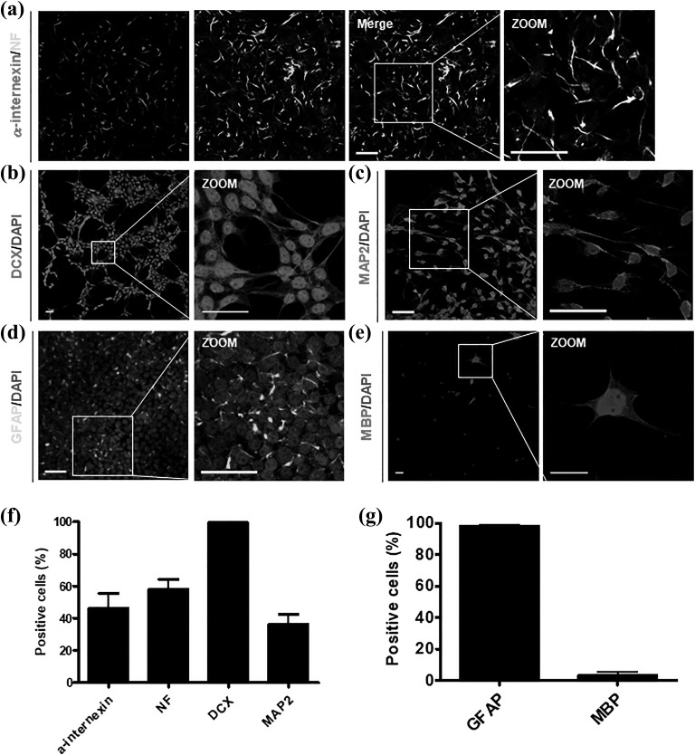
Neural stem cells (NSCs) are a prominent cell source for understanding neural pathogenesis and for developing therapeutic applications to treat neurodegenerative disease because of their regenerative capacity and multipotency. Recently, a variety of cellular reprogramming technologies have been developed to facilitate in vitro generation of NSCs, called induced NSCs (iNSCs). However, the genetic safety aspects of established virus-based reprogramming methods have been considered, and non-integrating reprogramming methods have been developed. Reprogramming with in vitro transcribed (IVT) mRNA is one of the genetically safe reprogramming methods because exogenous mRNA temporally exists in the cell and is not integrated into the chromosome. Here, we successfully generated expandable iNSCs from human umbilical cord blood-derived mesenchymal stem cells (UCB-MSCs) via transfection with IVT mRNA encoding SOX2 (SOX2 mRNA) with properly optimized conditions. We confirmed that generated human UCB-MSC-derived iNSCs (UM-iNSCs) possess characteristics of NSCs, including multipotency and self-renewal capacity. Additionally, we transfected human dermal fibroblasts (HDFs) with SOX2 mRNA. Compared with human embryonic stem cell-derived NSCs, HDFs transfected with SOX2 mRNA exhibited neural reprogramming with similar morphologies and NSC-enriched mRNA levels, but they showed limited proliferation ability. Our results demonstrated that human UCB-MSCs can be used for direct reprogramming into NSCs through transfection with IVT mRNA encoding a single factor, which provides an integration-free reprogramming tool for future therapeutic application.
Keywords: cellular reprogramming; direct conversion; neural stem cell; synthetic mRNA; umbilical cord blood.
Conflict of interest statement
Figures
Similar articles
-
SOX2 and SOX2-MYC Reprogramming Process of Fibroblasts to the Neural Stem Cells Compromised by Senescence.PLoS One. 2015 Nov 4;10(11):e0141688. doi: 10.1371/journal.pone.0141688. eCollection 2015. PLoS One. 2015. PMID: 26535892 Free PMC article.
-
Direct reprogramming of mouse and human fibroblasts into multipotent neural stem cells with a single factor.Cell Stem Cell. 2012 Jul 6;11(1):100-9. doi: 10.1016/j.stem.2012.05.018. Epub 2012 Jun 7. Cell Stem Cell. 2012. PMID: 22683203 Free PMC article.
-
mRNA-Driven Generation of Transgene-Free Neural Stem Cells from Human Urine-Derived Cells.Cells. 2019 Sep 6;8(9):1043. doi: 10.3390/cells8091043. Cells. 2019. PMID: 31489945 Free PMC article.
-
Methods of reactivation and reprogramming of neural stem cells for neural repair.Methods. 2018 Jan 15;133:3-20. doi: 10.1016/j.ymeth.2017.08.014. Epub 2017 Aug 31. Methods. 2018. PMID: 28864354 Review.
-
Concise review: the involvement of SOX2 in direct reprogramming of induced neural stem/precursor cells.Stem Cells Transl Med. 2013 Aug;2(8):579-83. doi: 10.5966/sctm.2012-0179. Epub 2013 Jul 1. Stem Cells Transl Med. 2013. PMID: 23817132 Free PMC article. Review.
Cited by
-
Evolving principles underlying neural lineage conversion and their relevance for biomedical translation.F1000Res. 2019 Aug 30;8:F1000 Faculty Rev-1548. doi: 10.12688/f1000research.18926.1. eCollection 2019. F1000Res. 2019. PMID: 31559012 Free PMC article. Review.
-
Direct cell-fate conversion of somatic cells: Toward regenerative medicine and industries.Proc Jpn Acad Ser B Phys Biol Sci. 2020;96(4):131-158. doi: 10.2183/pjab.96.012. Proc Jpn Acad Ser B Phys Biol Sci. 2020. PMID: 32281550 Free PMC article. Review.
-
Human iNSC-derived brain organoid model of lysosomal storage disorder in Niemann-Pick disease type C.Cell Death Dis. 2020 Dec 12;11(12):1059. doi: 10.1038/s41419-020-03262-7. Cell Death Dis. 2020. PMID: 33311479 Free PMC article.
-
Vascularization of iNSC spheroid in a 3D spheroid-on-a-chip platform enhances neural maturation.Biotechnol Bioeng. 2022 Feb;119(2):566-574. doi: 10.1002/bit.27978. Epub 2021 Nov 6. Biotechnol Bioeng. 2022. PMID: 34716703 Free PMC article.
-
SOX2 for Stem Cell Therapy and Medical Use: Pros or Cons?Cell Transplant. 2020 Jan-Dec;29:963689720907565. doi: 10.1177/0963689720907565. Cell Transplant. 2020. PMID: 32233795 Free PMC article.
References
-
- Fong CY, Gauthaman K, Bongso A. Teratomas from pluripotent stem cells: A clinical hurdle. J Cell Biochem. 2010;111:769–781. - PubMed
-
- Miura K, Okada Y, Aoi T, Okada A, Takahashi K, Okita K, Nakagawa M, Koyanagi M, Tanabe K, Ohnuki M, Ogawa D, Ikeda E, Okano H, Yamanaka S. Variation in the safety of induced pluripotent stem cell lines. Nat Biotechnol. 2009;27:743–745. - PubMed
-
- Giorgetti A, Marchetto MC, Li M, Yu D, Fazzina R, Mu Y, Adamo A, Paramonov I, Cardoso JC, Monasterio MB, Bardy C, Cassiani-Ingoni R, Liu GH, Gage FH, Izpisua Belmonte JC. Cord blood-derived neuronal cells by ectopic expression of Sox2 and c-Myc. Proc Natl Acad Sci USA. 2012;109:12556–12561. - PMC - PubMed
Publication types
MeSH terms
Substances
LinkOut - more resources
Full Text Sources
Other Literature Sources
