. 2018 Aug 7;28(2):282-288.e3.
doi: 10.1016/j.cmet.2018.05.022. Epub 2018 Jun 14.

Reversible De-differentiation of Mature White Adipocytes into Preadipocyte-like Precursors during Lactation

Affiliations

Reversible De-differentiation of Mature White Adipocytes into Preadipocyte-like Precursors during Lactation

Qiong A Wang et al. Cell Metab. .

Abstract

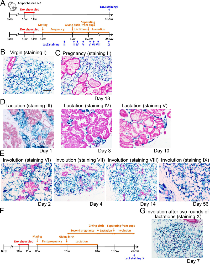
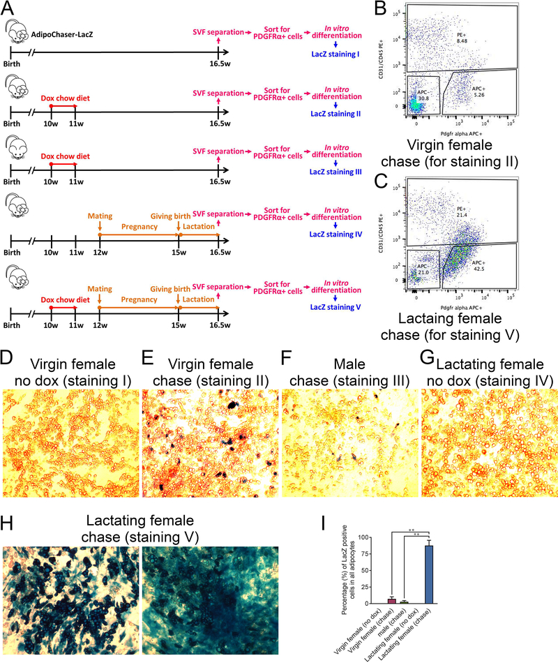
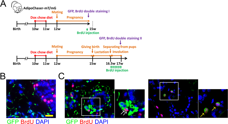
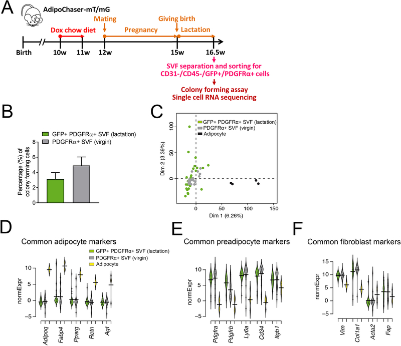
Adipose tissue in the mammary gland undergoes dramatic remodeling during reproduction. Adipocytes are replaced by mammary alveolar structures during pregnancy and lactation, then reappear upon weaning. The fate of the original adipocytes during lactation and the developmental origin of the re-appearing adipocyte post involution are unclear. Here, we reveal that adipocytes in the mammary gland de-differentiate into Pdgfrα+ preadipocyte- and fibroblast-like cells during pregnancy and remain de-differentiated during lactation. Upon weaning, de-differentiated fibroblasts proliferate and re-differentiate into adipocytes. This cycle occurs over multiple pregnancies. These observations reveal the potential of terminally differentiated adipocytes to undergo repeated cycles of de-differentiation and re-differentiation in a physiological setting.

Keywords: adipose tissue; de-differentiation; lactation; mammary gland; preadipocyte.


Conflict of interest statement

DECLARATION OF INTERESTS

The authors declare no competing interests.

Figures

Figure 1.
Figure 1.. Lineage tracing of adipocytes in the mammary gland during reproduction
(A) Experimental design: 10-week old AdipoChaser-LacZ female mice were put on a doxycycline chow diet for 7 days to ensure that LacZ expression is turned on in all mature adipocytes. After doxycycline treatment, mice were switched back to regular chow diet for a week to ensure washout of doxycycline. Thereafter, mice were kept as virgins or bred with male mice to undergo pregnancy and lactation. LacZ staining is performed at the indicated time points. (B-E) Representative β-gal staining of the mammary gland from female AdipoChaser-LacZ mice. (B) Virgin female: 7 weeks after doxycycline diet treatment. (C) During pregnancy: female mice were pregnant for 18 days. (D) During lactation: female mice were lactating pups at day 1, day 3 and day 10. (E) Involution: pups were taken away from lactating female mice at L10 for 2 days, 4 days, two weeks or 2 months (56 days) (F) Experimental design: 10-week old AdipoChaser-LacZ female mice were treated with doxycycline chow diet to label all mature adipocytes as described in panel A. Mice were then bred with male mice to undergo pregnancy and lactation for two rounds. LacZ staining is performed 7 days after weaning. (G) Representative β-gal staining of the mammary gland from female AdipoChaser-LacZ mice, after two rounds of lactations. n = 3–5 mice per group. Scale bar: 100 μm. This experiment is representative of three independent experiments.
Figure 2.
Figure 2.. In vitro isolation and differentiation of PDGFRα positive cells from the SVF of the lactating mammary gland
(A) Experimental design: 5 cohorts of 10-week old AdipoChaser-LacZ female or male mice were collected, and 5 experimental conditions were performed as indicated. At the end of each experiment, the mammary glands (subcutaneous adipose tissues) were used for SVF separation. The separated SVF were then flow sorted for CD31/CD45/PDGFRα+ cells. The isolated CD31/CD45/PDGFRα+ cells were plated in dishes and differentiated into adipocyte for LacZ staining. (B-C) Flow sorting images for condition II and V. (D-H) β-gal staining on the differentiated adipocytes. (D) Adipocytes differentiated from the CD31/CD45/PDGFRα+ cells from the mammary gland SVF of virgin females without any doxycycline treatment. (E) Adipocytes differentiated from the CD31/CD45/PDGFRα+ cells from the mammary gland SVF of virgin females with doxycycline pre-labeling. (F) Adipocytes differentiated from the CD31/CD45/PDGFRα+ cells from the sWAT SVF of male mice with doxycycline pre-labeling. (G) Adipocytes differentiated from the CD31/CD45/PDGFRα+ cells from the mammary gland SVF of lactating female without doxycycline pre-labeling. (H) adipocytes differentiated from the CD31/CD45/PDGFRα+ cells from the mammary gland SVF of lactating female with doxycycline pre-labeling. (I) Quantification of the percentage of LacZ positive adipocytes in the total adipocytes. n = 3 images, Virgin female (no dox), Virgin female (chase), male (chase) and Lactating female (no dox); n = 7 images, Lactating female (chase). **, P < 0.01 compared to Virgin female (chase) or male (chase). This experiment is representative of three independent experiments.
Figure 3.
Figure 3.. In vivo detection of proliferating de-differentiated adipocytes in the mammary gland upon weaning
(A) Experimental design: 10-week old AdipoChaser-mT/mG female mice were put on a doxycycline chow diet for 7 days to ensure that Tomato expression is turned on in all mature adipocytes. After doxycycline treatment, mice were switched back to regular chow diet for a week to ensure washout of doxycycline. Thereafter, mice were bred with male mice to undergo pregnancy and lactation. BrdU injection was performed at the indicated time points. (B-C) Representative Immunofluorescence staining shows GFP (green), BrdU (red), and DAPI (blue) in the mammary gland during pregnancy (day 20) (B) and involution (pups removed from lactating female on lactation day 10, 3 days after weaning) (C). White arrow: co-localization of GFP and BrdU in the same cell. Yellow arrow: dividing cell with GFP and BrdU co-localization. Scale bar: 50 μm. n = 3 mice per group. This experiment is representative of 4 independent experiments.
Figure 4.
Figure 4.. Characterization of de-differentiated adipocyte in the lactating mammary gland by single-cell RNA-seq
(A) Experimental design: 10-week old AdipoChaser-mT/mG female mice were put on a doxycycline chow diet for 7 days to ensure that GFP expression is turned on in all mature mammary adipocytes. After doxycycline treatment, mice were switched back to regular chow diet for a week to ensure the washout of doxycycline. For chasing mature mammary adipocyte in lactating female, mice were bred with male mice to undergo pregnancy and lactation. When the mice had been lactating for 10 days, the mammary glands were used for SVF separation. The separated SVF was then flow-sorted for CD31/CD45/PDGFRα+/GFP+ cells for single-cell RNA sequencing analysis. (B) Quantification of colony-forming unit of FACS purified CD31/CD45/PDGFRα+/GFP+ from the mammary gland SVFs of lactating female, comparing to the classic preadipocyte population (CD31/CD45/PDGFRα+ cells) from the mammary gland SVFs of virgin female. (D-G) Gene expressions of these de-differentiated mammary adipocytes (CD31/CD45/PDGFRα+/GFP+ cells) from the mammary gland SVFs of lactating female were compared to the classic preadipocyte population (CD31/CD45/PDGFRα+ cells) from the mammary gland SVFs of virgin female and an adipocyte cell line through single-cell RNA-seq. (C) Overview of single-cell grouping by principal component analysis. (D-F) Beanplots showing the distribution of normalized expression values (normExpression) across cells that belong to one of these three categories of common adipocyte markers (D), common preadipocyte markers (E) and common fibroblast markers (F). n = 26 cells for CD31/CD45/PDGFRα+/GFP+ SVF cells from lactating female, n = 20 cells for CD31/CD45/PDGFRα+ SVF cells from virgin female, n = 4 cells for brown adipocyte cell line.

Comment in

References

    1. Anders S, Pyl PT, and Huber W (2015). HTSeq--a Python framework to work with high-throughput sequencing data. Bioinformatics (Oxford, England) 31, 166–169. - PMC - PubMed
    1. Berry R, Jeffery E, and Rodeheffer, Matthew S (2014). Weighing in on Adipocyte Precursors. Cell Metabolism 19, 8–20. - PMC - PubMed
    1. Berry R, and Rodeheffer MS (2013). Characterization of the adipocyte cellular lineage in vivo. Nature cell biology 15, 302–308. - PMC - PubMed
    1. Chen W, Gardeux V, Meireles-Filho A, and Deplancke B (2017). Profiling of Single-Cell Transcriptomes. Current protocols in mouse biology 7, 145–175. - PubMed
    1. Cinti S (2007). The adipose organ In Adipose tissue and adipokines in health and disease (Springer; ), pp. 3–19.

Publication types