. 2018 Aug 9;174(4):982-998.e20.
doi: 10.1016/j.cell.2018.05.057. Epub 2018 Jun 18.

A Single-Cell Transcriptome Atlas of the Aging Drosophila Brain

Affiliations

A Single-Cell Transcriptome Atlas of the Aging Drosophila Brain

Kristofer Davie et al. Cell. .

Abstract

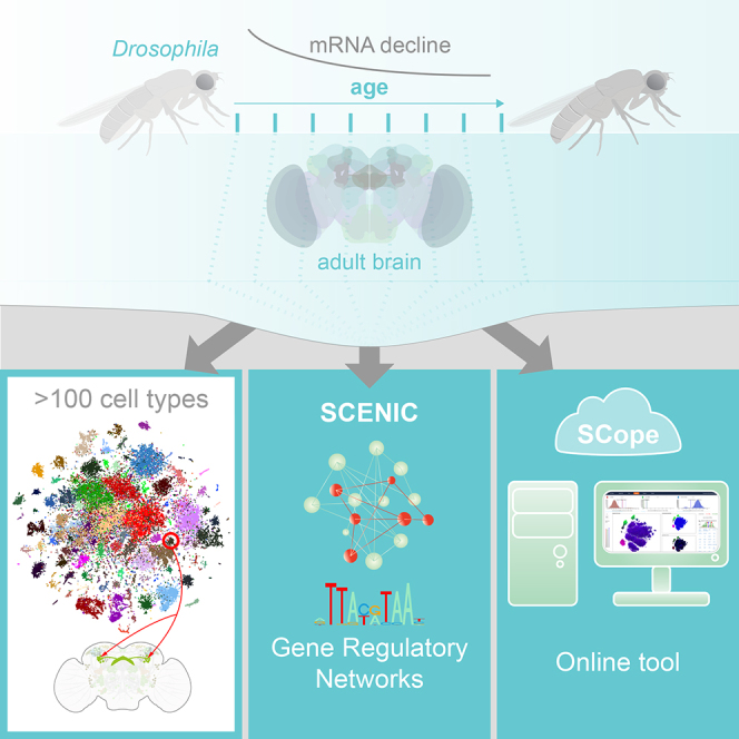
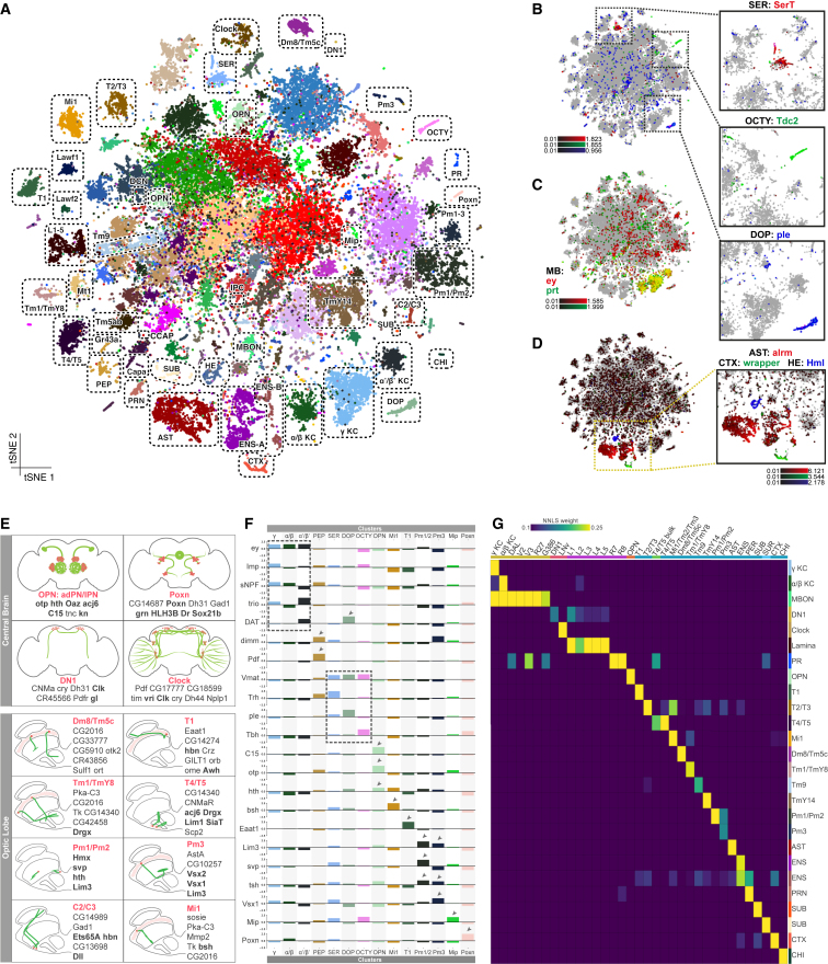
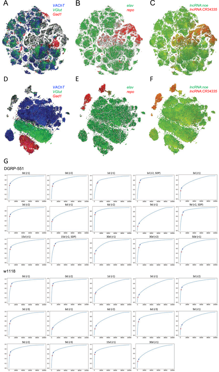
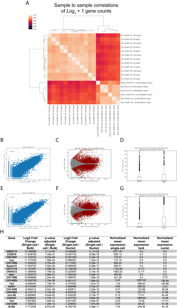
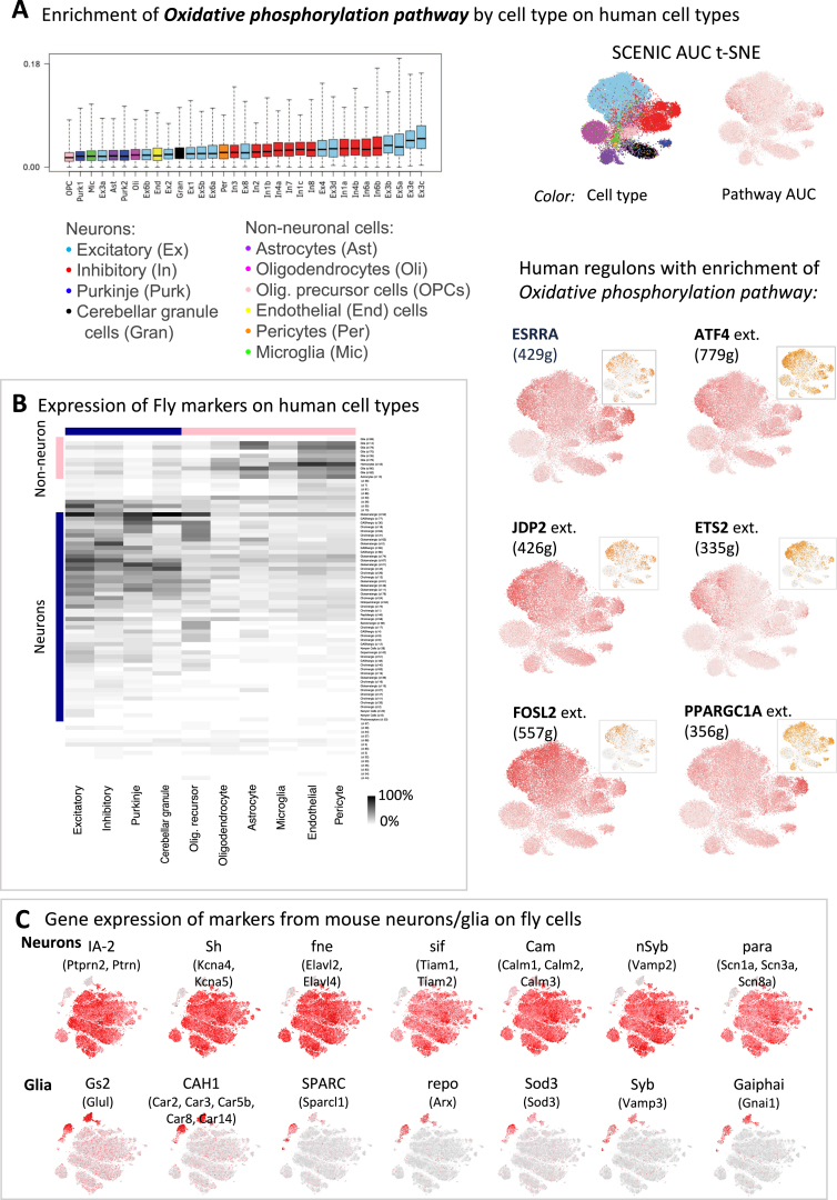
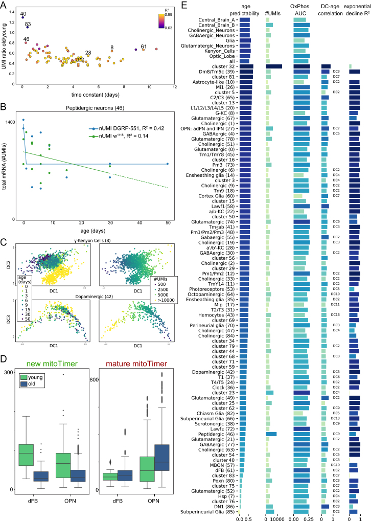
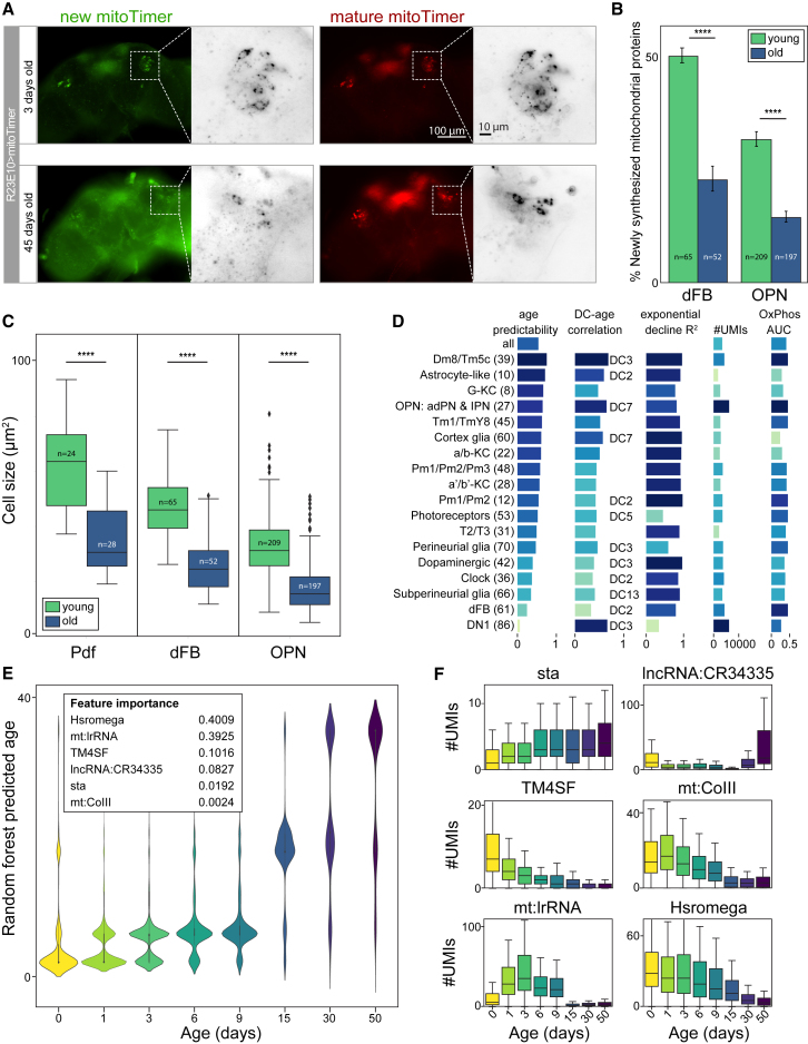
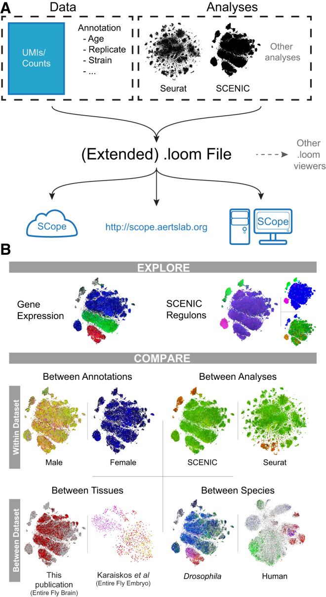
The diversity of cell types and regulatory states in the brain, and how these change during aging, remains largely unknown. We present a single-cell transcriptome atlas of the entire adult Drosophila melanogaster brain sampled across its lifespan. Cell clustering identified 87 initial cell clusters that are further subclustered and validated by targeted cell-sorting. Our data show high granularity and identify a wide range of cell types. Gene network analyses using SCENIC revealed regulatory heterogeneity linked to energy consumption. During aging, RNA content declines exponentially without affecting neuronal identity in old brains. This single-cell brain atlas covers nearly all cells in the normal brain and provides the tools to study cellular diversity alongside other Drosophila and mammalian single-cell datasets in our unique single-cell analysis platform: SCope (http://scope.aertslab.org). These results, together with SCope, allow comprehensive exploration of all transcriptional states of an entire aging brain.

Keywords: Drosophila; aging; brain; gene regulatory networks; mitochondria; neuronal subtypes; oxidative phosphorylation; single-cell RNA-seq; single-cell bioinformatics.


Figures

None
Graphical abstract
Figure 1
Figure 1
Diversity of the Cell Types Identified by Single-Cell RNA-Seq of the Adult Brain (A) Annotated cell types on the Seurat t-SNE of 57K cells. AST, astrocyte-like glia; CHI, chiasm glia; CTX, cortex glia; DCN, dorsal cluster neurons; DOP, dopaminergic neurons; ENS, ensheathing glia; HE, hemocytes; IPC, insulin-producing cells; KC, Kenyon cells; OCTY, octopaminergic-tyraminergic neurons; PEP, peptidergic neurons; PR, photoreceptors; PRN, perineurial glia; SER, serotonergic; SUB, subperineurial glia; SUR, surface glia; MBON, mushroom body output neurons. (B) Cells colored by expression of SerT (red), Tdc2 (green), and ple (blue) show SER, OCTY, and DOP clusters, respectively. (C) Cells colored by expression of ey (red) and prt (green) show MB KC clusters. (D) Cells colored by expression of alrm (red), wrapper (green), and Hml (blue) show AST, CTX, and HE clusters, respectively. (E) For a subset of the annotated cell types from the central brain and the optic lobe, cellular localizations (pink) and projections (green) are illustrated. Representative genes from Seurat markers are listed (see Table S3 for the full list); TFs are shown in bold. Only one neuron per cell type is illustrated for the optic lobe cells to show the morphology. (F) Expression levels for selected marker genes (shown by arrowheads and dashed lines) for several clusters. (G) Heatmap shows the mapping of publicly available bulk RNA-seq data on the clusters from Seurat analysis. The source datasets are color coded (yellow, Crocker et al., 2016; red, Abruzzi et al., 2017; purple, Tan et al., 2015; orange, Li et al., 2017; blue, Konstantinides et al., 2018; green, Pankova and Borst; 2016; light blue, DeSalvo et al., 2014). See also Figures S1 and S2 and Tables S1, S2, and S3.
Figure S1
Figure S1
Comparison of Two Different Filtering Cutoffs, Related to Figure 1 (A–C) SCENIC t-SNEs of the 157K dataset (lenient filtering) colored by (A) VAChT indicating cholinergic neurons in blue, VGlut indicating glutamatergic neurons in green and Gad1 indicating GABAergic neurons in red, (B) elav indicating neurons in green and repo indicating glia in red, (C) lncRNA:noe indicating neurons in green and lncRNA:CR34335 indicating glia in red. (D–F) SCENIC t-SNEs of the 57K dataset (stringent filtering), with aforementioned colors. (G) Plots per 10x Chromium run indicating the cumulative fraction of UMIs, red dots indicate Cell Ranger cutoffs used for the 57K dataset (note that additional filtering by Scater was applied after the Cell Ranger cutoff), blue dots indicate our less stringent cutoffs used for the 157K dataset
Figure S3
Figure S3
Bias Analysis of the Clusters and Kenyon Cell Subclusters, Related to Figure 2 (A) Bar plots showing UMI counts across the annotated and unannotated clusters at resolution 2. (B–F) Stacked bar plots showing biases across the Seurat clusters at resolution 2 for (B) sex (C) age (D) genotype (E) DGRP-551 replicates (F) w1118 replicates. (G and H) Groups are normalized for the number of cells (nCells) they contain. t-SNEs (left) and heatmaps (right) showing subclustering of the cells annotated as KCs consisting of the main clusters 8 (γ KC), 22 (α/β KC), and 28 (α’/β’ KC) (G) at resolution 0.2, which results in 4 clusters and (H) at resolution 2 which results in 16 clusters. Clusters were annotated by the expression of marker genes. (I) Stacked bar plots showing biases across the subclusters at resolution 0.2 (left) and 2 (right) for sex, age, genotype, and replicates. The groups are normalized for number of cells. Contribution of the cells from the main Seurat clusters 8, 22, and 28 is consistent with the cluster annotations. Resolution 0.2 doesn’t provide more information about subtypes of KCs and higher resolutions result in biased clustering toward the parameters shown.
Figure S2
Figure S2
Correlation between Single-Cell RNA-Seq and Bulk RNA-Seq Data of Adult Fly Brains, Related to Figure 1 (A) Sample to sample correlations of single-cell replicates (sum of cells) and bulk data. (B) Gene correlation between bulk and single-cell samples. (C) MA plot of all bulk and single-cell data. Green box highlights lowly expressed genes significantly expressed in bulk samples. (D) Boxplot showing median gene expression for significantly changing genes (padj ≤ 0.05, log2FC > = 2.0) between bulk and single-cell samples. (E) Gene correlation between nuclei and single-cell samples. (F) MA plot of all nuclei and single-cell data. Green box highlights lowly expressed, genes significantly expressed in bulk samples. (G) Boxplot showing median gene expression for significantly changing genes (padj ≤ 0.05, log2FC > = 2.0) between nuclei and single-cell samples. (H) Table of the top and bottom 10 genes differentially expressed between bulk/nuclei and single-cell samples.
Figure 2
Figure 2
Subclustering Reveals Presence of Rare Cell Types (A) Expression pattern of R23E10-Gal4 highlighting the dFBs in the adult brain. Scale bar, 50 μm. (B) Heatmap showing the mapping of FAC-sorted scRNA-seq on the Seurat clusters. (C) t-SNE showing seven subclusters within cluster 61. (D) Heatmap showing the mapping of FAC-sorted scRNA-seq on these subclusters. (E) t-SNE showing five subclusters within the lamina cluster. (F) Heatmap showing the mapping of the RNA-seq data of the lamina monopolar cells from Tan et al. (2015) on the lamina subclusters. (G) t-SNE showing ICIM clustering of SMART-Seq2 data from Li et al. (2017). Clusters are colored by co-expression of selected genes. (H) t-SNE showing ICIM subclustering of the OPN cluster. Clusters are colored by co-expression of selected genes. (I) t-SNE showing the subclustering of dopaminergic neurons, revealing the presence of the PAM cluster by expression of Fer2. (J) t-SNE showing the separation of octopaminergic and tyraminergic neurons in cluster 64 by expression of Tbh. (K) Subclusters of neurons colored by co-expression of neuropeptides. (L) Table comparing the number of cells present in our dataset with the estimated numbers from literature. See also Figure S3 and Table S2.
Figure 3
Figure 3
Gene Regulatory Networks Underlie Neuronal and Glial Cell Types (A) Seurat- and SCENIC-based t-SNEs showing the organization of Seurat clusters and the three main neurotransmitter types. (B) The C15 regulon, active in the olfactory projection neurons with the expression of four target genes as inset. Otp and Hth were added as co-regulatory factors because their motif is also enriched in the C15 regulon, through an analysis with iRegulon (Janky et al., 2014). See also Figure S4. (C) Various combinations of regulon activities highlighting the variety of cells with active gene regulatory networks. (D) All 150 regulons and the normalized expression score (NES) associated with their highest-scoring motif. Regulons for TFs with known brain phenotypes are indicated in red. Several relevant regulons have been indicated, with their highest-scoring motif shown. (E) DNA footprint of the Onecut binding-site predictions, using ATAC-seq. ATAC-seq signal of the best Onecut site near genes of the Onecut regulon (blue); the best Onecut sites near randomly selected genes (orange). In Figure S4A, we also compare this with background Tn5 insertions. See also Figure S4 and Table S4.
Figure S4
Figure S4
Validation of the Predicted Gene Regulatory Networks, Related to Figure 3 (A) Comparison of the Onecut transcription factor DNA footprint against the Tn5 transposase sequence bias. (B) The Dimmed regulon, active in peptidergic neurons and expression of dimm and several of its predicted target genes in these cells. (C) Heatmap of transcription factors found as markers of clusters by Seurat (values are row zscores, only markers with p < 1e-150 are shown). TFs with regulons found by SCENIC are indicated by a yellow bar, those found by Seurat only are marked by a blue bar.
Figure 4
Figure 4
SCENIC Uncovers a Neuronal Axis Guided by Oxidative Phosphorylation (A) Loadings for the top two principal components for each regulon. (B–D) SCENIC t-SNE showing expression of neuroblast “master regulators.” (E) Immunostaining of Pros and Imp-GFP showing a very strong disjoint expression of both factors, in agreement with the t-SNE. (F) Division of the t-SNE into three main parts based on expression of dati and Imp: central brain A (dati high), central brain B (dati low, Imp positive), and optic lobe (dati low). Glia were grouped separately, as were neurons that did not meet any of these thresholds. (G) GO analysis of differential expression between the three main neuronal groups reveals metabolic terms related to oxidative phosphorylation to be downregulated in central brain A. (H) Number of mitochondrial reads in each neuronal group showing an enrichment in central brain B and optic lobe. (I) SCENIC t-SNE showing non-overlapping expression of the Dati regulon (green) and a regulon enriched for oxidative phosphorylation (red). Motifs for both regulon are shown, where the OxPhos-enriched motif is the same as previously described in literature. (J) Boxplot showing the OxPhos expression measured by AUCell for all Seurat clusters. Clusters are colored by which compartment they are part of. (K) MitoTimer measurements on the R23E10-Gal4 dFBs, showing newly synthesized mitochondrial proteins in green and matured mitochondrial protein in red. (L) Percentage of newly synthesized mitochondrial proteins for six neuronal populations, colored by central brain compartment. Error bars show 95% confidence interval. Scale bars, 50 μm in half-brain images, 5 μm in zoom-ins. See also Figure S5.
Figure S5
Figure S5
Dati and Prospero Regulatory Networks and Glycolysis Levels among the Clusters, Related to Figure 4 (A) Predicted network for Dati and Prospero target genes. (B) Expression of predicted target genes positively and negatively correlated with Dati. (C) Dati, Jim, Hb and Rn share the same binding motif and are positively correlated with each other, suggesting some form of redundancy in the network. (D) Glycolysis activity for each Seurat cluster. Clusters are colored by main cell group.
Figure S6
Figure S6
Comparison of OxPhos Levels and Main Cell Types between Fly and Human/Mouse, Related to Figures 4 and 7 (A) Oxidative phosphorylation gene set on human brain cells (Lake et al., 2018). The boxplot shows the AUC scores of the OxPhos gene set per cell type (same approach as Figure 4J). The t-SNEs on the right show the pathway AUC, and regulons with enrichment of the pathway. For each regulon, the AUC (red) and TF expression (orange) are shown. (B) Comparison of human and fly cell types: The heatmap shows the percentage of human cells from each cell type in which the fly cluster signature (e.g., cluster markers) are selected as “active” with AUCell. (C) Expression of mouse glia/neuron markers on the fly cells.
Figure 5
Figure 5
Aged Cells Have Reduced Transcriptional Output but Retain Identity (A) The number of UMIs and genes show an exponential decline over aging for both DGRP-551 and w1118. (B) t-SNE of young and old cells, showing the stability of cell types manifested by the presence of each cluster in both t-SNEs. Cluster 23 (gap in the middle of old t-SNE) is likely an artifact because it occurs only in one replicate of young flies (Figures S3B–S3F). Pie plots show a change in composition, with a slight increase in relative amount of glia (5% to 11%). (C) Correlation of each gene with age. Most genes decline with age, especially those involved in oxidative phosphorylation, while ribosomal genes involved in translation are more protected. (D) Change in OxPhos activity, as measured by AUCell, for the three neuronal groups defined above. See also Figures S4 and S7.
Figure S7
Figure S7
Effects of Aging through Several Cell Types and Machine-Learning-Based Age Prediction, Related to Figures 5 and 6 (A) Scatterplot showing the ratio of the number of UMIs of old cells versus young cells against the time constant derived from the exponential decline model. Colors are a measure for the goodness of fit (R2). (B) Decline in number of UMIs for the peptidergic neurons. (C) Diffusion maps of the diffusion components mostly correlated with age for the mushroom body and dopaminergic neurons. Separation by age is mostly driven by the number UMIs. (D) Boxplots showing a decline in synthesis of mitochondrial proteins (new), with only a slight increase in old proteins (mature). (E) Performance of the Random Forest Regressor age prediction for the different Seurat clusters and distinct major groups. Possible confounding variables are shown. The diffusion component most correlated with age is shown; when no specification is made, DC1 is the major correlate.
Figure 6
Figure 6
Age Affects Mitochondrial Turnover and Cell Size and Can Be Accurately Predicted by the Transcriptome (A) MitoTimer for the dFBs (R23E10-Gal4), showing mitochondrial renewal over aging. Scale bars, 100 μm, 10 μm. (B) Bar plot showing a significant (t test, p < 0.0001) decline in synthesis of mitochondrial proteins. Error bars show SEM. (C) Boxplot showing a significant (t test, p < 0.0001) reduction in cell size for all measured neuronal types. (D) Random Forest Regressor prediction score for each Seurat cluster (age predict). The score is independent of number of UMIs, OxPhos activity, or correlation of diffusion components with age and seems to only correlate with the R2 value of the exponential fit. (E) Predictions of the Random Forest Model trained on all cells. Six features were found to be most influential. (F) Expression of the six most important features over time, displaying different patterns. See also Figure S7.
Figure 7
Figure 7
Overview of the Data Format Used and Features of SCope (A) A schematic of the data and analyses stored inside the loom files used by SCope. SCope can be accessed at http://scope.aertslab.org, by being installed on a local machine or server, or through use of a Singularity container usable on cloud-computing platforms. (B) On overview of some of the exploration and comparison options available in SCope. See also Figure S6.

Comment in

References

    1. Abruzzi K.C., Zadina A., Luo W., Wiyanto E., Rahman R., Guo F., Shafer O., Rosbash M. RNA-seq analysis of Drosophila clock and non-clock neurons reveals neuron-specific cycling and novel candidate neuropeptides. PLoS Genet. 2017;13:e1006613. - PMC - PubMed
    1. Aibar S., González-Blas C.B., Moerman T., Huynh-Thu V.A., Imrichova H., Hulselmans G., Rambow F., Marine J.-C., Geurts P., Aerts J. SCENIC: single-cell regulatory network inference and clustering. Nat. Methods. 2017;14:1083–1086. - PMC - PubMed
    1. Allada R., Chung B.Y. Circadian organization of behavior and physiology in Drosophila. Annu. Rev. Physiol. 2010;72:605–624. - PMC - PubMed
    1. Angerer P., Haghverdi L., Büttner M., Theis F.J., Marr C., Buettner F. destiny: diffusion maps for large-scale single-cell data in R. Bioinformatics. 2016;32:1241–1243. - PubMed
    1. Aronesty E. Expression Analysis; Durham, NC: 2011. ea-utils: Command-line tools for processing biological sequencing data.

Publication types

Substances