. 2018 Jul 9;28(13):2007-2017.e4.
doi: 10.1016/j.cub.2018.04.064. Epub 2018 Jun 14.

Reconfiguration of a Multi-oscillator Network by Light in the Drosophila Circadian Clock

Affiliations

Reconfiguration of a Multi-oscillator Network by Light in the Drosophila Circadian Clock

Abhishek Chatterjee et al. Curr Biol. .

Abstract

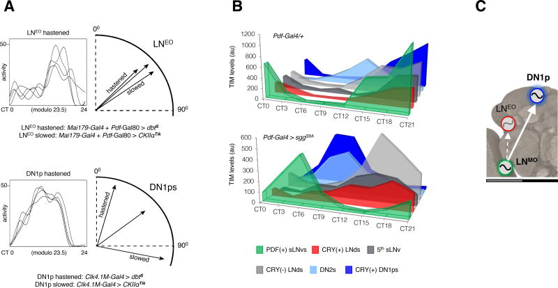
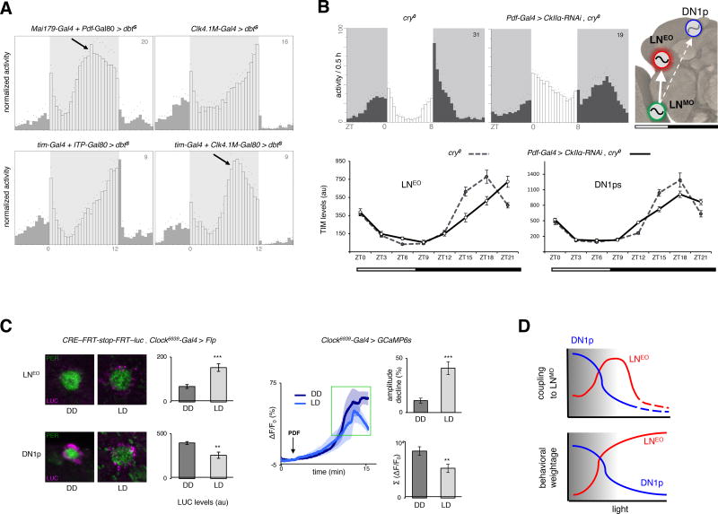
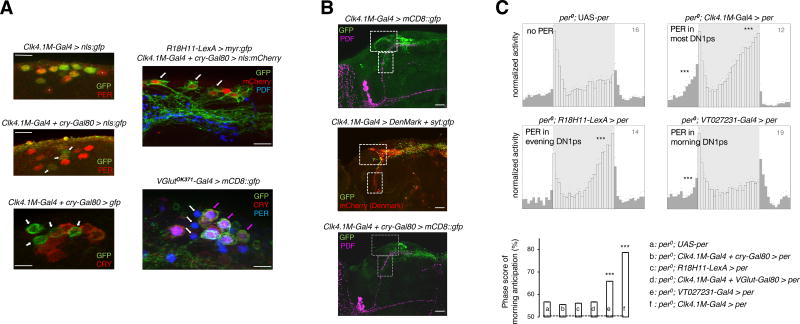
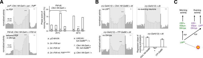
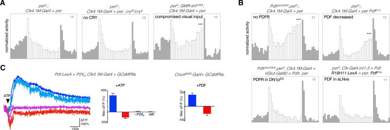
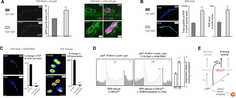
The brain clock that drives circadian rhythms of locomotor activity relies on a multi-oscillator neuronal network. In addition to synchronizing the clock with day-night cycles, light also reformats the clock-driven daily activity pattern. How changes in lighting conditions modify the contribution of the different oscillators to remodel the daily activity pattern remains largely unknown. Our data in Drosophila indicate that light readjusts the interactions between oscillators through two different modes. We show that a morning s-LNv > DN1p circuit works in series, whereas two parallel evening circuits are contributed by LNds and other DN1ps. Based on the photic context, the master pacemaker in the s-LNv neurons swaps its enslaved partner-oscillator-LNd in the presence of light or DN1p in the absence of light-to always link up with the most influential phase-determining oscillator. When exposure to light further increases, the light-activated LNd pacemaker becomes independent by decoupling from the s-LNvs. The calibration of coupling by light is layered on a clock-independent network interaction wherein light upregulates the expression of the PDF neuropeptide in the s-LNvs, which inhibits the behavioral output of the DN1p evening oscillator. Thus, light modifies inter-oscillator coupling and clock-independent output-gating to achieve flexibility in the network. It is likely that the light-induced changes in the Drosophila brain circadian network could reveal general principles of adapting to varying environmental cues in any neuronal multi-oscillator system.

Keywords: Drosophila; circadian clock; hormone receptor-like in 38; light; morning and evening oscillators; oscillator coupling; pigment-dispersing factor; posterior dorsal neurons 1; rest-activity rhythms; visual system.


Figures

Figure 1
Figure 1. Coupling between the period-determining s-LNvs and phase-determining DN1ps is the main axis of network operation in the absence of light
(A) (Left) Representative waveforms of locomotor activity of a single fly over first 4 DD days are depicted in each box. (Right) Using the trough of the waveform as the phase-marker, phase vectors are constructed on circular plot on a 24-hour dial. Only the relevant part of the plot is depicted here for flies in which either LNEO or DN1p oscillator underwent speed change. Unlike the LNEO, DN1p oscillator triggered dramatic phase changes (significant at α=0.05 by Watson's non-parametric two-sample U2 statistic) upon alteration of its endogenous pace. (see Table 1) (B) Immunostaining of TIM protein at eight different time-points on the fourth day of DD shows oscillations for each neuronal group. Synchrony across subsets (except the DN2s) occurred in Pdf-Gal4/+ flies but was dismantled in Pdf-Gal4 > sggS9A (faster pace of the PDF+ LNv oscillator) flies. Comparison of the TIM cycling profiles of different subgroups of PDF(−) oscillators of Pdf-Gal4 > sggS9A flies reveals that no other subgroup within the PDF(−) clock neurons could follow the PDF(+) s-LNvs as much as the CRY(+) DN1ps did (see Figure S2). (C) The model shows the dominant axis of coupling in DD within the multi-oscillator network. See also Figure S1.
Figure 2
Figure 2. Opportunistic swap of the coupled partner in the presence of light
(A) DBTS-mediated acceleration of the LNEO and DN1p oscillators, or all oscillators except LNEO/DN1p under 12:12 low-light LD cycles (See Figure S3 for quantifications). Low light is indicated by grey shading on daytime. (B) (Upper panel) Under short photoperiod (8:16) LD cycles, the LNMO was decelerated (30h period in DD) through knockdown of CkIIα in cry−/− background, and the resulting delay in the evening output produced by the PDF(−) oscillators was assessed. (Lower panel). Differential changes in the clock program of the LNEO (left) and DN1p (right) oscillators under such conditions, with the former showing stronger coupling to the LNMO. Each point in the line graph represents the average of at least 30 cells from at least 10 brain hemispheres. Cosinor analysis on the TIM cycling pattern reveals a >1.5 hr phase-delay in LNEO and a <0.5 h delay in DN1p, enforced by the slower-running LNMO. The model shows the dominant axis of coupling under LD cycles. (C) (Left panel) CRE-luc staining in LNEO is higher under LD (day 4, ZT3–4), while higher in DN1ps under DD (day 4, CT3–4). In the bar graph showing LUC staining intensity, n from left to right are 18, 8 for the LNEO and 51, 30 for the DN1ps. (Right panel) GCaMP6s fluorescence in DN1ps after bath-application of 30µM PDF under DD and LD cycles. The traces are averages of 5 representative responses. n=24 for the two bar plots, recorded during ZT/CT6–9 on day 4–5 of LD and DD. **p<0.01, *** p<0.0001 after unpaired two-tailed Student’s t-test. (D) The working model posits that with increased light, the LNMO switches coupling from DN1ps to LNEO, thereby optimizing its influence on the behavioral phase set by the PDF(−) oscillators. In excess light, the LNEO takes the lead for controlling behavior and liberates from the pacesetting influence of the LNMO (see Table S1). The number on the top-right corner of the activity plots shows the sample size of analyzed flies for a single run of the behavioral experiment. Error bars represent the s.e.m.
Figure 3
Figure 3. Functional subdivision of the DN1p cluster
(A) (Left panel) About 4–5 DN1ps lacking CRY protein expression, examples of which are marked with white arrow, were labelled with cytosolic-GFP and nls-GFP driven by the intersectional driver Clk4.1M-Gal4 + cry-Gal80. Clk4.1M-Gal4 alone drove GFP expression in about 10 of the DN1ps including all the 6 CRY(+) cells. The few DN1p neurons that were not labeled by the Clk4.1M-Gal4 driver are marked with white asterisks. (Right panel, top) Expression pattern of the DN1p-restricted R18H11-LexA which drives evening anticipation like the CRY(−) DN1ps, has extensive overlap with the latter subgroup, as marked with white arrows. (Right panel, bottom) Most of the CRY(+) DN1ps co-expressed VGlutOK371-Gal4-driven CD8::GFP demonstrating the convergence of VGlut and cry expression in the morning subset of the DN1ps, marked with colored arrows. Evening DN1p cells that were both VGlut(−) as well as CRY(−), are marked with white arrows (See Figure S4 for further characterization of the subgroups). All PER stainings were carried out at ZT0–2. (B) Projection patterns of the Clk4.1M-Gal4-expressing DN1ps (top) with dendritic arborization recognized by the DenMark marker (middle) labeled with white boxes. The CRY(−) evening DN1ps (bottom) lack the afferent fibers in the lateral and ventral protocerebrum. (C) Averaged locomotor activity profiles over 24-hour LD days reveal that an oscillator in the R18H11-LexA labeled, CRY(−) or VGlut(−) DN1ps (see Figure S4) was unable to elicit morning anticipation but could evoke evening anticipation. Another subset of the DN1p oscillator, identified by the Gal4 line VT027231 covering the VGlut(+) DN1ps (Figure S4), was sufficient for morning anticipation but not for evening anticipation. Significance of anticipatory activity was ascertained by Spearman’s non-parametric rank-correlation test (to measure the strength and direction of putatively monotonic association between the ranked variables activity-count and time-interval); **p<0.01, *** p<0.001. The column chart depicts mean ± s.e.m of the 3h/6h activity ratio prior to the light-on transition, i.e., an estimate of the amplitude of morning anticipation. *** p<0.0001, by one-way ANOVA followed by Tukey’s post-hoc test. Light intensity during the 12 hour of photoperiod was 50 lux, for all the eductions shown.
Figure 4
Figure 4. Distinct logic of circuit organization for morning and evening activity
(A) (Left panel) Morning and evening anticipatory activity were differentially affected when the DN1p clock had no access to the PDF neuropeptide. The averaged activity profile showed no significant morning anticipation (p=0.87), but persistent evening anticipation (*** p<0.0001) based on Spearman’s non-parametric rank-correlation test. Restoring PDF signaling onto the DN1ps of LNv-less flies brought back the morning peak (*** p<0.0001). (Right panel) Impact on morning anticipation of expressing membrane-tethered PDF, i.e., t-PDFML, or its scrambled analog, i.e., scr-PDF, or an inactive control peptide µO-MrVIA, in the DN1ps of LNv-less flies (Pdf-dti) in the absence or presence of PDFR or silencing the DN1ps by adult-specific expression of the Kir2.1 channel. For the column chart, n from left to right are 16, 16, 15, 9, 15, 11, and 16. *** p<0.0001, by one-way ANOVA followed by Tukey’s post-hoc test. (B) Evening activity in flies that lack the LNEO and/or the DN1ps (see Figure S4). The column plot shows mean ± s.e.m of the slope of a linear regression fitted on the last four hours of activity prior to the evening peak, which is a measure of the strength of the evening peak. Light intensity during the 12 hours of photoperiod was 50 lux, for all the eductions shown. (C) Scheme showing the LNMO and CRY(+) DN1pMO working in series to build the morning activity, while the LNEO and CRY(−) DN1pEO working in parallel to produce the evening activity. PDF is required for morning activity and influences the phasing of LNEO generated evening activity. The effect of genotype was significant by one-way ANOVA at α=0.0001. The number on the top-right corner of the activity plots shows the sample size of analyzed flies for a single run of the behavioral experiment. Error bars represent the s.e.m.
Figure 5
Figure 5. Direct gating of the DN1pEO output by visual light inputs and PDF
Evening peak under high light (1000 lux) conditions in flies with working oscillator confined to the DN1ps (See Figure S5 for quantifications). Status of the DN1p-made evening peak when (A) different modes of light input were compromised (B) PDF/PDFR signaling was manipulated. (C) Different patterns of calcium response in DN1p cells on activation of the LNv neurons (left), and bath application of 0.1mM PDF (right). The representative traces depict signal changes from four different cells of a single brain. Note the presence of a group, marked by shades of red, mounting a response consistent with physiological inhibition. 5mM ATP causes significant (p<0.05 by Kruskal-Wallis multiple comparisons test followed by Dunn’s post-hoc analysis) increase (bluish hues) or decrease (reddish hues) in GCaMP6 signal, compared to P2X2-non-expressing (−P2X2) and vehicle (veh) controls. 89 of the 131 recorded DN1ps elicited excitatory response, while 38 of them elicited inhibitory response. n=14,14,4,4 brains for the ATP/P2X2 bar plot (left) and n=8,8 for the PDF bar plot (right). Recordings were carried out at ZT6–9 (See Figure S6 for bioluminescence-based live imaging of intracellular calcium in DN1p).
Figure 6
Figure 6. Ambient light-intensity is encoded in Hr38-dependent Pdf transcription
(A) Expression of the active-zone marker BRP (BRUCHPILOT) (left) or the transcription-based CaLexA-GFP reporter (right) in the s-LNvs under low and high light intensities at ZT13–14. (B) Comparison of the levels of PDF peptide in the axon terminals and cell bodies of the s-LNvs under different light intensities at ZT13–14 indicates that the physiological output from the s-LNv neurons is promoted by high light. The column plot shows mean ± s.e.m of the slope of a linear regression fitted on the last four hours of activity prior to the evening peak. (C) Light-induction of PDF levels in s-LNv terminals in wild type flies or flies with downregulated Hr38 in the LNvs (left). Light-induction of a Tomato-based transcriptional reporter of Pdf in the s-LNv nuclei of wild-type flies and flies with downregulated Hr38 in the LNvs. % changes are from low-to-high light. Labelings are done at ZT13–14. (D) High light LD activity profiles of flies with a functional oscillator in the evening DN1ps in a wild type (1) or downregulated Hr38 (2) background. (E) Scheme showing that visually estimated ambient light-intensity changes PDF levels in the s-LNv cells. PDF suppresses the output of the CRY(−) DN1ps that produce evening activity. Each column in immunostaining experiments of (d), (e), and (f) represents mean ± s.e.m. of at least 8 brain hemispheres. *** p<0.0001 by unpaired two-tailed Student’s t-test, Fisher’s exact test was used for comparing % changes in (f). Representative stained images are pseudocolored, such that red-shifted colors denote stronger signal intensity. The number on the top-right corner of the activity plots shows the sample size of analyzed flies for a single run of the behavioral experiment. See also Figure S6.

Similar articles

Cited by

References

    1. Beckwith EJ, Ceriani MF. Communication between circadian clusters: The key to a plastic network. FEBS Lett. 2015;589:3336–3342. - PubMed
    1. Evans JA. Collective timekeeping among cells of the master circadian clock. J Endocrinol. 2016;230:R27–49. - PMC - PubMed
    1. Duffy JF, Czeisler CA. Effect of Light on Human Circadian Physiology. Sleep Med Clin. 2009;4:165–177. - PMC - PubMed
    1. Yoshii T, Rieger D, Helfrich-Forster C. Two clocks in the brain: An update of the morning and evening oscillator model in Drosophila. Prog Brain Res. 2012;199:59–82. - PubMed
    1. Lucas RJ. Mammalian inner retinal photoreception. Curr Biol. 2013;23:R125–33. - PubMed

Publication types

LinkOut - more resources