. 2018 Jun 21;70(6):1101-1110.e4.
doi: 10.1016/j.molcel.2018.05.011. Epub 2018 Jun 14.

A Mechanism for the Activation of the Influenza Virus Transcriptase

Affiliations

A Mechanism for the Activation of the Influenza Virus Transcriptase

Itziar Serna Martin et al. Mol Cell. .

Erratum in

Abstract

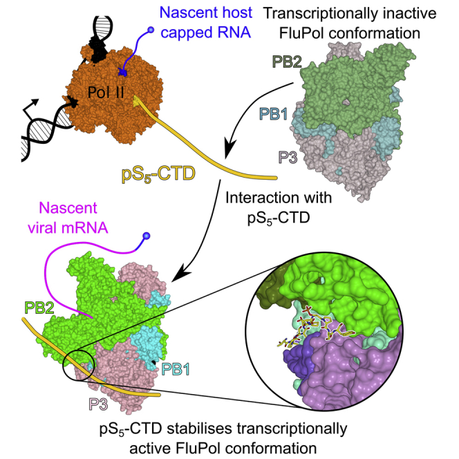
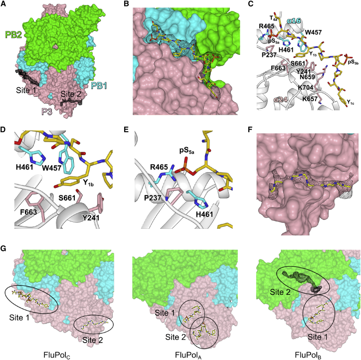
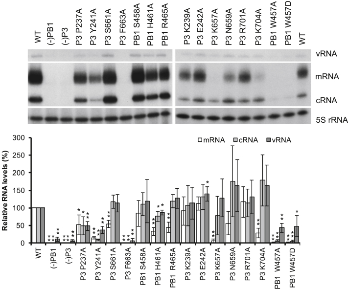
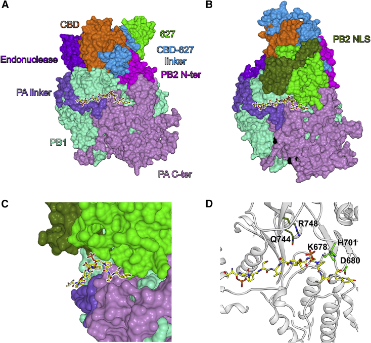
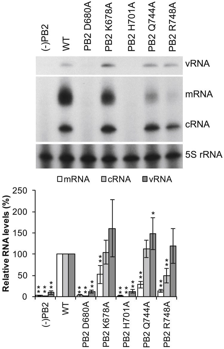
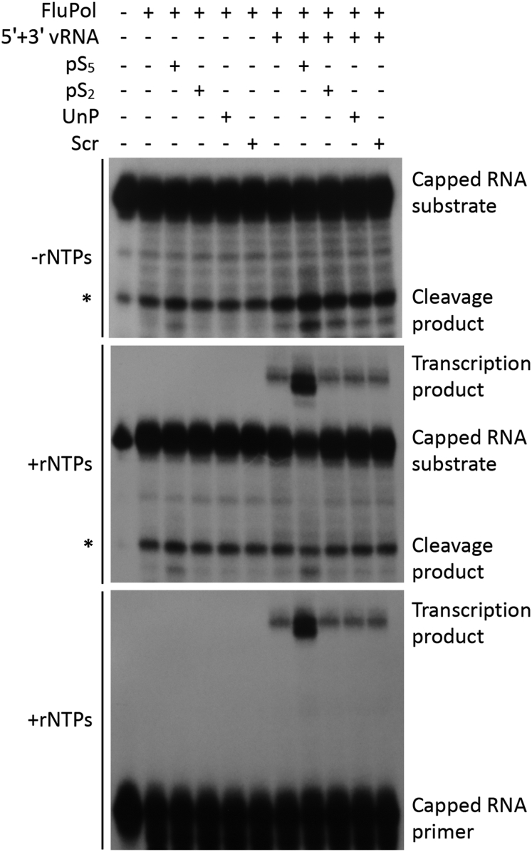
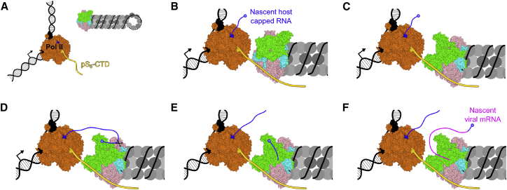
Influenza virus RNA polymerase (FluPol), a heterotrimer composed of PB1, PB2, and PA subunits (P3 in influenza C), performs both transcription and replication of the viral RNA genome. For transcription, FluPol interacts with the C-terminal domain (CTD) of RNA polymerase II (Pol II), which enables FluPol to snatch capped RNA primers from nascent host RNAs. Here, we describe the co-crystal structure of influenza C virus polymerase (FluPolC) bound to a Ser5-phosphorylated CTD (pS5-CTD) peptide. The position of the CTD-binding site at the interface of PB1, P3, and the flexible PB2 C-terminal domains suggests that CTD binding stabilizes the transcription-competent conformation of FluPol. In agreement, both cap snatching and capped primer-dependent transcription initiation by FluPolC are enhanced in the presence of pS5-CTD. Mutations of amino acids in the CTD-binding site reduce viral mRNA synthesis. We propose a model for the activation of the influenza virus transcriptase through its association with pS5-CTD of Pol II.

Keywords: CTD; Pol II; RNA polymerase; cap snatching; influenza virus; replication; transcriptase; transcription.


Figures

None
Graphical abstract
Figure 1
Figure 1
Structure of FluPolC Bound to Pol II CTD Peptide (A) Surface representation of FluPolC crystal structure with electron density maps shown at CTD-binding sites 1 and 2 (site 1: Sigma-A-weighted 2Fo-Fc, 0.9σ, black mesh; site 2: Sigma-A-weighted Fo-Fc, 2.6σ, black mesh). (B) Binding site 1 close up with the pS5-CTD peptide (yellow) modeled in the Sigma-A-weighted 2Fo-Fc electron density map (0.9σ). (C) Detailed view of the pS5-CTD peptide interaction at site 1 with key amino acids highlighted. (D) Detailed view of the hydrophobic pocket that accommodates Y1b. (E) Detailed view of the pocket that accommodates pS5a. (F) Binding site 2 close up with poly-alanine chain (yellow) backbone shown in the difference electron density map. (G) Comparison of the Pol II CTD-binding sites on FluPolC, FluPolA (PDB: 5M3H), and FluPolB (PDB: 5M3J). Site 2 in FluPolB is shown as difference electron density map (Sigma-A-weighted Fo-Fc, 1.9σ, black mesh). See also Table 1 and Figures S1, S2, and S3.
Figure 2
Figure 2
Effect of Mutations in CTD-Binding Site 1 on FluPolC Function The effect of single-amino-acid mutations in CTD-binding site 1 was analyzed by RNP reconstitution assays. vRNA, mRNA, and cRNA levels were analyzed by primer extension and quantitated by phosphorimage analysis with 5S rRNA as a loading control. RNA levels generated by the wild-type (WT) polymerase were set to 100%. RNP reconstitutions without PB1 (−PB1) or P3 (−P3) served as negative controls. The mean of three independent experiments is shown with error bars representing SD. Asterisks indicate a significant difference from WT (two-tailed one-sample t test) as follows: p < 0.05 and ∗∗p < 0.01. See also Figures S2 and S3.
Figure 3
Figure 3
Cryo-EM Analysis of FluPolC Bound to vRNA Promoter Structure of the vRNA promoter-bound FluPolB in a transcription pre-initiation conformation (PDB: 4WSA) fit into the cryo-EM density map of FluPolC bound to vRNA promoter. See also Figure S4.
Figure 4
Figure 4
Modeled FluPolC in the Transcription Pre-initiation Conformation Bound to Pol II CTD Peptide (A) Surface representation of pS5-CTD-bound FluPolC crystal structure in a transcriptionally inactive conformation with highlighted subunit domain arrangement. pS5-CTD is shown in yellow. (B) Surface representation of modeled pS5-CTD-bound FluPolC in a transcription pre-initiation conformation with highlighted subunit domain arrangement. The vRNA promoter is shown in black. (C) Detailed view of the PB2 627 and NLS domains packing above the pS5-CTD peptide in the transcription pre-initiation conformation of FluPolC. (D) Detailed view of the pS5-CTD peptide interaction at site 1 in the modeled transcription pre-initiation conformation of FluPolC with key amino acid residues in the PB2 627 and NLS domains highlighted. See also Figure S5.
Figure 5
Figure 5
Effect of Mutations in the PB2 627 and NLS Domains on FluPolC Function The effect of single-amino-acid mutations in the PB2 627 and NLS domains proximal to CTD-binding site 1 was analyzed by RNP reconstitution assays. vRNA, mRNA, and cRNA levels were analyzed by primer extension and quantitated by phosphorimage analysis with 5S rRNA as a loading control. RNA levels generated by the WT polymerase were set to 100%. RNP reconstitutions without PB2 (−PB2) served as negative controls. The mean of five independent experiments is shown with error bars representing SD. Asterisks indicate a significant difference from WT (two-tailed one-sample t test) as follows: p < 0.05 and ∗∗p < 0.01. See also Figure S5.
Figure 6
Figure 6
Effect of Pol II CTD Peptides on FluPolC Endonuclease and Transcriptional Activity In vitro endonuclease and cap-dependent transcription initiation assays were performed using purified FluPolC in the presence of pS5, pS2, un-phosphorylated (UnP), or scrambled (Scr) Pol II CTD peptides. Assays were carried out in the absence or presence of 5′ and 3′ vRNA promoter RNAs. The absence or presence of rNTP substrates is indicated. Top panel: capped RNA cleavage assay is shown; middle panel: capped RNA cleavage and transcription initiation assay are shown; bottom panel: cap-dependent transcription initiation assay is shown. The position of a non-specific cleavage product that partially overlaps with the viral polymerase-specific cleavage products is indicated by a star (). See also Figure S6.
Figure 7
Figure 7
Model for Viral Transcription Activation Induced by Binding to Pol II CTD (A) Prior to association with pS5-CTD of Pol II, multiple conformations of vRNP-associated FluPol exist in a dynamic equilibrium. However, most FluPol is likely present in a transcriptionally inactive conformation, in agreement with its low transcriptional activity. (B) FluPol binds to the pS5-CTD of Pol II. (C) Binding to the CTD stabilizes FluPol in the transcription-competent conformation as the PB2 627 and NLS domains interact with pS5-CTD. (D) In the transcription pre-initiation conformation, with the PB2 cap-binding and PA endonuclease domains reconfigured for cap snatching, FluPol binds the nascent-capped RNA produced by Pol II. (E) Cap snatching occurs, resulting in a short, capped RNA primer, which inserts into the FluPol active site. (F) Transcription initiation takes place, and the capped RNA primer is elongated by FluPol to produce viral mRNA.

References

    1. Adams P.D., Afonine P.V., Bunkóczi G., Chen V.B., Davis I.W., Echols N., Headd J.J., Hung L.W., Kapral G.J., Grosse-Kunstleve R.W. PHENIX: a comprehensive Python-based system for macromolecular structure solution. Acta Crystallogr. D Biol. Crystallogr. 2010;66:213–221. - PMC - PubMed
    1. Arranz R., Coloma R., Chichón F.J., Conesa J.J., Carrascosa J.L., Valpuesta J.M., Ortín J., Martín-Benito J. The structure of native influenza virion ribonucleoproteins. Science. 2012;338:1634–1637. - PubMed
    1. Beaton A.R., Krug R.M. Selected host cell capped RNA fragments prime influenza viral RNA transcription in vivo. Nucleic Acids Res. 1981;9:4423–4436. - PMC - PubMed
    1. Biasini M., Bienert S., Waterhouse A., Arnold K., Studer G., Schmidt T., Kiefer F., Gallo Cassarino T., Bertoni M., Bordoli L., Schwede T. SWISS-MODEL: modelling protein tertiary and quaternary structure using evolutionary information. Nucleic Acids Res. 2014;42:W252–W258. - PMC - PubMed
    1. Bieniossek C., Imasaki T., Takagi Y., Berger I. MultiBac: expanding the research toolbox for multiprotein complexes. Trends Biochem. Sci. 2012;37:49–57. - PMC - PubMed

Publication types

MeSH terms

LinkOut - more resources