

Review
doi: 10.1161/JAHA.117.007146.
Patent Foramen Ovale Closure for Stroke Prevention and Other Disorders
Affiliations
- PMID: 29910192
- PMCID: PMC6220531
- DOI: 10.1161/JAHA.117.007146
Item in Clipboard
Review
Patent Foramen Ovale Closure for Stroke Prevention and Other Disorders
J Am Heart Assoc.
.
No abstract available
Keywords: migraine; patent foramen ovale; patent foramen ovale closure; shunt; stroke.
Figures
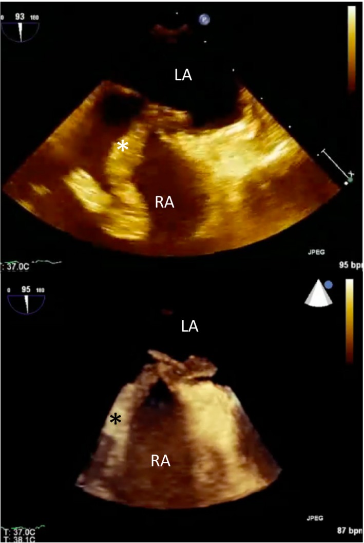
Clot in transit. Transesophageal echocardiogram images (2D and 3D) showing a large thrombus (*) in the right atrium (RA ) traversing a patent foramen ovale (PFO ) into the left atrium (LA ). The patient suffered a fatal stroke.
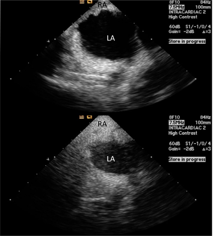
Intracardiac echo (ICE ) showing a positive “bubble study” confirming a right‐to‐left shunt across a patent foramen ovale (PFO ). Agitated saline is injected intravenously and bubbles are seen opacifying the right atrium (RA ). Subsequently, bubbles are seen in the left atrium (LA ) in less than 3 cardiac cycles.
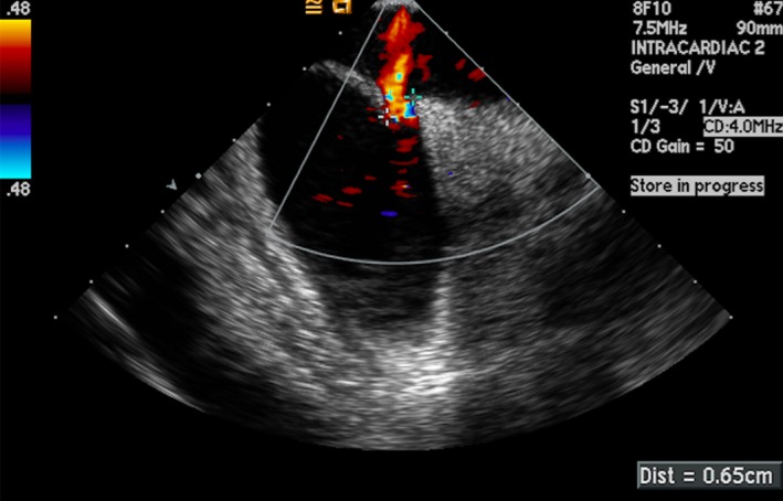
Intracardiac echocardiogram (ICE ) image showing a PFO measuring 6.5 mm using color Doppler.
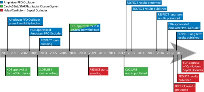
Timeline showing important dates of patent foramen ovale (PFO ) closure trials and US Food and Drug Administration (FDA ) milestones in the United States. HDE indicates Humanitarian Device Exemption.
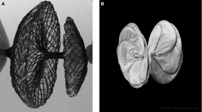
A, The Amplatzer PFO Occluder (courtesy of Abbott. ©2018 Abbott. All rights reserved) and B, the Gore Cardioform Septal Occluder (courtesy of W.L. Gore and Associates, Inc).
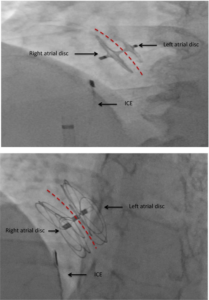
Fluoroscopic image of an Amplatzer PFO Occluder (top) and Gore Cardioform Septal Occluder (bottom) demonstrating a stable position in the atrial septum after release. The intracardiac echo (ICE ) is seen in the right atrium. Red dotted lines represent the interatrial septum in this anteroposterior view.
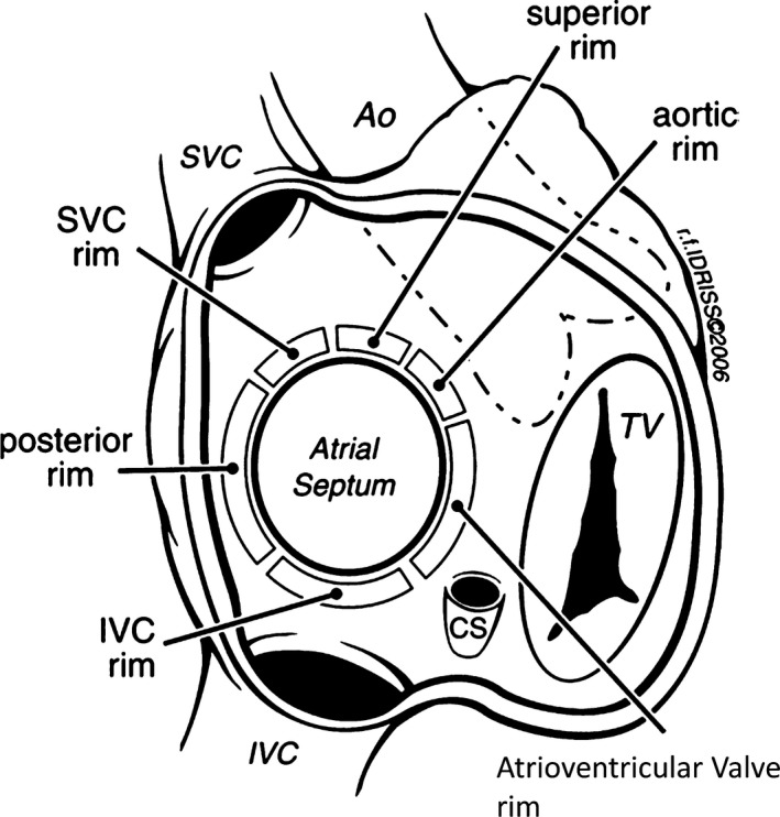
Rims of the atrial septum as seen from the right atrium. Ao indicates aorta; SVC , superior vena cava; IVC , inferior vena cava; TV , tricuspid valve. Reprinted from Amin et al59 with permission. Copyright ©2006, John Wiley & Sons.

Intracardiac echocardiogram showing a patent foramen ovale (PFO ) with a tunnel length of 1.15 cm.
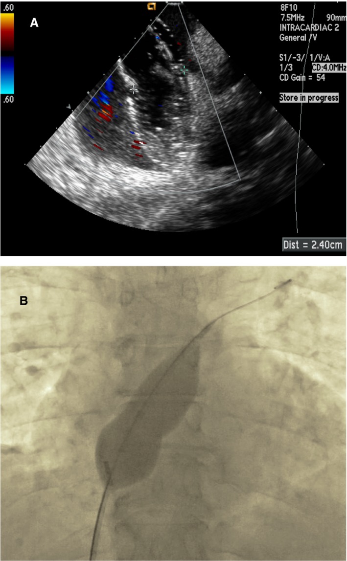
A, Intracardiac image of balloon sizing using stop‐flow technique and (B) fluoroscopic image of an inflated sizing balloon across the patent foramen ovale (PFO ).
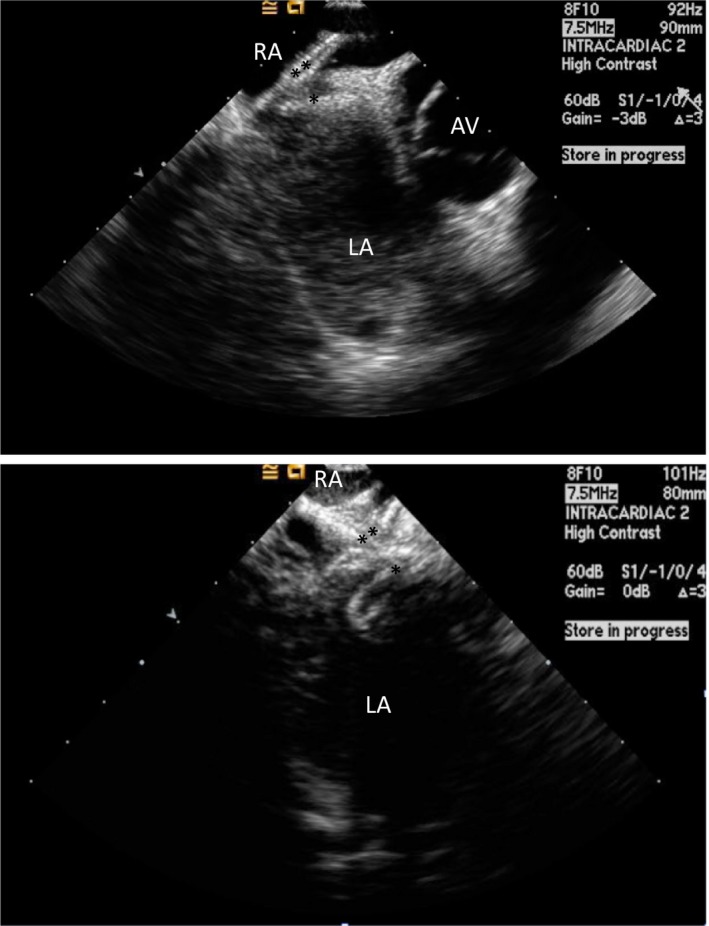
Intracardiac echocardiogram image of the right (**) and left atrial disc (*) of an Amplatzer PFO Occluder (top) and Gore Cardioform Septal Occluder (bottom) before the release of the device. Note the splaying of the discs at the aortic rim. AV indicates aortic valve; LA, left atrium; RA, right atrium.
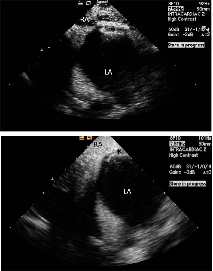
Intracardiac echocardiogram (ICE ) image of a released Amplatzer PFO Occluder (top) (*) and Gore Cardioform Septal Occluder (bottom) (*). Agitated saline is seen in the right atrium (RA ), but not in the left atrium (LA ), confirming the absence of right to left shunt after device deployment.
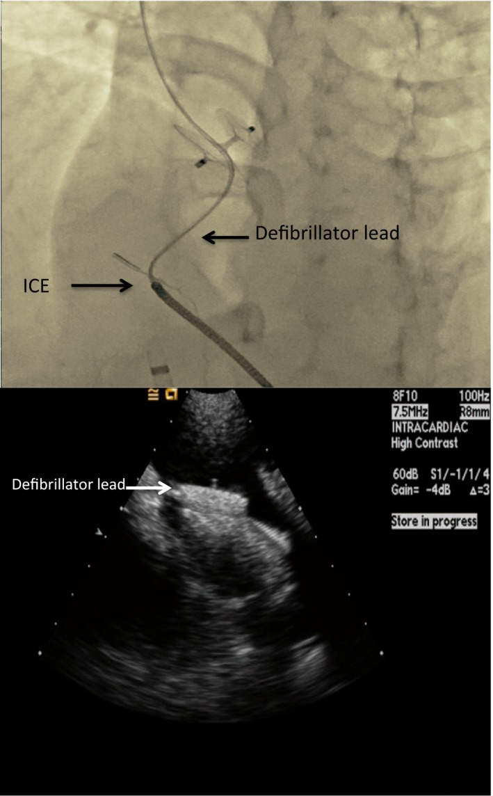
A, Fluoroscopic and (B) ICE images showing a trapped defibrillator lead in between the right atrial disc and the interatrial septum. ICE indicates intracardiac echocardiogram.
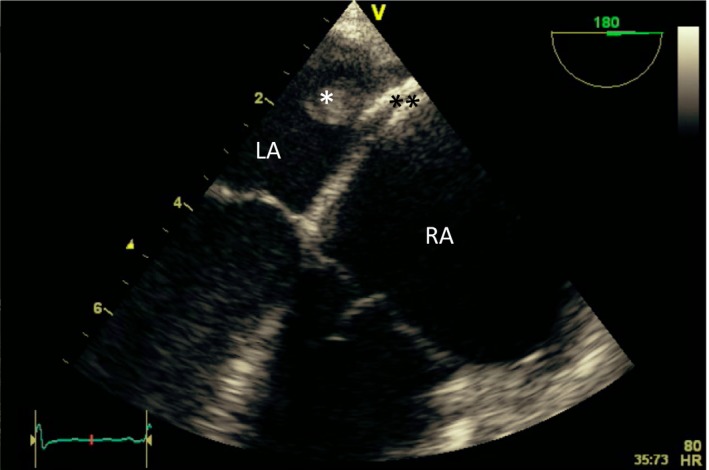
Transesophageal echocardiogram (midesophageal at 180 degrees) showing a thrombus (*) attached to the left atrial disc of an Amplatzer PFO occluder (**). LA indicates left atrium; RA, right atrium.
References
-
- Benjamin EJ, Blaha MJ, Chiuve SE, Cushman M, Das SR, Deo R, de Ferranti SD, Floyd J, Fornage M, Gillespie C, Isasi CR, Jimenez MC, Jordan LC, Judd SE, Lackland D, Lichtman JH, Lisabeth L, Liu S, Longenecker CT, Mackey RH, Matsushita K, Mozaffarian D, Mussolino ME, Nasir K, Neumar RW, Palaniappan L, Pandey DK, Thiagarajan RR, Reeves MJ, Ritchey M, Rodriguez CJ, Roth GA, Rosamond WD, Sasson C, Towfighi A, Tsao CW, Turner MB, Virani SS, Voeks JH, Willey JZ, Wilkins JT, Wu JH, Alger HM, Wong SS, Muntner P; American Heart Association Statistics Committee and Stroke Statistics Subcommittee . Heart disease and stroke statistics—2017 update: a report from the American Heart Association. Circulation. 2017;135:e146–e603. - PMC - PubMed
-
- Writing Group Members , Mozaffarian D, Benjamin EJ, Go AS, Arnett DK, Blaha MJ, Cushman M, Das SR, de Ferranti S, Despres JP, Fullerton HJ, Howard VJ, Huffman MD, Isasi CR, Jimenez MC, Judd SE, Kissela BM, Lichtman JH, Lisabeth LD, Liu S, Mackey RH, Magid DJ, McGuire DK, Mohler ER III, Moy CS, Muntner P, Mussolino ME, Nasir K, Neumar RW, Nichol G, Palaniappan L, Pandey DK, Reeves MJ, Rodriguez CJ, Rosamond W, Sorlie PD, Stein J, Towfighi A, Turan TN, Virani SS, Woo D, Yeh RW, Turner MB; American Heart Association Statistics Committee; Stroke Statistics Subcommittee . Heart disease and stroke statistics—2016 update: a report from the American Heart Association. Circulation. 2016;133:e38–e360. - PubMed
-
- Adams HP Jr, Bendixen BH, Kappelle LJ, Biller J, Love BB, Gordon DL, Marsh EE III. Classification of subtype of acute ischemic stroke. Definitions for use in a multicenter clinical trial. TOAST. Trial of Org 10172 in Acute Stroke Treatment. Stroke. 1993;24:35–41. - PubMed
-
- Hagen PT, Scholz DG, Edwards WD. Incidence and size of patent foramen ovale during the first 10 decades of life: an autopsy study of 965 normal hearts. Mayo Clin Proc. 1984;59:17–20. - PubMed
Publication types
MeSH terms
LinkOut - more resources
Full Text Sources
Other Literature Sources
Medical
