

Granulocytic anaplasmosis in 2 dogs from Quebec
- PMID: 29910483
- PMCID: PMC5949946
Granulocytic anaplasmosis in 2 dogs from Quebec
Abstract
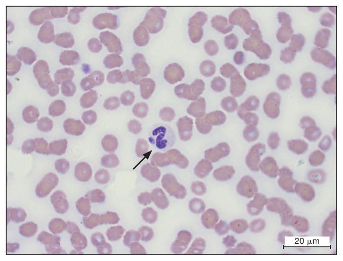
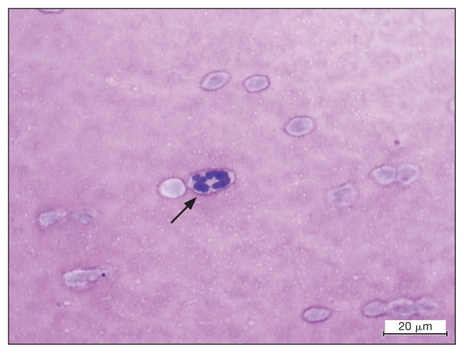
Two dogs from Quebec were diagnosed with granulocytic anaplasmosis. They both displayed fever, lethargy, and anorexia. Other clinical signs included vomiting, uveitis, polyarthritis, hepatomegaly, and splenomegaly. Thrombocytopenia, anemia, and lymphopenia were identified in both cases. Cytoplasmic inclusions were observed within neutrophils, and Anaplasma phagocytophilum infection was confirmed by polymerase chain reaction in both dogs.
Anaplasmose granulocytaire chez deux chiens au Québec. Deux chiens originaires du Québec ont été diagnostiqués avec une anaplasmose granulocytaire. Les chiens ont manifesté de façon aiguë de la fièvre, un abattement et de l’anorexie. D’autres signes cliniques ont été observés incluant vomissement, uvéite, polyarthrite, hépatomégalie et splénomagalie. Une thrombocytopénie, une anémie et une lymphopénie ont été détectées chez les deux chiens. Des inclusions intracytoplasmques étaient également présentent dans les neutrophiles et l’infection à Anaplasma phagocytophilum a été confirmée par réaction d’amplification en chaîne par la polymérase chez les deux chiens.(Traduit par les auteurs).
Figures
Similar articles
-
Granulocytic anaplasmosis in three dogs from Saskatoon, Saskatchewan.Can Vet J. 2009 Aug;50(8):835-40. Can Vet J. 2009. PMID: 19881921 Free PMC article.
-
Granulocytic anaplasmosis in captive ring-tailed lemur (Lemur catta) in Poland.BMC Vet Res. 2021 Mar 12;17(1):118. doi: 10.1186/s12917-021-02827-8. BMC Vet Res. 2021. PMID: 33712007 Free PMC article.
-
A lifelong study of a pack Rhodesian ridgeback dogs reveals subclinical and clinical tick-borne Anaplasma phagocytophilum infections with possible reinfection or persistence.Parasit Vectors. 2018 Apr 12;11(1):238. doi: 10.1186/s13071-018-2806-8. Parasit Vectors. 2018. PMID: 29650038 Free PMC article.
-
Human Granulocytic Anaplasmosis.Infect Dis Clin North Am. 2022 Sep;36(3):639-654. doi: 10.1016/j.idc.2022.02.008. Infect Dis Clin North Am. 2022. PMID: 36116840 Review.
-
Molecular diagnosis of human granulocytic anaplasmosis.Expert Rev Mol Diagn. 2004 Jul;4(4):559-69. doi: 10.1586/14737159.4.4.559. Expert Rev Mol Diagn. 2004. PMID: 15225103 Review.
Cited by
-
Epidemiological and Clinicopathological Features of Anaplasma phagocytophilum Infection in Dogs: A Systematic Review.Front Vet Sci. 2021 Jun 23;8:686644. doi: 10.3389/fvets.2021.686644. eCollection 2021. Front Vet Sci. 2021. PMID: 34250067 Free PMC article. Review.
-
Strategies for the Diagnosis of Granulocytic Anaplasmosis in Two Naturally Infected Dogs.Animals (Basel). 2023 Dec 22;14(1):49. doi: 10.3390/ani14010049. Animals (Basel). 2023. PMID: 38200780 Free PMC article.
-
The Impact of Tick-Borne Diseases on the Bone.Microorganisms. 2021 Mar 23;9(3):663. doi: 10.3390/microorganisms9030663. Microorganisms. 2021. PMID: 33806785 Free PMC article. Review.
References
-
- Carrade DD, Foley JE, Borjesson DL, Sykes JE. Canine granulocytic anaplasmosis: A review. J Vet Intern Med. 2009;23:1129–1141. - PubMed
-
- Kohn B, Galke D, Beelitz P, Pfister K. Clinical features of canine granulocytic anaplasmosis in 18 naturally infected dogs. J Vet Intern Med. 2008;22:1289–1295. - PubMed
-
- Egenvall A, Bjöersdorff A, Lilliehöök I, et al. Early manifestations of granulocytic ehrlichiosis in dogs inoculated experimentally with a Swedish Ehrlichia species isolate. Vet Rec. 1998;143:412–417. - PubMed
Publication types
MeSH terms
Substances
LinkOut - more resources
Full Text Sources