. 2018 Jun:90:119-160.
doi: 10.1016/j.simyco.2017.12.002. Epub 2018 Jan 2.

Zombie-ant fungi across continents: 15 new species and new combinations within Ophiocordyceps. I. Myrmecophilous hirsutelloid species

Affiliations

Zombie-ant fungi across continents: 15 new species and new combinations within Ophiocordyceps. I. Myrmecophilous hirsutelloid species

J P M Araújo et al. Stud Mycol. 2018 Jun.

Abstract

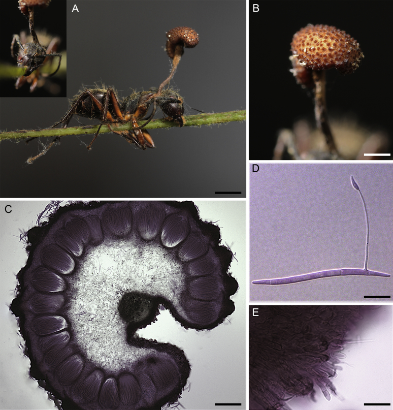
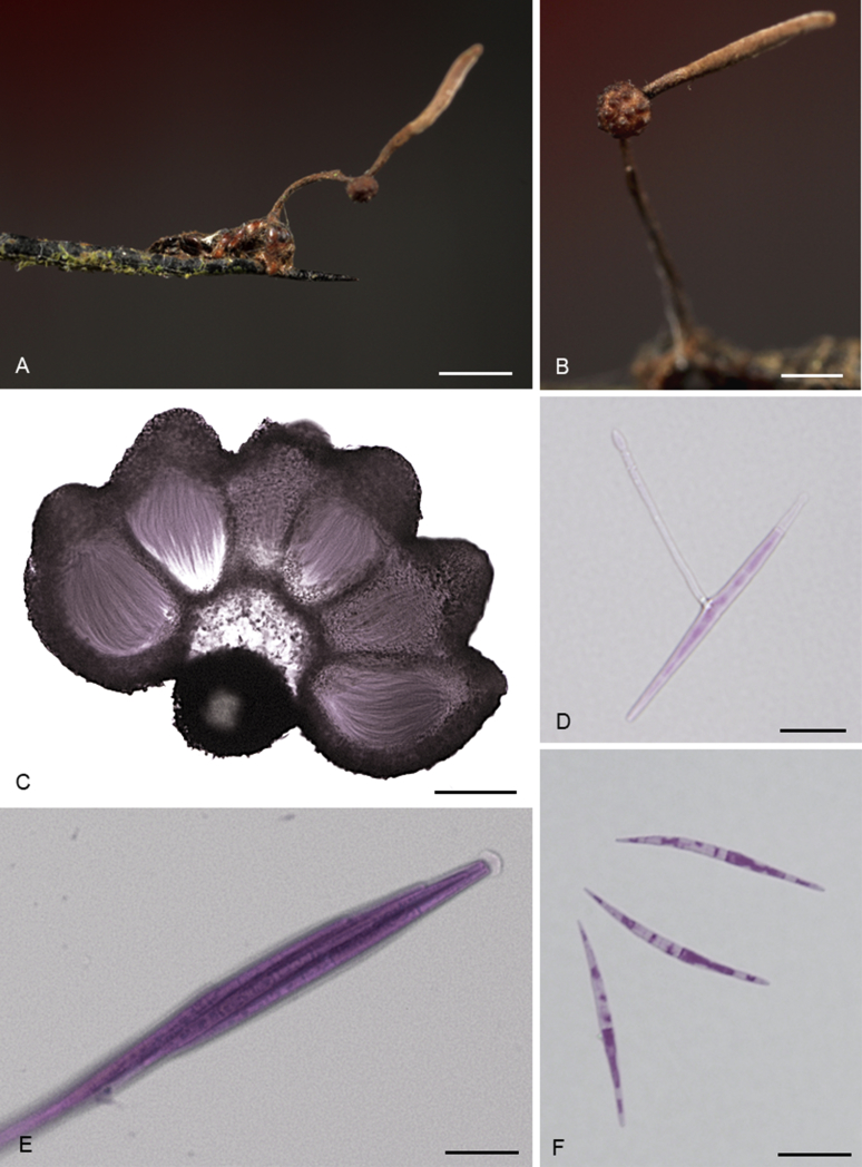
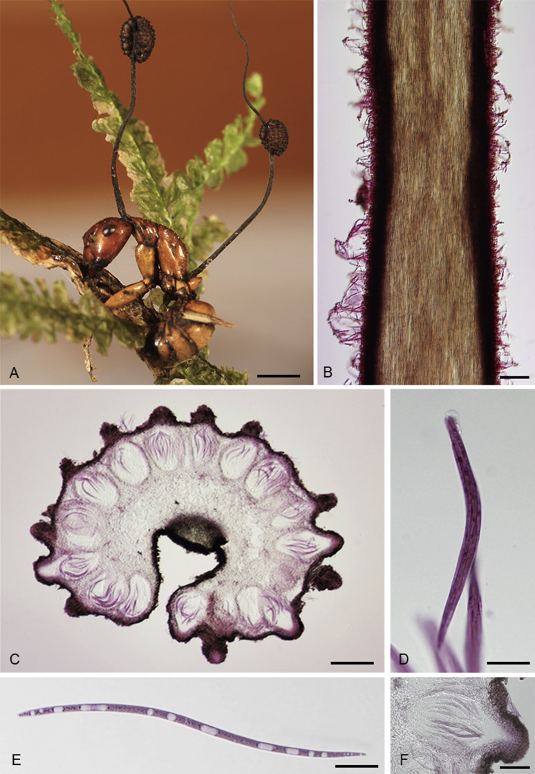
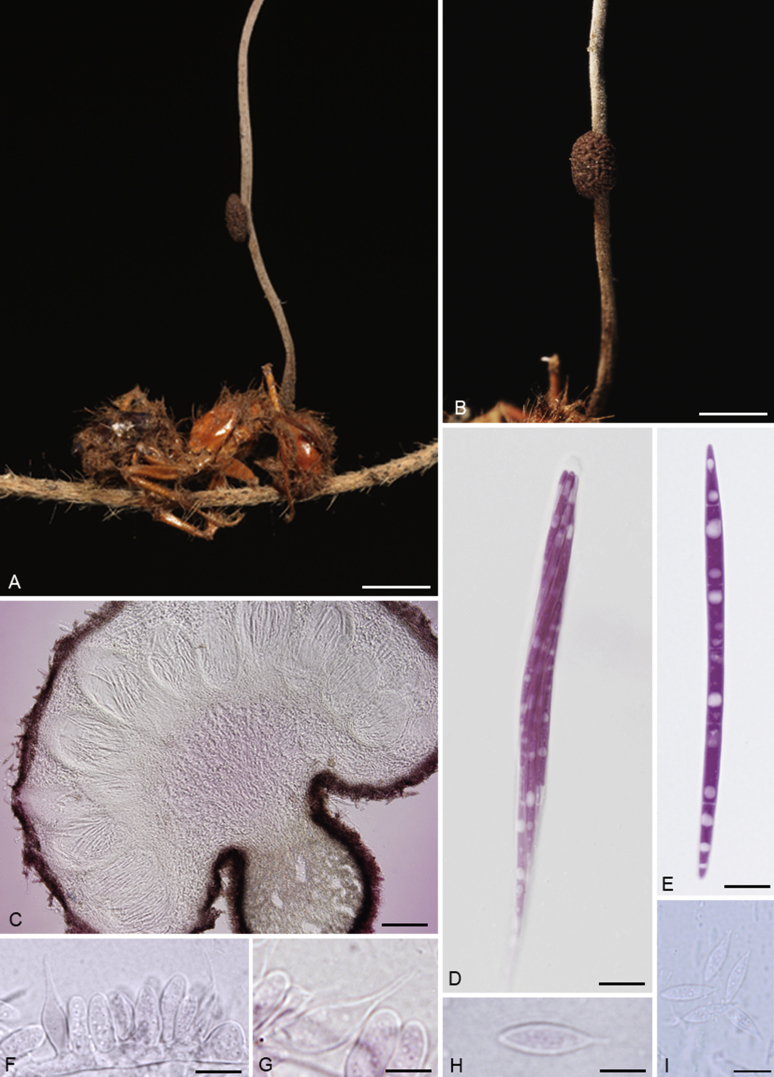
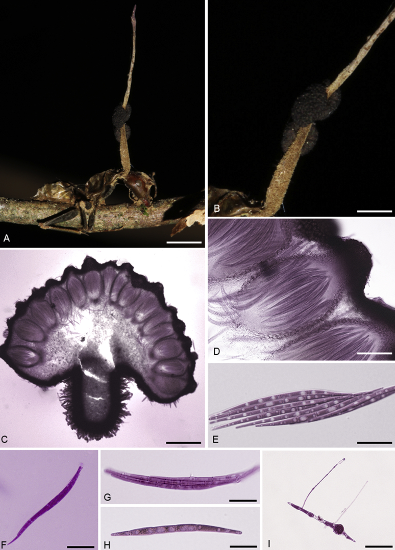
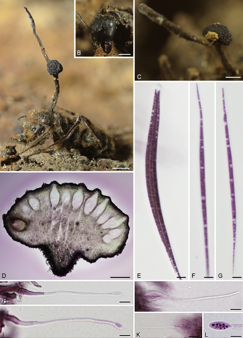
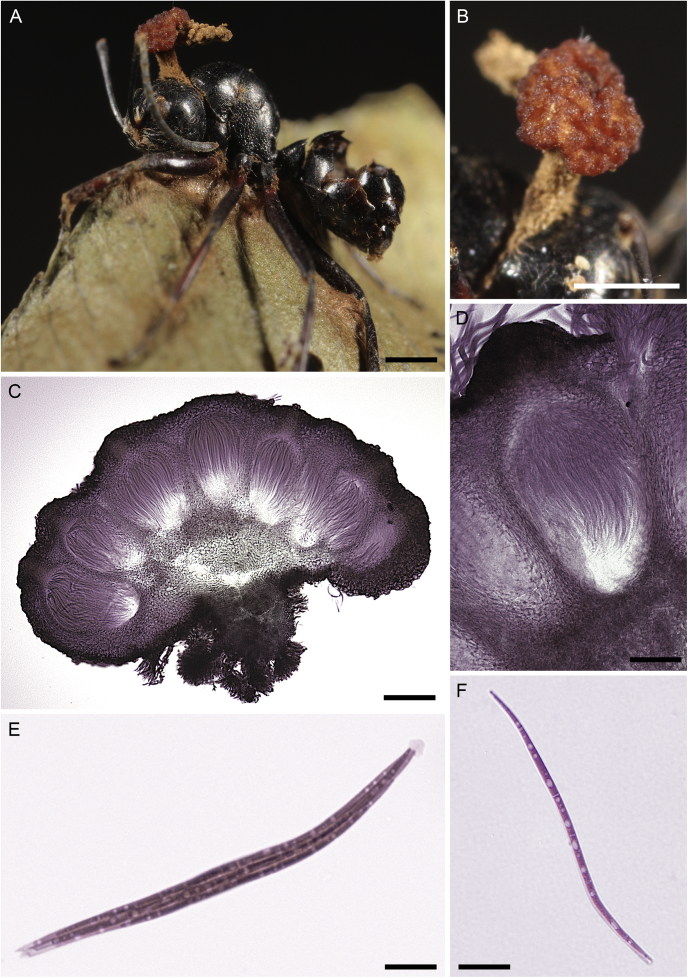
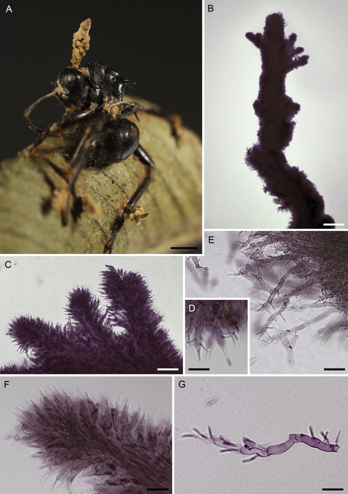
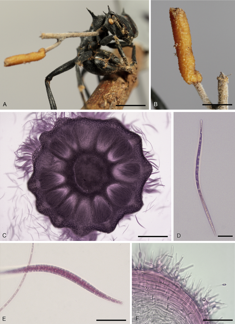
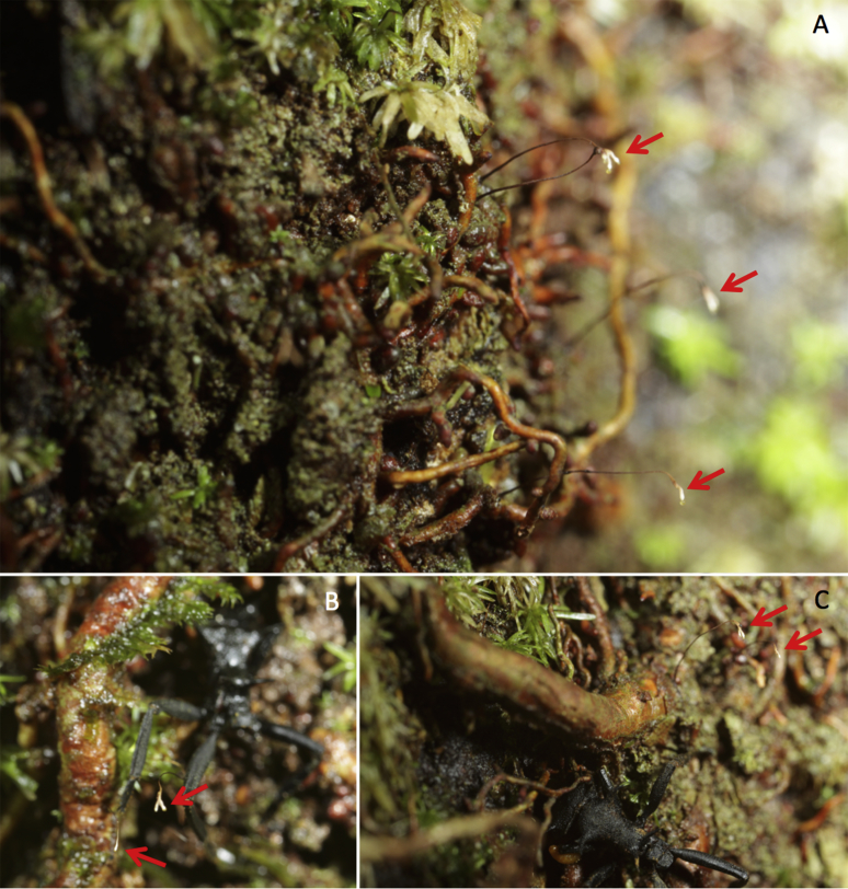
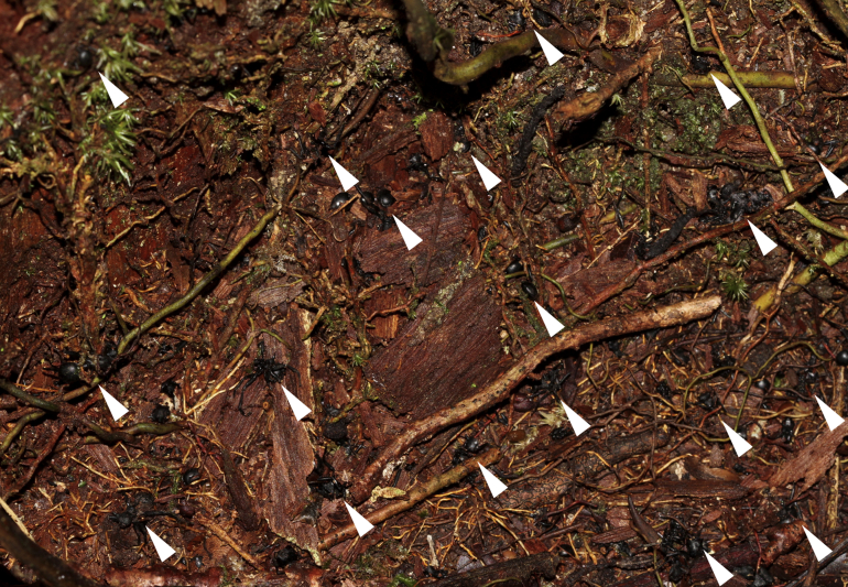
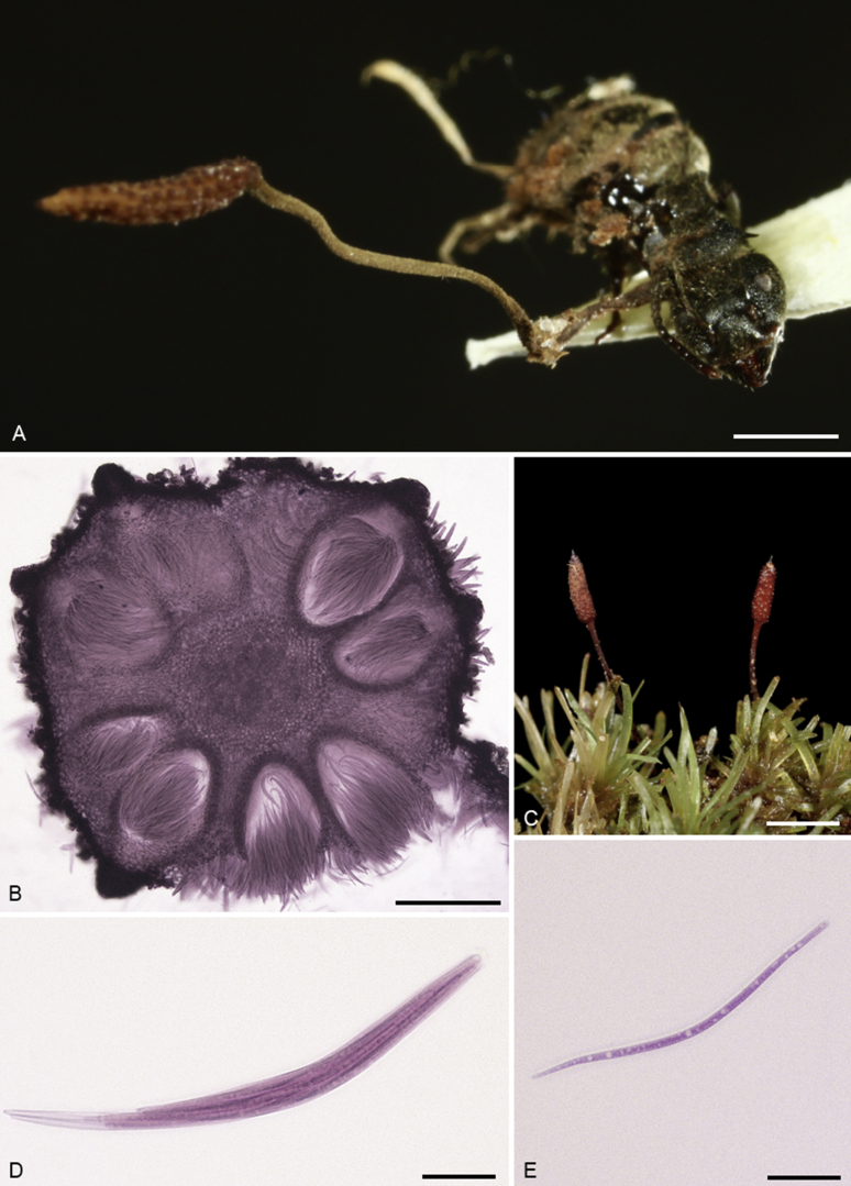
Ophiocordyceps species infecting ants - the so-called zombie-ant fungi - comprise one of the most intriguing and fascinating relationships between microbes and animals. They are widespread within tropical forests worldwide, with relatively few reports from temperate ecosystems. These pathogens possess the ability to manipulate host behaviour in order to increase their own fitness. Depending on the fungal species involved the infected ants are manipulated either to leave the nest to ascend understorey shrubs, to die biting onto vegetation, or descend from the canopy to die at the base of trees. Experimental evidence has demonstrated that the behavioural change aids spore dispersal and thus increases the chances of infection, because of the existing behavioural immunity expressed inside ant colonies that limits fungal development and transmission. Despite their undoubted importance for ecosystem functioning, these fungal pathogens are still poorly documented, especially regarding their diversity, ecology and evolutionary relationships. Here, we describe 15 new species of Ophiocordyceps with hirsutella-like asexual morphs that exclusively infect ants. These form a monophyletic group that we identified in this study as myrmecophilous hirsutelloid species. We also propose new combinations for species previously described as varieties and provide for the first time important morphological and ecological information. The species proposed herein were collected in Brazil, Colombia, USA, Australia and Japan. All species could readily be separated using classic taxonomic criteria, in particular ascospore and asexual morphology.

Keywords: Behaviour manipulation; Camponotini; Entomopathogenic fungi; Host association; Hypocreales; Insect pathogen; Multigene phylogeny; O. albacongiuae Araújo, H.C. Evans & D.P. Hughes; O. blakebarnesii Araújo, H.C. Evans & D.P. Hughes; O. camponoti-chartificis Araújo, H.C. Evans & D.P. Hughes; O. camponoti-femorati Araújo, H.C. Evans & D.P. Hughes; O. camponoti-floridani Araújo, H.C. Evans & D.P. Hughes; O. camponoti-hippocrepidis Araújo, H.C. Evans & D.P. Hughes; O. camponoti-nidulantis Araújo, H.C. Evans & D.P. Hughes; O. camponoti-renggeri Araújo, H.C. Evans & D.P. Hughes; O. camponoti-sexguttati Araújo, H.C. Evans & D.P. Hughes; O. daceti Araújo, H.C. Evans & D.P. Hughes; O. kimflemingiae Araújo, H.C. Evans & D.P. Hughes; O. monacidis (H.C. Evans & Samson) Araújo, H.C. Evans & D.P. Hughes; O. naomipierceae Araújo, H.C. Evans & D.P. Hughes; O. oecophyllae Araújo, S. Abell, T. Marney, R. Shivas H.C. Evans & D.P. Hughes; O. ootakii Araújo, H.C. Evans & D.P. Hughes.; O. satoi Araújo, H.C. Evans & D.P. Hughes; Ophiocordyceps; Ophiocordyceps dolichoderi (H.C. Evans & Samson) Araújo, H.C. Evans & D.P. Hughes; Ophiocordyceps unilateralis; Zombie-ant fungi.


Figures

Fig. 1
Fig. 1
Ophiocordyceps daceti. A. Infected Daceton armigerum on the leaf litter. B. Cross-section of the synnema. C. Close-up of synnema showing the Hirsutella hymenium. D–F. Verrucose phialides. G. Phialides at early developmental stage. H. Close-up of the hymenium of verrucose Hirsutella phialides. Scale bars: A = 5 mm, B = 200 μm, C = 50 μm, D–H = 10 μm.
Fig. 2
Fig. 2
A.Oecophylla smaragdina infected and biting the main vein of a leaf. B. Leg joints with phialides. C–D. Phialides. E. Phialides and conidia. Scale bars: A = 1 mm, B = 0.4 mm, C–E = 10 μm.
Fig. 3
Fig. 3
Ophiocordyceps camponoti–sexguttati. A.Camponotus sexguttatus biting into vegetation with the long stroma arising from its dorsal pronotum. B. Close-up of the ascoma. C. Section through ascoma showing the perithecial arrangement. D. Close-up of perithecium. E. Long ascospores with the straight capilliconidiophore bearing an apical capilliconidium. F. Ascus. Scale bars: A = 5 mm, B = 1 mm, C = 100 μm, D = 50 μm, E–F = 20 μm.
Fig. 4
Fig. 4
Ophiocordyceps camponoti–renggeri. A.Camponotus renggeri, dead and attached to bryophytes on the base of trees. B. Close-up of the fertile part (ascoma). C. Section through ascoma showing the perithecial arrangement. D. Close-up of perithecium. E. Asci. F. Ascospores. Scale bars: A = 5 mm, B = 1 mm, C = 250 μm, D = 50 μm, E = 70 μm, F = 20 μm.
Fig. 5
Fig. 5
Ophiocordyceps camponoti–renggeri (asexual morph). A. Ant biting into the moss carpet with the antennae raised, showing detail of the sporodochia. B. Phialides (hirsutella B-type). Scale bars: A = 1 mm, B = 30 μm.
Fig. 6
Fig. 6
Ophiocordyceps camponoti–chartificis. A.Camponotus chartifex biting onto a palm leaf. B. Close-up of the ascoma. C. Cross section of the ascoma showing the perithecial arrangement. D. Close-up of the perithecium. E. Ascus with ascospores arranged within. F. Non-germinated ascospore. G. Ascospore with long capilliconidia. H. Hirsutella A-type phialides on the stroma. Scale bars. A = 5 mm, B = 1 mm, C = 200 μm, D = 30 μm, E–F = 5 μm, G–H = 10 μm.
Fig. 7
Fig. 7
Ophiocordyceps camponoti–nidulantis (sexual morph). A.Camponotus nidulans infected and biting into a leaf (with fly larvae on the stroma). B. Close-up of the ascoma. C. Section through ascoma showing the perithecial arrangement. D. Close-up of perithecium. E. Asci. F. Ascospore with capilliconidium. Scale bars: A = 3 mm, B = 1 mm, C = 200 μm, D = 75 μm, E=F = 20 μm.
Fig. 8
Fig. 8
Ophiocordyceps camponoti–nidulantis (asexual morph). A. Leg joints with hirsutella-like phialides. B. Close-up of leg phialides. C. Close-up of antenna covered with Hirsutella phialides. D. Antenna covered with phialides. E. Typical antennal display exhibited by the ant after being killed by O. camponoti-nidulantis, exposing the Hirsutella hymenium. Scale bars: A = 1 mm, B = 40 μm, C = 100 μm, D = 0.5 mm, E = 0.5 mm.
Fig. 9
Fig. 9
Ophiocordyceps camponoti–femorati. A.Camponotus femoratus biting a palm leaf. B. Close-up of the ascoma. C. Cross section showing the perithecial arrangement. D. Ascospore with capilliconidia E. Hirsutella A-type phialide on the stroma. Scale bars: A = 1 mm, B = 0.3 mm, C = 200 μm, D = 20 μm, E = 5 μm.
Fig. 10
Fig. 10
Ophiocordyceps camponoti-hippocrepidis. A. Minute Camponotus hippocrepis (ca. 1.5 mm) biting onto a palm spine. B. Close-up of the ascoma. C. Cross-section of the ascoma showing the perithecial arrangement. D. Ascospore with capilliconidiophore with verrucose apical portion. E. Ascus. F. Ascospores just after being released . Scale bars: A = 1 mm, B = 0.5 mm, C = 100 μm, D–E = 20 μm, F = 20 μm.
Fig. 11
Fig. 11
Ophiocordyceps albacongiuae. A.Camponotus sp. with two fruiting bodies emerging from its dorsal pronotum and petiole. B. Sterile synnema with its hairy surface. C. Cross-section of the ascoma. D. Ascus. E. Ascospore. F. Close-up of the perithecium. Scale bars: A = 0.2 mm, B = 100 μm, C = 200 μm, D = 20 μm, E = 10 μm, F = 30 μm.
Fig. 12
Fig. 12
Ophiocordyceps camponoti–floridani. A.Camponotus floridanus infected, biting into a plant. B. Close-up of the disc-shape ascoma attached to the stroma. C. Cross-section of the ascoma. D. Ascus. E. Ascospore. F–G.Hirsutella phialides. H–I. Limoniform conidia. Scale bars: A = 2 mm, B = 1 mm, C = 100 μm, D–F = 10 μm, G–I = 5 μm.
Fig. 13
Fig. 13
Ophiocordyceps kimflemingiae. A.Camponotus castaneus biting a twig. B. Close-up of the stroma showing two ascomatal plates attached on it. C. Ascoma section and Perithecia arranged on its surface. D. Perithecium. E. Cluster of ascospores. F–G. Ascus. H. Ascospore. I. Ascospore after 2–5 d on agar, exhibiting a swollen section and two capilliconidiophores. Scale bars: A = 2 mm, B = 0.5 mm, C = 300 μm, D = 100 μm, E–F = 20 μm, G = 10 μm, H = 20 μm, I = 40 μm.
Fig. 14
Fig. 14
Ophiocordyceps blakebarnesii. A.Camponotus cf. chromaoides with the stroma arising from the dorsal pronotum. B. Close-up showing the biting behaviour inside the log. C. Stroma. D. Cross-section of the ascoma. E. Ascus. F–G. Ascospores. H–K. Phialides. L. Multi guttulate conidia. Scale bars: A = 2 mm, B = 0.5 mm, C = 0.3 mm, D = 200 μm, E–G = 5 μm, H–K = 10 μm, L = 5 μm.
Fig. 15
Fig. 15
Ophiocordyceps naomipierceae (sexual morph). A.Polyrhachis sp. biting the edge of a leaf. B. Close-up of the orange ascoma. C. Cross-section of the ascoma. D. Perithecium. E. Ascus. F. Ascospore. Scale bars: A = 0.5 mm, B = 1 mm, C = 100 μm, D = 50 μm, E–F = 20 μm.
Fig. 16
Fig. 16
Ophiocordyceps naomipierceae (asexual morph). A.Polyrhachis sp. with stromata arising from leg joints and dorsal pronotum. B. Synnema. C. Close-up of synnema. D. Close-up phialides E. Phialides. F. Close-up synnema showing apical phialides. G. Individual long phialide with multiple verrucose necks. Scale bars: A = 0.5 mm, B = 100 μm, C = 20 μm, D = 15 μm, E–G = 10 μm.
Fig. 17
Fig. 17
Ophiocordyceps ootakii. A.Polyrhachis moesta biting on a leaf edge. B. Close-up showing two ascomatal plates attached to the stroma. C. Cross-section of ascoma. D. Ascus. E. Ascospore. F. Hirsutella A-type phialides on stroma. Scale bars: A–B = 1 mm, C = 250 μm, D–E = 20 μm, F = 10 μm.
Fig. 18
Fig. 18
Ophiocordyceps satoi. A.Polyrhachis lamellidens. with three stromata arising from its body. B. Close-up stroma with two ascomatal cushions. C. Cross-section of the ascoma, showing the perithecial arrangement. D. Close-up perithecium. E. Ascus. F. Ascospore germinating on agar plate after 3–5 d. G. Capilliconidium. H. Ascospore with two capilliconidiophores bearing one capilliconidium at their apices. Scale bars: A = 1 mm, B = 0.5 mm, C = 100 μm, D = 40 μm, E–F = 20 μm, G = 2 μm, H = 20 μm.
Fig. 19
Fig. 19
Maximum Likelihood tree of Ophiocordyceps obtained with a combined dataset of SSU, LSU, tef, RPB1 and RPB2 based on Bayesian/RAxML analysis with only >0.95/70 shown. Species proposed in this study are highlighted. Ant figures correspond to the ant genera infected by each clade within hirsutelloid Ophiocordyceps. At the top right a round phylogeny showing the whole analyses with the entire dataset used in this study, which included Cordycipitaceae, Clavicipitaceae and Ophiocordycipitaceae species, with Ophiocordyceps highlighted. (ant images from www.AntWeb.org and the photographers: Oecophylla, Camponotus, Dolichoderus, Cephalotes, Paraponera and Daceton: April Nobile, Polyrhachis: Will Ericson.
Fig. 20
Fig. 20
Comparison of the phialide morphology for the species within the O. kniphofioides clade (A-phialides and conidia). A.O. kniphofioides sensu stricto. B.O. monacidis. C.O. ponerinarumD.O. kniphofioides var. gnamptogenyos. E.O. daceti sp. nov. (A–D. Evans & Samson 1984; E. This study). Scale bar = 10 μm.
Fig. 21
Fig. 21
Ophiocordyceps kniphofioides sensu stricto. A.Cephalotes atratus with the stroma arising laterally from pronotum. B. Close-up of the ascoma. C. Section of the ascoma showing the immersed perithecia. D. Ascospore. E. Ascus. F. Hirsutella-like phialides, present along the stroma (stalk). Scale bar: A = 2 mm. B = 1 mm. C = 200 μm. D–F = 20 μm.
Fig. 22
Fig. 22
A. Synnemata of the asexual morph (Hirsutella type-C. arising from the moss/trunk which remain attached, even after removal of the corpse. B–C. Close-up of the infected ants with synnemata on the surrounding substrate.
Fig. 23
Fig. 23
Base of the tree, with corpses of infected Cephalotes atratus removed from the trunk by the activity of other workers (Araújo & Hughes 2017).
Fig. 24
Fig. 24
Ophiocordyceps monacidis. A.Dolichoderus (Monacis) bispinosus infected by O. monacidis. B. Cross-section of the ascoma. C. Ascomata arising from a carpet of moss. D. Ascus. E. Ascospore. Scale bars: A = 1 mm, B = 200 μm, C = 3 mm, D = 20 μm, E = 30 μm.
Fig. 25
Fig. 25
A–B.Ophiocordyceps monacidis stromata among the moss Octoblepharum albidum. The ants die hidden underneath the moss carpet just exposing the fungal structures (white arrows). C–D.O. albidum on the tree surface with its orange sporophytes.
Fig. 26
Fig. 26
Daceton armigerum infected by Ophiocordyceps daceti. A. Infected ant attached on the leaf petiole, in its original upside–down position. B. Close-up showing the attachment exclusively by the host legs with no apparent fungal attachment structures. C. Close-up showing the early stages of O. daceti emerging from the host’s dorsal pronotum.
Fig. 27
Fig. 27
Different behavioural manipulation within the O. unilateralis complex. A–C. Dead O. camponoti-renggeri as they are typically found, among moss at the base of trees. D–E. Smaller ants (e.g. O. camponoti–bispinosi, O. camponoti–hippocrepidis and O. camponoti–femorati) die often at the very tip of palm spines and epiphytes where water droplets form, providing continuous water resource.

References

    1. Andersen S.B., Gerritsma S., Yusah K.M. The life of a dead ant: the expression of an adaptive extended phenotype. American Naturalist. 2009;174:424–433. - PubMed
    1. Araújo J.P.M., Evans H.C., Geiser D.M. Unravelling the diversity behind the Ophiocordyceps unilateralis (Ophiocordycipitaceae) complex: three new species of zombie-ant fungi from the Brazilian Amazon. Phytotaxa. 2015;220:224–238.
    1. Araújo J.P.M., Hughes D.P. Diversity of entomopathogenic fungi: which groups conquered the insect body? Advances in Genetics. 2016;94:1–39. - PubMed
    1. Araújo J.P.M., Hughes D.P. The fungal spore: myrmecophilous Ophiocordyceps as a case study. In: Dighton J., White J.M., editors. The Fungal Community: Its Organization and Role in the Ecosystem. CRC Press; USA: 2017.
    1. Castlebury L.A., Rossman A.Y., Sung G.-H. Multigene phylogeny reveals new lineage for Stachybotrys chartarum, the indoor air fungus. Mycological Research. 2004;108:864–872. - PubMed

LinkOut - more resources