Review
. 2018 Jun 1:9:656.
doi: 10.3389/fphys.2018.00656. eCollection 2018.

Squeezing for Life - Properties of Red Blood Cell Deformability

Affiliations
Review

Squeezing for Life - Properties of Red Blood Cell Deformability

Rick Huisjes et al. Front Physiol. .

Abstract

Deformability is an essential feature of blood cells (RBCs) that enables them to travel through even the smallest capillaries of the human body. Deformability is a function of (i) structural elements of cytoskeletal proteins, (ii) processes controlling intracellular ion and water handling and (iii) membrane surface-to-volume ratio. All these factors may be altered in various forms of hereditary hemolytic anemia, such as sickle cell disease, thalassemia, hereditary spherocytosis and hereditary xerocytosis. Although mutations are known as the primary causes of these congenital anemias, little is known about the resulting secondary processes that affect RBC deformability (such as secondary changes in RBC hydration, membrane protein phosphorylation, and RBC vesiculation). These secondary processes could, however, play an important role in the premature removal of the aberrant RBCs by the spleen. Altered RBC deformability could contribute to disease pathophysiology in various disorders of the RBC. Here we review the current knowledge on RBC deformability in different forms of hereditary hemolytic anemia and describe secondary mechanisms involved in RBC deformability.

Keywords: deformability; enzymopathies; hemolysis; hereditary spherocytosis; hydration; sickle cell anemia; thalassemia; vesiculation.


Figures

FIGURE 1
FIGURE 1
Osmotic gradient ektacytometry as a tool to measure red blood cell (RBC) deformability. The technique is discussed in detail by Clark et al. (1983); Lazarova et al. (2017), and Da Costa et al. (2016). Briefly, during osmotic gradient ektacytometry, the RBC is subjected to an osmotic gradient (range ≈50 mOsmol/kg H2O – 650 mOsmol/kg H2O) under constant shear stress, while the elongation index (EI) is measured. The EI corresponds to the deformability at various osmotic conditions. (A) the left graph depicts an osmotic gradient ektacytometry curve from a healthy control with various intersection points: the EImax reflects the maximal deformability of the RBC, Omin reflects the osmotic fragility and Ohyper reflects the (de)hydration state (or intracellular viscosity) of the RBC. (B) In the right graph osmotic gradient curves of patients with sickle cell disease (black line), β-thalassemia (green line), hereditary xerocytosis (blue line) and hereditary spherocytosis (red line) and several individual healthy controls (gray lines, n = 20) are depicted.
FIGURE 2
FIGURE 2
schematic overview of RBC ion pumps (Sachs, 2003; Tiffert et al., 2003), symporters and antiporters (Bernhardt et al., 1988; Wolfersberger, 1994) and ion channels (Bouyer et al., 2012; Kaestner, 2011, 2015).
FIGURE 3
FIGURE 3
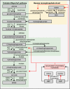
Glycolytic pathway of RBCs is essential for energy metabolism as RBCs lack mitochondria and a nucleus. Enzyme deficiencies can lead to decreased intracellular ATP levels, which possibly reduce RBC deformability. Depicted here are the Embden–Meyerhof pathway, hexose monophosphate shunt and the influence of a pyrimidine 5′-nucleotidase deficiency. Red arrows illustrate negative feedback or reduced production. This illustration is a merged figure adapted from both van Wijk and van Solinge (2005) and Zanella et al. (2006).
FIGURE 4
FIGURE 4
cAMP production by adenylyl cyclase (AC). AC is activated after stimulation by G-protein coupled receptors (GPCR) and converts ATP to cAMP. Examples of GPCRs 2-adrenergic receptor, the lysophosphatidic β in RBCs are the erythrocyte acid (LPA) receptor and the purinergic (P2Y) receptor. cAMP acts as a second messenger in RBCs and among other things stimulate protein kinase A (PKA). Under low ATP concentrations, AMP-activated protein kinase (AMPK) becomes active and can inactivate or activate yet not fully understood processes in RBCs.
FIGURE 5
FIGURE 5
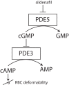
effect of PDE-5 inhibitors, such as sildenafil, on RBC deformability. PDE-5 inhibitors elevate intracellular concentrations of cGMP, thereby indirectly inhibiting PDE3. Inhibition of PDE3 does raise the intracellular concentration of cAMP. Depending on the degree of this intracellular cAMP elevation, RBC deformability can be either increased or decreased.
FIGURE 6
FIGURE 6
RBC dehydration in sickle cell disease. The Na-K pump (Na, K-ATPase) is more active in sickle RBCs, leading to dehydration as extrusion of 3 Na+ ions leads to the influx of 2 K+ ions. Dehydration of sickle cell is initiated by PIEZO1 activation, likely because PIEZO1 is stretch-activation after deoxygenation (Ma et al., 2012). Dehydration in sickle cell disease is also initiated by the upregulation of the NMDA receptor. Both the NMDA receptor and PIEZO1 activation result in Ca2+ influx that leads to KCNN4 activation (KCCN4 in RBCs is better known as the Gardos channel). Gardos channel activation in sickle cell disease is also achieved by signaling cascades which respond to increased levels of cytokines. This leads to K+ and water efflux by aquaporins (AQ1). In sickle cell disease, the K-Cl cotransporter is also activated at low oxygen tensions, causing efflux of K+ and subsequently loss of water by aquaporins (AQ1), while under normal conditions KCC is only active at normal pO2. Although water can be cotransported through KCC1 (Zeuthen and Macaulay, 2012), water efflux in RBCs mainly driven by aquaporins (AQ1).
FIGURE 7
FIGURE 7
Red blood cell vesiculation is driven by increased concentrations of intracellular Ca2+. Phosphatidylserine (PS) is normally presented in the inner-leaflet of the RBC. Elevated intracellular Ca2+ levels activate scramblase and inhibit flippase, leading to phosphatidylserine (PS) externalization and redistribution of membrane and cytoskeletal proteins. PS externalization and protein redistribution leads to vesicle formation and loss of RBC deformability (Alaarg et al., 2013).

References

    1. Adderley S. P., Sprague R. S., Stephenson A. H., Hanson M. S. (2010). Regulation of cAMP by phosphodiesterases in erythrocytes. Pharmacol. Rep. 62 475–482. 10.1016/s1734-1140(10)70303-0 - DOI - PMC - PubMed
    1. Adderley S. P., Thuet K. M., Sridharan M., Bowles E. A., Stephenson A. H., Ellsworth M. L., et al. (2011). Identification of cytosolic phosphodiesterases in the erythrocyte: a possible role for PDE5. Med. Sci. Monit. 17 CR241–CR247 10.12659/msm.881763 - DOI - PMC - PubMed
    1. Adragna N. C., Di Fulvio M., Lauf P. K. (2004). Regulation of K-Cl cotransport: from function to genes. J. Membr. Biol. 201 109–137. 10.1007/s00232-004-0695-6 - DOI - PubMed
    1. Alaarg A., Schiffelers R. M., van Solinge W. W., van Wijk R. (2013). Red blood cell vesiculation in hereditary hemolytic anemia. Front. Physiol. 4:365 10.3389/fphys.2013.00365 - DOI - PMC - PubMed
    1. Alapan Y., Little J. A., Gurkan U. A. (2015). Heterogeneous red blood cell adhesion and deformability in sickle cell disease. Sci. Rep. 4:7173 10.1038/srep07173 - DOI - PMC - PubMed

LinkOut - more resources