Review
. 2018 Jun 1:9:1190.
doi: 10.3389/fmicb.2018.01190. eCollection 2018.

Extracellular Vesicles in Chagas Disease: A New Passenger for an Old Disease

Affiliations
Review

Extracellular Vesicles in Chagas Disease: A New Passenger for an Old Disease

Luis M de Pablos Torró et al. Front Microbiol. .

Abstract

Extracellular vesicles (EVs) are small lipid vesicles released by prokaryotic and eukaryotic cells containing nucleic acids, proteins, and small metabolites essential for cellular communication. Depending on the targeted cell, EVs can act either locally or in distant tissues in a paracrine or endocrine cell signaling manner. Released EVs from virus-infected cells, bacteria, fungi, or parasites have been demonstrated to perform a pivotal role in a myriad of biochemical changes occurring in the host and pathogen, including the modulation the immune system. In the past few years, the biology of Trypanosoma cruzi EVs, as well as their role in innate immunity evasion, has been started to be unveiled. This review article will present findings on and provide a coherent understanding of the currently known mechanisms of action of T. cruzi-EVs and hypothesize the implication of these parasite components during the acute and chronic phases of Chagas disease.

Keywords: Leishmania spp.; Trypanosoma brucei; Trypanosoma cruzi; ectosome; exosome; kinetoplastids; microvesicle; pathogen.


Figures

FIGURE 1
FIGURE 1
The life cycle of Trypanosoma cruzi. (A) Bloodstream trypomastigotes (green) are ingested with the bloodmeal by the reduviid vector. (B) Those forms will transform into epimastigotes (purple) and intermediate spheromastigotes forms (red) in the insect midgut. (C) New developmental differentiation into metacyclic trypomastigotes (MTs) (orange) will occur in the insect rectum before those forms are released with the feces, infecting the mammalian host. (D) The MTs (1st round of intracellular replication) or bloodstream trypomastigotes (green) parasites will then invade a wide range of nucleated cells in an intracellular cycle composed of up to four steps: (1) Attachment, invasion, and parasitophorous vacuole formation (0–4 h). (2) Differentiation into intracellular amastigote forms and release from parasitophorous vacuole (12–24 h). (3) Amastigote replication by binary fission (24–72 h). (4) Differentiation into bloodstream trypomastigotes and release from the infected host cell (72–120 h). The bloodstream forms are then ready to either invade new cells or been taken by the insect vector. Parasite life cycle stage abbreviations: AMA, amastigotes; META, metacyclic trypomastigotes; TRYPO, bloodstream trypomastigote; EPI, epimastigotes; SPHERO, spheromastigotes.
FIGURE 2
FIGURE 2
Types of EV transfer and interaction in acute and chronic phases of Chagas disease. (A) EVs could be released by Trypomastigote or Amastigote forms during Acute and/or Chronic phase of the disease. This secretion could be mediated between parasites, parasites and its host cells or between infected and uninfected cells mediating Autocrine, Yuxtacrine, or Paracrine type of signaling. EVs could be also released to the bloodstream or lymphatic fluids for distant endocrine targeting. During Chronic phase endocrine signaling may mediate changes for tissue re-education, colonization, and further inflammation. (B) Scanning electron microscopy of a MT invading a host cell. The right figure shows a magnified image of the MT point of contact with the host cell membrane. The gray arrows indicate the EVs of different sizes that may derive from both parasite and host cell. This picture corresponds to an infection of Vero Cells with MTs (strain PAN4 DTU Ia) for 2 h and belongs to the same series of scanning microscopic pictures published in Díaz Lozano et al. (2017).
FIGURE 3
FIGURE 3
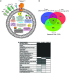
Trypanosoma cruzi EV protein composition. (A) Schematic representation of protein components described in T. cruzi EVs. Note that for the purpose of the figure, the different components were not represented in the scale or density of molecules. Only representative proteins from each protein group (surface, peptidases, metabolic enzymes, conserved cytosqueletal, antioxidant, RNA and DNA related proteins) are represented. (B) Venn diagram of T. cruzi (n = 237) (Bayer-Santos et al., 2013), Leishmania major (n = 261) (Silverman et al., 2010), and Trypanosoma brucei (n = 191) (Szempruch et al., 2016) EVs proteomic datasets (hypothetical proteins has been excluded from the analysis). (C) List of T. cruzi, L. major, and T. brucei EV proteins also found among the top 100 most frequently found vesicular proteins in mammalian EVs (Choi et al., 2013).
FIGURE 4
FIGURE 4
The new passengers. EVs could either target surrounding cells (EVs) or be released to the cell body fluids where they will be targeted by antibodies forming EV-ICs, which will influence the mechanisms of uptake into the targeted cells, specially by immune cells expressing Fc-gamma receptors on their surfaces.

References

    1. Akers J. C., Gonda D., Kim R., Carter B. S., Chen C. C. (2013). Biogenesis of extracellular vesicles (EV): exosomes, microvesicles, retrovirus-like vesicles, and apoptotic bodies. J. Neurooncol. 113 1–11. 10.1007/s11060-013-1084-8 - DOI - PMC - PubMed
    1. Ali S., Arbab M. J. (2014). When seed and soil theory meets chicken or egg theory in cancer metastasis. Biochem. Physiol. 4:1000e131. 10.4172/2168-9652.1000e131 - DOI - PMC - PubMed
    1. Alvarez V. E., Niemirowicz G. T., Cazzulo J. J. (2012). The peptidases of Trypanosoma cruzi: digestive enzymes, virulence factors, and mediators of autophagy and programmed cell death. Biochim. Biophys. Acta 1824 195–206. 10.1016/j.bbapap.2011.05.011 - DOI - PubMed
    1. Bayer-Santos E., Aguilar-Bonavides C., Rodrigues S. P., Cordero E. M., Marques A. F., Varela-Ramirez A., et al. (2013). Proteomic analysis of Trypanosoma cruzi secretome: characterization of two populations of extracellular vesicles and soluble proteins. J. Proteome Res. 12 883–897. 10.1021/pr300947g - DOI - PubMed
    1. Bermejo D. A., Amezcua Vesely M. C., Khan M., Acosta Rodríguez E. V., Montes C. L., Merino M. C., et al. (2011). Trypanosoma cruzi infection induces a massive extrafollicular and follicular splenic B-cell response which is a high source of non-parasite-specific antibodies: B-cell response to T. cruzi. Immunology 132 123–133. 10.1111/j.1365-2567.2010.03347.x - DOI - PMC - PubMed