Review
. 2018 Jun 1:9:1230.
doi: 10.3389/fimmu.2018.01230. eCollection 2018.

A Review of Notch Processing With New Insights Into Ligand-Independent Notch Signaling in T-Cells

Affiliations
Review

A Review of Notch Processing With New Insights Into Ligand-Independent Notch Signaling in T-Cells

Martin Peter Steinbuck et al. Front Immunol. .

Abstract

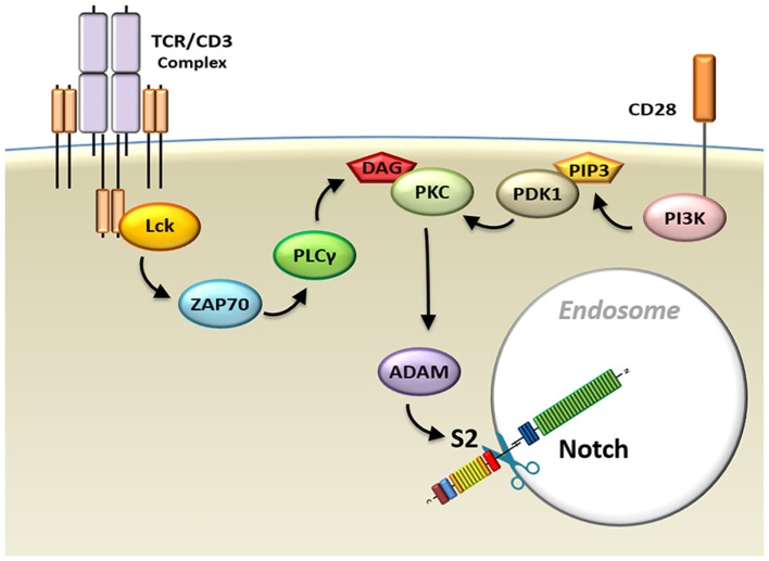
The Notch receptor is an evolutionarily highly conserved transmembrane protein essential to a wide spectrum of cellular systems, and its deregulation has been linked to a vast number of developmental disorders and malignancies. Regulated Notch function is critical for the generation of T-cells, in which abnormal Notch signaling results in leukemia. Notch activation through trans-activation of the receptor by one of its ligands expressed on adjacent cells has been well defined. In this canonical ligand-dependent pathway, Notch receptor undergoes conformational changes upon ligand engagement, stimulated by a pulling-force on the extracellular fragment of Notch that results from endocytosis of the receptor-bound ligand into the ligand-expressing cell. These conformational changes in the receptor allow for two consecutive proteolytic cleavage events to occur, which release the intracellular region of the receptor into the cytoplasm. It can then travel to the nucleus, where it induces gene transcription. However, there is accumulating evidence that other pathways may induce Notch signaling. A ligand-independent mechanism of Notch activation has been described in which receptor processing is initiated via cell-internal signals. These signals result in the internalization of Notch into endosomal compartments, where chemical changes existing in this microenvironment result in the conformational modifications required for receptor processing. This review will present mechanisms underlying both canonical ligand-dependent and non-canonical ligand-independent Notch activation pathways and discuss the latter in the context of Notch signaling in T-cells.

Keywords: Notch; T-cell; T-cell receptor; endocytosis; ligand-independent; protein kinase C.


Figures

Figure 1
Figure 1
Structure of Notch receptor family and its ligands. (A) Structure of Notch receptors on signal-receiving cells and Notch ligands on signal-sending cells. In humans and mice, there are four Notch receptors, and five Notch ligands within two families, the Jagged and the DLL family. *However, DLL3 is expressed exclusively in intracellular compartments. (B) Locations of the three cleavage sites on the Notch receptor (S1–3), and mutation hotspot regions in the HD and PEST domains formula image
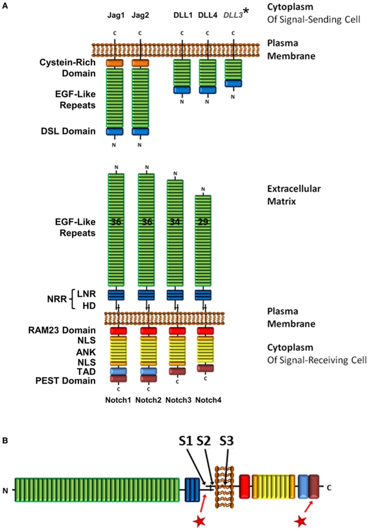
. Abbreviations: NRR, negative regulatory region; LNR, cysteine-rich Lin12/Notch repeats; HD, heterodimerization domain; NLS, nuclear localization sequence; TAD, transcriptional activation domain; DSL, Delta/Serrate/Lag-2; Jag, jagged; DLL, delta-like.
Figure 2
Figure 2
Mechanisms underlying ligand-induced Notch processing. Upon ligand engagement, the signal-sending cell exerts a pulling force on the Notch receptor by internalizing the ligand and Notch extracellular domain. Simultaneously, the residual portion of the Notch receptor is endocytosed into the signal-receiving cell via Ras-related protein 5 (Rab5)-positive vesicles, where it is cleaved by ADAMs (red scissors). This first cleavage event primes the Notch receptor for a subsequent cleavage by γ-secretase complex (blue scissors) in the endosome, which releases intracellular Notch (ICN) from the membrane and allows it to transmigrate to the nucleus. Alternatively, the Notch receptor can be escorted by endosomal sorting complexes required for transport proteins (ESCRT) to the lysosome where it is degraded.
Figure 3
Figure 3
Model of endocytosis in ligand-independent Notch activation. (1) Deltex facilitates ligand-independent Notch receptor internalization into clathrin-coated vesicles (CCV) (2) that fuse with Ras-related protein 5-positive early endosomes (EE). (3) Deltex forms a complex with adaptor protein 3 (AP-3) and homotypic fusion and vacuole protein sorting (HOPS) that directs Notch to Rab7-positive late endosomes (LE) and (4) targets it to the limiting membrane of the multivesicular body (MVB). (5) This protects intracellular Notch (ICN) from lysosomal degradation and allows its release into the cytosol upon γ-secretase complex-mediated processing. (6) Alternatively, Su(dx) can redirect Notch into the intraluminal vesicles, where the Notch receptor will be proteolyzed when the MVB fuses with a lysosome.
Figure 4
Figure 4
Possible mechanisms of Notch processing in the endosome. Upon delivery of Notch to the multivesicular body, there are three possible ways in which the endocytic environment may prepare the Notch receptor to allow the release of intracellular Notch from the limiting membrane. (A) Lysozymes (red dots) present in this compartment may proteolyze the intraluminal region of Notch and therefore allow γ-secretase complex (γSec) cleavage to proceed. (B) The change in ion concentrations and decreasing pH may cause the negative regulatory region (NRR) of Notch to unravel, mimicking a ligand-mediated pull, which opens the S2 site for access by ADAM (red scissors), followed by γSec cleavage (blue scissors). (C) The lysosomal environment may cause the Notch extracellular domain (ECD) to dissociate entirely, in which case γSec may directly process the 70aa juxtamembrane stub or rely on ADAM proteases (light red because of uncertain requirement) to increase its affinity for the S3 site through processing the juxtamembrane stub to 12aa.
Figure 5
Figure 5
Model schematic diagram of TCR/CD28-induced Notch activation in T-cells. Signals from the TCR–CD3 complex, as well as CD28 co-receptors, activate the Notch cleavage machinery and induce endocytosis of the Notch receptor. Abbreviations: Lck, lymphocyte-specific protein tyrosine kinase; ZAP70, z-chain associated protein kinase 70 kDa; TCR, T-cell receptor.

References

    1. Andersson ER, Sandberg R, Lendahl U. Notch signaling: simplicity in design, versatility in function. Development (2011) 138:3593–612.10.1242/dev.063610 - DOI - PubMed
    1. Pui JC, Allman D, Xu L, DeRocco S, Karnell FG, Bakkour S, et al. Notch1 expression in early lymphopoiesis influences B versus T lineage determination. Immunity (1999) 11:299–308.10.1016/S1074-7613(00)80105-3 - DOI - PubMed
    1. Radtke F, Wilson A, Stark G, Bauer M, van Meerwijk J, MacDonald HR, et al. Deficient T cell fate specification in mice with an induced inactivation of Notch1. Immunity (1999) 10:547–58.10.1016/S1074-7613(00)80054-0 - DOI - PubMed
    1. Radtke F, Wilson A, Ernst B, Macdonald H. The role of Notch signaling during hematopoietic lineage commitment. Immunol Rev (2002) 187:65–74.10.1034/j.1600-065X.2002.18706.x - DOI - PubMed
    1. Allman D, Punt JA, Izon DJ, Aster JC, Pear WS. An invitation to T and more: Notch signaling in lymphopoiesis. Cell (2002) 109:1–11.10.1016/S0092-8674(02)00689-X - DOI - PubMed

Publication types