. 2016 Feb 1;7(2):1440-1446.
doi: 10.1039/c5sc02902c. Epub 2015 Nov 17.

N6-Methyladenine hinders RNA- and DNA-directed DNA synthesis: application in human rRNA methylation analysis of clinical specimens

Affiliations

N6-Methyladenine hinders RNA- and DNA-directed DNA synthesis: application in human rRNA methylation analysis of clinical specimens

Shaoru Wang et al. Chem Sci. .

Abstract

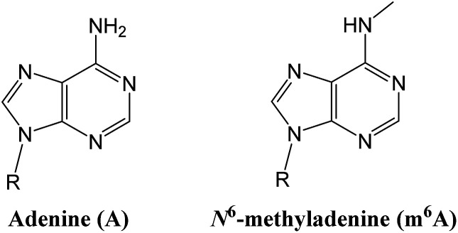
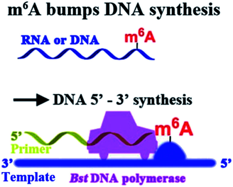
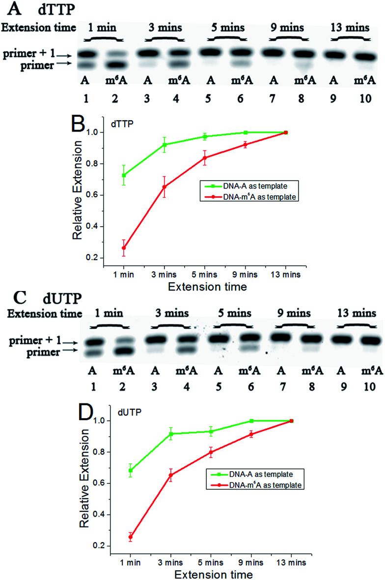
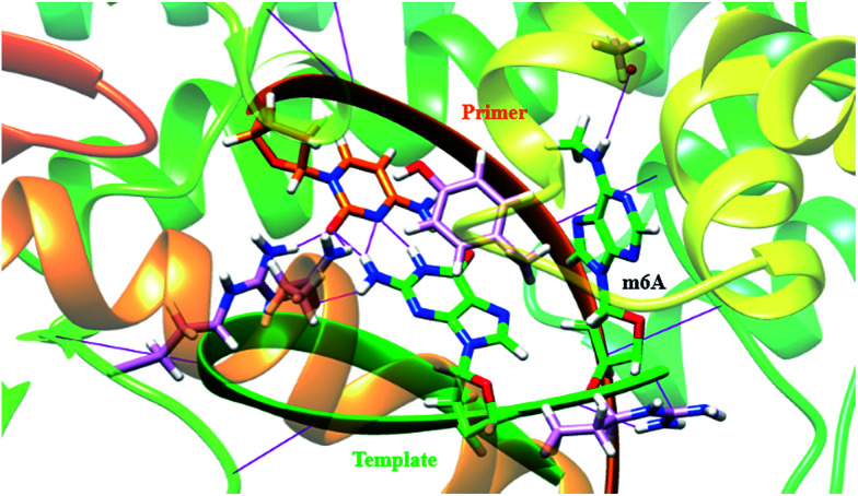
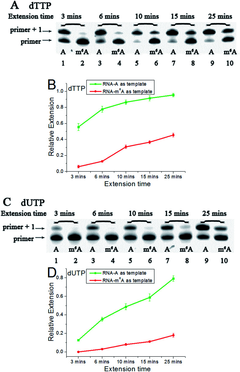
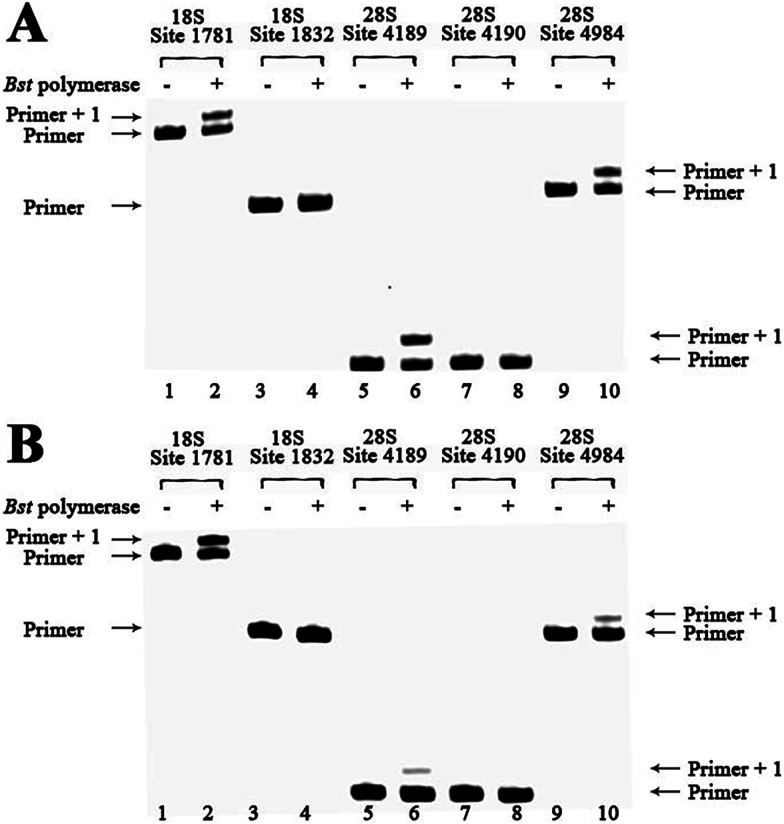
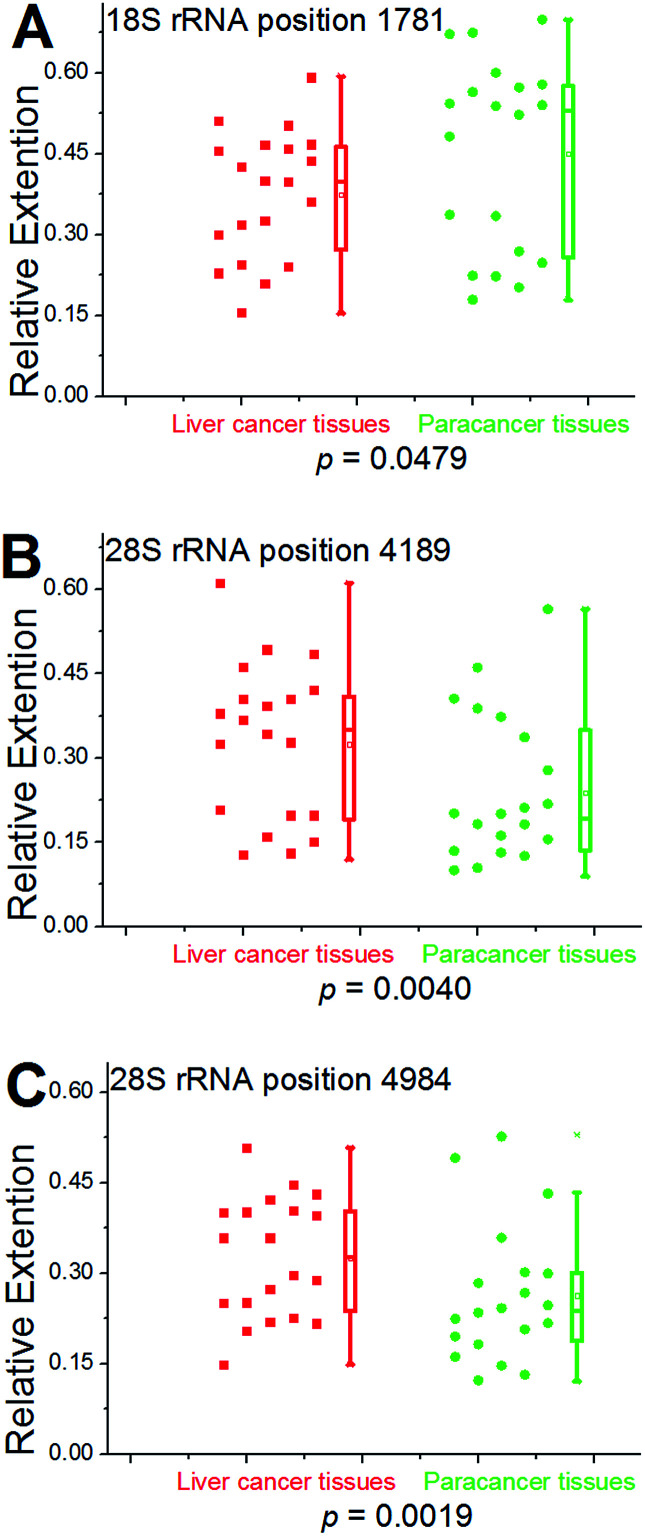
N6-Methyladenine (m6A) is the most abundant internal modification on mammalian mRNA. Very recently, m6A has been reported as a potentially important 'epigenetic' mark in eukaryotes. Until now, site-specific detection of m6A is technically very challenging. Here, we first reveal that m6A significantly hinders DNA- and RNA-directed DNA synthesis. Systematic investigations of 5'-triphosphates of a variety of 5-substituted 2'-deoxyuridine analogs in primer extension have been performed. In the current study, a quantitative analysis of m6A in the RNA or DNA context has been achieved, using Bst DNA polymerase catalyzed primer extension. Molecular dynamics study predicted that m6A in template tends to enter into and be restrained in the MGR region of Bst DNA polymerase, reducing conformational flexibility of the DNA backbone. More importantly, a site-specific determination of m6A in human ribosomal RNA (rRNA) with high accuracy has been afforded. Through a cumulative analysis of methylation alterations, we first reveal that significantly cancer-related changes in human rRNA methylation were present in patients with hepatocellular carcinoma.


Figures

Fig. 1
Fig. 1. Structure illustration of adenine and N6-methyladenine (m6A).
Fig. 2
Fig. 2. m6A hinders DNA- or RNA-directed DNA synthesis.
Fig. 3
Fig. 3. The analysis displays the difference of nucleotide incorporation opposite a templating A or m6A. Lane 1, 3, 5, 7, 9, DNA-A was used as a template; lane 2, 4, 6, 8, 10, DNA-m6A was used as a template. (A) and (C), representative gel image showing incorporation of dTTP or dUTP; (B) and (D), all data are presented as the means ± SEM from three independent experiments.
Fig. 4
Fig. 4. A template containing m6A forms a stable complex with primer in the Bst DNA polymerase active site place. The structure is superposed with the template–primer (green/orange) duplex. Key residues in the MGR region of enzyme (pink) are displayed, including Tyr 714 (766), Arg 615 (668) and Gln 797 (849). The protein–DNA interface in the MGR region is stabilized by hydrogen bonds (H-bonds, violet) and the stacking interaction between the template base and the corresponding residue [Tyr 714 (766)]. The torsion angles were significantly changed by m6A in the template.
Fig. 5
Fig. 5. The analysis displays the difference of nucleotide incorporation opposite a templating A or m6A. Lane 1, 3, 5, 7, 9, RNA-A used as a template; lane 2, 4, 6, 8, 10, RNA-m6A used as a template. (A) and (C), representative gel image showing incorporation of dTTP or dUTP; (B) and (D), all data are presented as the means ± SEM from three independent experiments.
Fig. 6
Fig. 6. Methylation analysis of 18S and 28S rRNA for hepatic cancerous or paracancerous tissue from a same patient with hepatocellular carcinoma. dTTP is incorporated for analysis. Lanes 1, 3, 5, 7 and 9, control samples without addition of Bst DNA polymerase; lanes 2, 4, 6, 8 and 10, 0.1 U Bst DNA polymerase was used. (A) Analysis of hepatic cancerous tissues of patient 1 with hepatocellular carcinoma; (B) analysis of hepatic paracancerous tissues of the same patient.
Fig. 7
Fig. 7. Statistical analysis for 18S and 28S rRNA methylation in clinical specimens. Left scatter plots indicate the RE values of all tested cases obtained from hepatic cancerous or paracancerous tissues in patients with hepatocellular carcinoma. Right box plots represent the distribution of the data. The median value is identified by a line inside the box. The length of the box represents the interquartile range. The P-value is calculated by Wilcoxon rank-sum test.

References

    1. Jaenisch R. Bird A. Nat. Genet. 2003;33:245–254. doi: 10.1038/ng1089. - DOI - PubMed
    1. Wion D. Casadesus J. Nat. Rev. Microbiol. 2006;4:183–192. doi: 10.1038/nrmicro1350. - DOI - PMC - PubMed
    1. Jia G. Fu Y. He C. Trends Genet. 2013;29:108–115. doi: 10.1016/j.tig.2012.11.003. - DOI - PMC - PubMed
    1. Fang G. Munera D. Friedman D. I. Mandlik A. Chao M. C. Banerjee O. Feng Z. Losic B. Mahajan M. C. Jabado O. J. Deikus G. Clark T. A. Luong K. Murray I. A. Davis B. M. Keren-Paz A. Chess A. Roberts R. J. Korlach J. Turner S. W. Kumar V. Waldor M. K. Schadt E. E. Nat. Biotechnol. 2012;30:1232–1239. doi: 10.1038/nbt.2432. - DOI - PMC - PubMed
    1. Fu Y. Luo G. Z. Chen K. Deng X. Yu M. Han D. Hao Z. Liu J. Lu X. Dore L. C. Weng X. Ji Q. Mets L. He C. Cell. 2015;161:879–892. doi: 10.1016/j.cell.2015.04.010. - DOI - PMC - PubMed