

Transducin β-Subunit Can Interact with Multiple G-Protein γ-Subunits to Enable Light Detection by Rod Photoreceptors
- PMID: 29911170
- PMCID: PMC6001135
- DOI: 10.1523/ENEURO.0144-18.2018
Transducin β-Subunit Can Interact with Multiple G-Protein γ-Subunits to Enable Light Detection by Rod Photoreceptors
Abstract
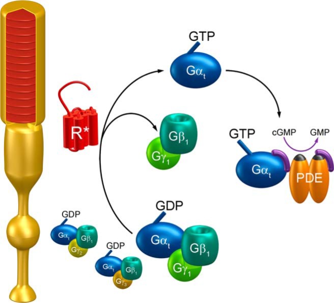
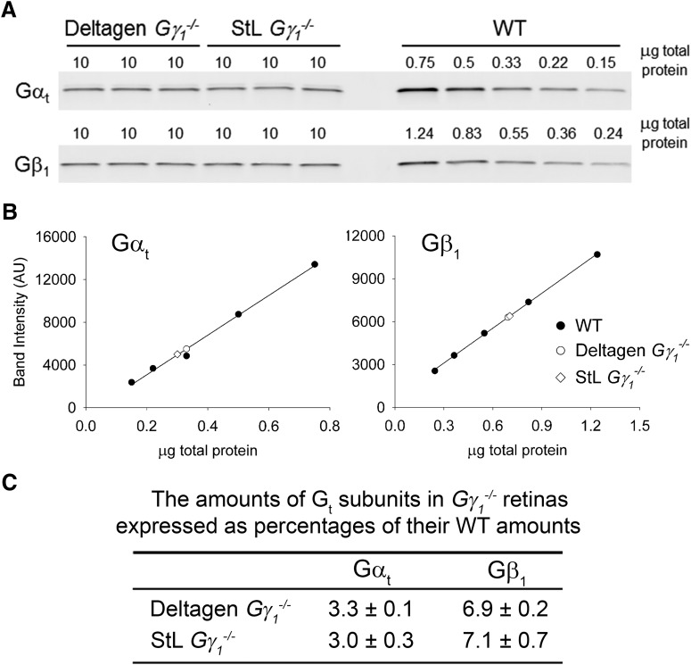
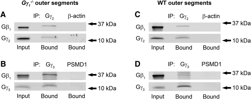
The heterotrimeric G-protein transducin mediates visual signaling in vertebrate photoreceptor cells. Many aspects of the function of transducin were learned from knock-out mice lacking its individual subunits. Of particular interest is the knockout of its rod-specific γ-subunit (Gγ1). Two studies using independently generated mice documented that this knockout results in a considerable >60-fold reduction in the light sensitivity of affected rods, but provided different interpretations of how the remaining α-subunit (Gαt) mediates phototransduction without its cognate Gβ1γ1-subunit partner. One study found that the light sensitivity reduction matched a corresponding reduction in Gαt content in the light-sensing rod outer segments and proposed that Gαt activation is supported by remaining Gβ1 associating with other Gγ subunits naturally expressed in photoreceptors. In contrast, the second study reported the same light sensitivity loss but a much lower, only approximately sixfold, reduction of Gαt and proposed that the light responses of these rods do not require Gβγ at all. To resolve this controversy and elucidate the mechanism driving visual signaling in Gγ1 knock-out rods, we analyzed both mouse lines side by side. We first determined that the outer segments of both mice have identical Gαt content, which is reduced ∼65-fold from the wild-type (WT) level. We further demonstrated that the remaining Gβ1 is present in a complex with endogenous Gγ2 and Gγ3 subunits and that these complexes exist in wild-type rods as well. Together, these results argue against the idea that Gαt alone supports light responses of Gγ1 knock-out rods and suggest that Gβ1γ1 is not unique in its ability to mediate vertebrate phototransduction.
Keywords: G-protein; phototransduction; retinal degeneration; transducin.
Figures
Similar articles
-
Regulation of rod photoreceptor function by farnesylated G-protein γ-subunits.PLoS One. 2022 Aug 8;17(8):e0272506. doi: 10.1371/journal.pone.0272506. eCollection 2022. PLoS One. 2022. PMID: 35939447 Free PMC article.
-
Probing the mechanism by which the retinal G protein transducin activates its biological effector PDE6.J Biol Chem. 2024 Feb;300(2):105608. doi: 10.1016/j.jbc.2023.105608. Epub 2023 Dec 28. J Biol Chem. 2024. PMID: 38159849 Free PMC article.
-
Signs and symptoms to determine if a patient presenting in primary care or hospital outpatient settings has COVID-19.Cochrane Database Syst Rev. 2022 May 20;5(5):CD013665. doi: 10.1002/14651858.CD013665.pub3. Cochrane Database Syst Rev. 2022. PMID: 35593186 Free PMC article.
-
Photoreceptor degeneration induces homeostatic rewiring of rod bipolar cells.Curr Biol. 2025 Jul 7;35(13):3263-3268.e2. doi: 10.1016/j.cub.2025.05.057. Epub 2025 Jun 25. Curr Biol. 2025. PMID: 40570846
-
Blue-light filtering intraocular lenses (IOLs) for protecting macular health.Cochrane Database Syst Rev. 2018 May 22;5(5):CD011977. doi: 10.1002/14651858.CD011977.pub2. Cochrane Database Syst Rev. 2018. PMID: 29786830 Free PMC article.
Cited by
-
AAA+ Protein-Based Technologies to Counter Neurodegenerative Disease.Biophys J. 2019 Apr 23;116(8):1380-1385. doi: 10.1016/j.bpj.2019.03.007. Epub 2019 Mar 22. Biophys J. 2019. PMID: 30952364 Free PMC article. Review.
-
Probing Proteostatic Stress in Degenerating Photoreceptors Using Two Complementary In Vivo Reporters of Proteasomal Activity.eNeuro. 2020 Jan 8;7(1):ENEURO.0428-19.2019. doi: 10.1523/ENEURO.0428-19.2019. Print 2020 Jan/Feb. eNeuro. 2020. PMID: 31826915 Free PMC article.
-
Regulation of rod photoreceptor function by farnesylated G-protein γ-subunits.PLoS One. 2022 Aug 8;17(8):e0272506. doi: 10.1371/journal.pone.0272506. eCollection 2022. PLoS One. 2022. PMID: 35939447 Free PMC article.
-
Comparative Proteome Analysis Indicates The Divergence between The Head and Tail Regeneration in Planarian.Cell J. 2021 Nov;23(6):640-649. doi: 10.22074/cellj.2021.7689. Epub 2021 Nov 23. Cell J. 2021. PMID: 34939757 Free PMC article.
-
Expression of a CARD Slows the Retinal Degeneration of a Geographic Atrophy Mouse Model.Mol Ther Methods Clin Dev. 2019 Jun 12;14:113-125. doi: 10.1016/j.omtm.2019.06.001. eCollection 2019 Sep 13. Mol Ther Methods Clin Dev. 2019. PMID: 31334304 Free PMC article.
References
-
- Cali JJ, Balcueva EA, Rybalkin I, Robishaw JD (1992) Selective tissue distribution of G protein γ subunits, including a new form of the γ subunits identified by cDNA cloning. J Biol Chem 267:24023–24027. - PubMed
-
- Calvert PD, Krasnoperova NV, Lyubarsky AL, Isayama T, Nicoló M, Kosaras B, Wong G, Gannon KS, Margolskee RF, Sidman RL, Pugh EN Jr, Makino CL, Lem J (2000) Phototransduction in transgenic mice after targeted deletion of the rod transducin α-subunit. Proc Natl Acad Sci U S A 97:13913–13918. 10.1073/pnas.250478897 - DOI - PMC - PubMed
Publication types
MeSH terms
Substances
Grants and funding
LinkOut - more resources
Full Text Sources
Other Literature Sources
Molecular Biology Databases
Research Materials