. 2018 Jun 14;5(3):ENEURO.0186-18.2018.
doi: 10.1523/ENEURO.0186-18.2018. eCollection 2018 May-Jun.

Mammalian TRIM67 Functions in Brain Development and Behavior

Affiliations

Mammalian TRIM67 Functions in Brain Development and Behavior

Nicholas P Boyer et al. eNeuro. .

Erratum in

Abstract

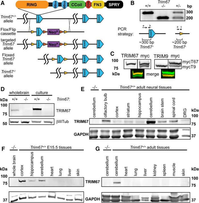
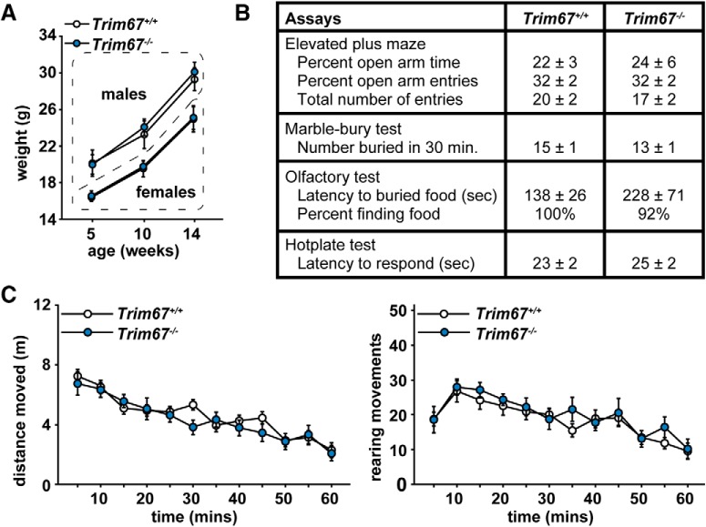
Class I members of the tripartite motif (TRIM) family of E3 ubiquitin ligases evolutionarily appeared just prior to the advent of neuronal like cells and have been implicated in neuronal development from invertebrates to mammals. The single Class I TRIM in Drosophila melanogaster and Caenorhabditis elegans and the mammalian Class I TRIM9 regulate axon branching and guidance in response to the guidance cue netrin, whereas mammalian TRIM46 establishes the axon initial segment. In humans, mutations in TRIM1 and TRIM18 are implicated in Opitz Syndrome, characterized by midline defects and often intellectual disability. We find that although TRIM67 is the least studied vertebrate Class I TRIM, it is the most evolutionarily conserved. Here we show that mammalian TRIM67 interacts with both its closest paralog TRIM9 and the netrin receptor DCC and is differentially enriched in specific brain regions during development and adulthood. We describe the anatomical and behavioral consequences of deletion of murine Trim67. While viable, mice lacking Trim67 exhibit abnormal anatomy of specific brain regions, including hypotrophy of the hippocampus, striatum, amygdala, and thalamus, and thinning of forebrain commissures. Additionally, Trim67-/- mice display impairments in spatial memory, cognitive flexibility, social novelty preference, muscle function, and sensorimotor gating, whereas several other behaviors remain intact. This study demonstrates the necessity for TRIM67 in appropriate brain development and behavior.

Keywords: DCC; TRIM67; commissure; hippocampus; knock-out; striatum.


Figures

Figure 1.
Figure 1.
TRIM67 is evolutionarily conserved. A, Phylogeny of Class I TRIM proteins across animal phyla based on protein sequences, showing the six members in vertebrate species (cyan, TRIM9; red, TRIM67; green, TRIM1/MID2; pink, TRIM18/MID1; purple, TRIM36; orange, TRIM46) and the one to two members in nonvertebrate animal phyla (blue, TRIM1/18/36/46-like; brown, TRIM9/67-like). Dashed red line separates TRIM9/67-like proteins from TRIM1/18/36/46-like proteins. Bootstrap values are shown at major group branches. B, Phylogenetic tree of invertebrate TRIM9/67-like proteins alongside several vertebrate TRIM9 and TRIM67 homologs (all proteins above dashed red line in A). Vertebrate TRIM67 exhibits higher sequence similarity than TRIM9 to invertebrate Class I TRIMs. Numbers denote bootstrapping values.
Figure 2.
Figure 2.
Generation of Trim67−/− mouse and TRIM67 brain localization. A, Diagram of targeting strategy for the Trim67 gene showing Cre-lox mediated excision of exon 1. This excision leads to the next 16 intronic and 4 exonic ATG codons being out of frame. LoxP sites are yellow, FLIP sites are orange, and neomycin resistance gene (NeoR) is purple. B, Agarose gel separation of genotyping PCR products, demonstrating deletion of both copies of the first exon of Trim67 (-/-) or one copy in a heterozygote (±). Diagrams show PCR products from wild-type and knock-out alleles. C, Duplicate Western blottings of lysate from HEK293T cells expressing both myc-TRIM9 and myc-TRIM67, were probed with both the indicated TRIM antibody (left, red) and myc (right, green), and spectrally distinct secondary antibodies. The newly generated TRIM67 polyclonal antibody recognizes TRIM67 but not TRIM9; the polyclonal TRIM9 antibody recognizes TRIM9, but not TRIM67, as can be seen in the merged images below. D, Western blotting of whole-brain or 2 d in vitro dissociated cortical neuron lysate from Trim67+/+ and Trim67−/− E15.5 embryos probed for TRIM67 and βIII-tubulin as a loading control. EG, Western blotting detects TRIM67 expression in various adult (E) and embryonic (F) neural tissues, but not in the examined tissues outside the nervous system (G). GAPDH is a loading control. Age-matched Trim67−/− lysate from indicated tissue shown in left most lane of each blot.
Figure 3.
Figure 3.
TRIM67 is present in multiple murine brain regions. A, Low-magnification sagittal sections of Trim67+/+ and Trim67−/− E15.5 brains stained for TRIM67 (green), β-III-tubulin (magenta), and nuclei (DAPI, blue). TRIM67 is present in cell bodies in the developing hippocampus (B) and cortex (C) at E15.5. D, TRIM67 expression is evident in the peduncular hypothalamus (5), diencephalon (1), and reticular complex (2), but not in the prethalamic eminence (3), zona incerta complex (4), or subpallium (6).
Figure 4.
Figure 4.
TRIM67 is expressed in developing cortex and interacts with TRIM9 and DCC. A, Western blottings of cortical lysate from indicated embryonic (E) and postnatal (P) ages reveal expression patterns of TRIM67 and the three isoforms of TRIM9. B, Western blotting of TRIM67 immunoprecipitates from Trim67+/+ and Trim67−/−mouse cortical neuronal lysate probed for TRIM9 and TRIM67. Coimmunoprecipitation of TRIM9 occurred in the presence of TRIM67. C, A similar immunoprecipitation of TRIM9 from embryonic brain lysates coimmunoprecipitates TRIM67 in wild-type cortical lysate, but not in the absence of TRIM9. D, HEK293T cells were cotransfected with HA-tagged DCC and either myc-tagged TRIM9 or TRIM67 lacking the RING domain (ΔRING). Immunoprecipitation of either myc-TRIMΔRING coprecipitated HA-DCC. E, The top of the blot shown in B, probed for DCC, showing that endogenous DCC is enriched over background levels in TRIM67 immunoprecipitates from wild-type embryonic brain lysate. The TRIM67 IP is the same as in B. F, Photomicrographs of neurites and growth cones from Trim67+/+ and Trim67−/− cultured embryonic cortical neurons show DCC immunostaining in the same cells as TRIM67. Residual TRIM67 staining in Trim67−/− is due to nonspecific binding of the antibody. In merged image, phalloidin is red, DCC is green, and TRIM67 is cyan.
Figure 5.
Figure 5.
Trim67 deletion reduces the thickness of certain fiber tracts. A, Photomicrographs of black-gold stained coronal brain sections, the fiber tracts measured are indicated. B, The corpus callosum was thinner in Trim67−/− mice (p = 0.00000909), with most significant individual measures toward the caudal end of the tract. The hippocampal commissure, including both the fornix and dorsal hippocampal commissure, was reduced in thickness in Trim67−/− mice (p = 0.000215). C, GFP staining in the corpus callosum of Nex-Cre:Taulox-STOP-lox-GFP:Trim67+/+ and Nex-Cre:Taulox-STOP-lox-GFP:Trim67fl/fl brains. The corpus callosum was thinner (p = 0.0304) in Trim67fl/fl mice (*p < 0.05 at this position).
Figure 6.
Figure 6.
Reduction in total brain weight and hypotrophy of multiple brain areas occurs with loss of Trim67. A, Photograph of representative Trim67+/+ (left) and Trim67−/− (right) brains from five-week-old mice. Total weight of Trim67−/− brains was reduced by ∼10% when compared to littermate controls (p = 0.0108, Wilcoxon signed-rank test). B, Photomicrographs of black-gold stained coronal sections of Trim67+/+ and Trim67−/− brains at 1.3 mm posterior to bregma. Outlines delineate regions used for measures in this figure. C, Deletion of Trim67 had no effect on the thickness of the cortex (p = 0.069), measured from the cingulum to the cortical surface (average distance between the dashed black lines in B). D, Area of the hippocampal gray matter (including both the dentate gyrus and Ammon’s horn), reported as the average of both individual hemispheres from its first emergence through the fimbriae to 2.7 mm posterior to bregma. The hippocampus was smaller in Trim67−/− brains (p = 0.00000889), with significant individual measures toward the rostral portion. E, Area of the amygdala, reported as the average of both individual hemispheres. The amygdala was significantly smaller in Trim67−/− brains (p = 0.000110). F, Area of the lateral ventricles reported as the average of both individual hemispheres, from the first section posterior to the anterior commissure to the last section with visible ventricle. Trim67−/− lateral ventricles were smaller (p = 0.000128; *p < 0.05 at this position).
Figure 7.
Figure 7.
Mouse growth, sensory ability and general locomotion are not affected by Trim67 deletion. A, Weights of mice over the course of behavioral assays, showing no effect of Trim67 loss on overall growth. B, Results of elevated plus maze and several sensory assays. Units are indicated. C, Locomotor results in the open field assay. None of these assays show significant results by ANOVA.
Figure 8.
Figure 8.
Loss of Trim67 leads to impairments in spatial learning and memory. A, Time (s) for mice to find a hidden platform in the Morris water maze, with a threshold for learning set at a group average of 15 s (dashed line). Trim67−/− mice fail to reach this threshold out to the maximum of 9 d of training and have overall significantly higher latencies to find the platform (*p < 0.05; all trials p = 0.0225). B, Time spent in target and opposite quadrants and number of times crossing the previous platform position in a probe trial following acquisition day 9. Whereas Trim67+/+ mice spent a higher amount of time in the quadrant previously occupied by the platform (*p = 0.0003), Trim67−/− mice failed to show this preference and spent a lower time in the target quadrant than Trim67+/+ mice (#p = 0.0343). C, Subsequent reversal trials demonstrated an increased latency to find the platform in the Trim67−/− cohort compared to Trim67+/+ littermates (*p < 0.05; all trials p = 0.0149). D, In a probe trial following reversal day 7, only Trim67+/+ mice spent more time in the new target quadrant (*p = 0.0335).
Figure 9.
Figure 9.
Trim67−/− mice display normal sociability, but impaired social novelty preference. A, Measures of total time spent in (top) and entries into (bottom) side chambers of a three-chamber socialization assay, showing mice of both genotypes exhibited a significant preference for proximity to stranger mouse 1 (***p < 0.0001). B, Total time spent in and entries into side chambers in subsequent three chamber social novelty assay. Trim67−/− mice show a lack of preference for the novel stranger, whereas Trim67+/+ mice show a preference (*p = 0.0295).
Figure 10.
Figure 10.
Prepulse inhibition of acoustic startle is impaired by loss of Trim67. A, Startle amplitude (in arbitrary units) in response to no stimulus (NoS) or 120-dB stimulus (AS), with varying prepulse levels 100 ms before stimulus. B, Prepulse inhibition is reported as % inhibition compared to AS. Test 1 took place at 10–11 weeks of age and test 2 at 17–19 weeks. Trim67−/− mice showed a decrease in prepulse inhibition in the second trial (p = 0.0088) but not in the first.
Figure 11.
Figure 11.
Muscle function, but not motor learning, is impaired in Trim67−/− mice. A, Mouse gait was measured by footprint analysis on a straight track, with forepaws in red and hindpaws in blue. B, There was no significant difference in base width (forepaws, p = 0.775; hindpaws, p = 0.229) or stride length (p = 0.252) during normal walking between Trim67−/− and Trim67+/+mice. C, Latency to fall from an accelerating rotarod in three trials separated by 45 s (1, 2, 3), followed by two additional trials 48 h later separated by 45 s (4, 5). Deletion of Trim67 led to a decrease in fall latency during the first day of trials (p = 0.0074); however, there was no difference during the retest trials. D, Muscle tone measured during a four-paw rolling wire hang assay, reported as maximum and average impulse (weight × time) over three trials. Trim67−/− mice have lower maximum (p = 0.0387) and average (p = 0.0339) hanging impulse than Trim67+/+mice; *p < 0.05.
Figure 12.
Figure 12.
Malformation of CPu in Trim67−/− mice. A, Photomicrographs of black-gold stained coronal brain sections showing the area used for quantification of CPu parameters. B, The area of the CPu, reported as the average of both individual hemispheres. Yellow box denotes position of white matter measurements. The CPu was smaller in Trim67−/− brains (p = 0.00000876), with most significantly different individual positions toward the anterior. C, Binary masks of white matter (black regions) in the CPu after semi-automated segmentation. D, Deletion of Trim67 resulted in a decrease in the area of the internal capsule, reported as the average of both hemispheres (p = 0.000138; *p <0.05 at this position).
Figure 13.
Figure 13.
Deletion of Trim67 is associated with a reduction in thalamus size. A, Coronal brain sections with the thalamus, habenula, stria terminalis, and stria medullaris regions outlined as used for measurements. B, Trim67 deletion resulted in a decrease in the area of the thalamus in the region under the anterior hippocampus (p = 0.000174). C, The area of the habenula was not affected by deletion of Trim67 (p = 0.299). D, Both the stria terminalis and stria medullaris were reduced in cross-sectional area in Trim67−/− brains (p = 0.0222 and p = 0.00152, respectively; *p < 0.05 at this position).

Comment in

References

    1. Alexander M, Selman G, Seetharaman A, Ming Chan KK, D’Souza SA, Byrne AB, Roy PJ (2010) MADD-2, a homolog of the opitz syndrome protein MID1, regulates guidance to the midline through UNC-40 in Caenorhabditis elegans . Dev Cell 18:961–972. 10.1016/j.devcel.2010.05.016 - DOI - PubMed
    1. Anuppalle M, Maddirevula S, Huh TL, Rhee M (2013) Ubiquitin proteasome system networks in the neurological disorders. Animal Cells Syst (Seoul) 17:383–387. 10.1080/19768354.2013.855256 - DOI
    1. Arganda-Carreras I, Kaynig V, Rueden C, Eliceiri KW, Schindelin J, Cardona A, Seung HS (2017) Trainable Weka segmentation: a machine learning tool for microscopy pixel classification. Bioinformatics 33:2424–2426. 10.1093/bioinformatics/btx180 - DOI - PubMed
    1. Atkin G, Paulson H (2014) Ubiquitin pathways in neurodegenerative disease. Front Mol Neurosci 7:63 10.3389/fnmol.2014.00063 - DOI - PMC - PubMed
    1. Beecham GW, Martin ER, Li YJ, Slifer MA, Gilbert JR, Haines JL, Pericak-Vance MA (2009) Genome-wide association study implicates a chromosome 12 risk locus for late-onset Alzheimer disease. Am J Hum Genet 84:35–43. 10.1016/j.ajhg.2008.12.008 - DOI - PMC - PubMed

Publication types

MeSH terms