. 2018 Aug 8;18(8):4803-4811.
doi: 10.1021/acs.nanolett.8b01374. Epub 2018 Jul 5.

Programmable Multivalent DNA-Origami Tension Probes for Reporting Cellular Traction Forces

Affiliations

Programmable Multivalent DNA-Origami Tension Probes for Reporting Cellular Traction Forces

Palash K Dutta et al. Nano Lett. .

Abstract

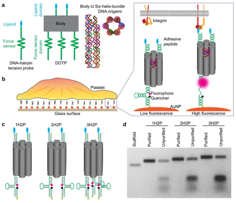
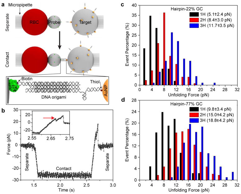
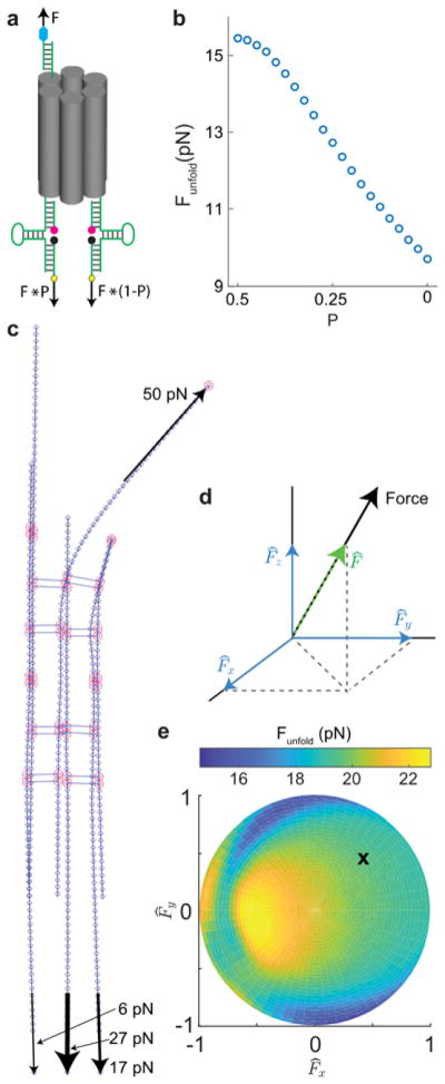
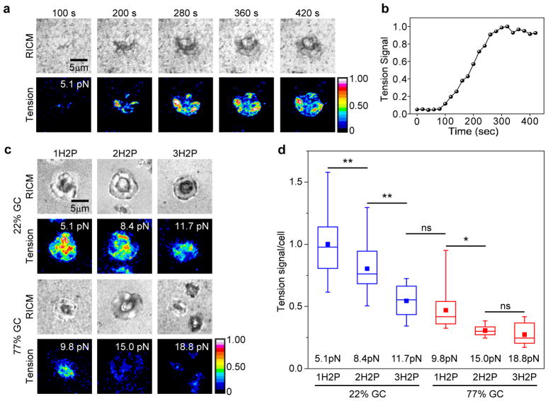
Mechanical forces are central to most, if not all, biological processes, including cell development, immune recognition, and metastasis. Because the cellular machinery mediating mechano-sensing and force generation is dependent on the nanoscale organization and geometry of protein assemblies, a current need in the field is the development of force-sensing probes that can be customized at the nanometer-length scale. In this work, we describe a DNA origami tension sensor that maps the piconewton (pN) forces generated by living cells. As a proof-of-concept, we engineered a novel library of six-helix-bundle DNA-origami tension probes (DOTPs) with a tailorable number of tension-reporting hairpins (each with their own tunable tension response threshold) and a tunable number of cell-receptor ligands. We used single-molecule force spectroscopy to determine the probes' tension response thresholds and used computational modeling to show that hairpin unfolding is semi-cooperative and orientation-dependent. Finally, we use our DOTP library to map the forces applied by human blood platelets during initial adhesion and activation. We find that the total tension signal exhibited by platelets on DOTP-functionalized surfaces increases with the number of ligands per DOTP, likely due to increased total ligand density, and decreases exponentially with the DOTP's force-response threshold. This work opens the door to applications for understanding and regulating biophysical processes involving cooperativity and multivalency.

Keywords: DNA origami; biomembrane force probe; cellular traction forces; platelets.


Conflict of interest statement

Notes

The authors declare no competing financial interest.

Figures

Figure 1
Figure 1
Design and characterization of DNA origami-based tension probes (DOTPs). (a) Schematic showing three components of a DOTP: a ligand presenting domain, an origami body, and a force sensing domain. The body is comprised of a six-helix-bundle DNA origami (side and top view), in which six parallel double-helices are packed on a honeycomb lattice. (b) Illustration of a platelet spreading on a glass surface functionalized with DOTPs. The zoomed-in scheme shows the mechanism of tension-to-fluorescence transduction. The origami constructs are conjugated to gold nanoparticle (AuNP)-coated glass surfaces utilizing thiol–Au binding. Upon receptor (integrin) engagement to the adhesive peptide (cRGDfk) and application of sufficient tension the hairpin unfolds, separating the fluorophore from the AuNP and organic quencher and dequenching the dye. (c) Schematic of DOTPs with two adhesive peptides (blue) on the ligand presenting domain (top end) and one, two or three hairpin(s) on the force sensor domain (bottom end), denoted 1H2P, 2H2P and 3H2P, respectively. (d) Agarose gel electrophoresis of purified and unpurified 1H2P, 2H2P and 3H2P. The scaffold band contains the 425 nucleotide scaffold strand.
Figure 2
Figure 2
DOTP calibration with BFP single-molecule force spectroscopy. (a) Schematic showing BFP setup. A micropipette-aspirated red blood cell (RBC) is affixed to a streptavidin (STV, green)-coated probe bead. A DOTP-coated target bead, which itself is aspirated by another micropipette, is brought into contact with the STV-coated bead. The ligand-presenting domains each present one biotin (purple sphere), resulting in biotin-STV binding between the two beads. The target bead is then retracted to apply tension to the DOTP. Zoom-in shows the assembled DOTP between the two beads. (b) Representative trace of a single molecule unfolding event showing unfolding of a 1H-77% GC DOTP. Zoom-in of the trace (inset) shows the unfolding event. The red arrow indicates the opening of the hairpin at ~10 pN. (c) Histogram of unfolding events for 1H-22% GC (black, n = 50), 2H-22% GC (red, n = 100) and 3H-22% GC (blue, n = 90) DOTPs. (d) Histogram of unfolding events for 1H-77% GC (black, n = 42), 2H-77% GC (red, n = 230) and 3H-77% GC (blue, n = 98). The legends in (c) and (d) show the corresponding Funfold with standard deviation for each probe.
Figure 3
Figure 3
Uneven distribution of tension between hairpins influences Funfold in an orientation-dependant manner. (a) Visual representation of force imbalance, where total force F is distributed between two hairpins according to constant P. (b) Results of Monte Carlo simulations showing that Funfold decreases as P decreases from 0.5 to 0 due to increasing tension imbalance. (c) Snapshot of a 3H DOTP rendered as a finite element structure. Elements (short blue lines) are connected by nodes (blue diamonds). Nodes at crossover positions are circled in red. A 50 pN force applied at the site of the ligand, as well as resulting 27 pN, 17 pN, and 6 pN forces experienced at the hairpins, are denoted as black arrows. (d) Coordinate system showing force vector (black arrow), unit force vector (, a vector of magnitude 1 that is parallel to the force denoted by the green dashed arrow), and the x, y, and z components of unit force vector (blue arrows). (e) Plot showing simulated Funfold as a function of force orientation, as denoted by the x and y components of the unit force vector. The z component of the unit vector is related to radial position on plot. Black x denotes the orientation shown in c.
Figure 4
Figure 4
Platelet mechanics measured by DOTPs. (a) Representative time-lapse images of platelet spreading imaged in the reflection interference contrast microscopy (RICM) channel and tension signal in total internal reflection fluorescence (TIRF) channel on a glass surface coated with 1H2P-22% GC origami tension sensors. Scale bar: 5 μm. (b) Plot of whole-cell tension signal as a function of time for the platelet shown in (a). (c) Representative platelet adhesion and corresponding tension signal of platelets activated on surfaces coated with DNA origami tension probes with one (1H2P), two (2H2P) and three (3H2P) hairpin(s) of 22% GC content and 77% GC content. For each tension signal image the Funfold of the corresponding DOTP is shown in the top-right corner. (d) Comparison of the tension signal from platelets activated on DNA origami tension probes with one, two and three hairpin(s) of 22% GC content and 77% GC content. Mean tension signal for 1H2P with 22% GC was normalized to 1 and all the others were calculated relative to it. Each mean value (solid square) represents averaged signal from more than 20 individual cells (whiskers indicate the range of the data, while the line and box represent the median ± quartile). Corresponding BFP-calibrated Funfold values are shown. (* p < 0.05; ** p < 0.01; ns: non-significant (p > 0.05); ANOVA).

Similar articles

Cited by

References

    1. Evans EA, Calderwood DA. Science. 2007;316(5828):1148–53. - PubMed
    1. Levental KR, Yu H, Kass L, Lakins JN, Egeblad M, Erler JT, Fong SF, Csiszar K, Giaccia A, Weninger W, Yamauchi M, Gasser DL, Weaver VM. Cell. 2009;139(5):891–906. - PMC - PubMed
    1. Kim ST, Takeuchi K, Sun ZY, Touma M, Castro CE, Fahmy A, Lang MJ, Wagner G, Reinherz EL. Journal of Biological Chemistry. 2009;284(45):31028–31037. - PMC - PubMed
    1. Qiu Y, Brown AC, Myers DR, Sakurai Y, Mannino RG, Tran R, Ahn B, Hardy ET, Kee MF, Kumar S, Bao G, Barker TH, Lam WA. Proc Natl Acad Sci U S A. 2014;111(40):14430–5. - PMC - PubMed
    1. Neuman KC, Nagy A. Nat Methods. 2008;5(6):491–505. - PMC - PubMed

Publication types