. 2018 Jun 18;10(6):334.
doi: 10.3390/v10060334.

Biochemical and Functional Characterization of Mouse Mammary Tumor Virus Full-Length Pr77Gag Expressed in Prokaryotic and Eukaryotic Cells

Affiliations

Biochemical and Functional Characterization of Mouse Mammary Tumor Virus Full-Length Pr77Gag Expressed in Prokaryotic and Eukaryotic Cells

Akhil Chameettachal et al. Viruses. .

Abstract

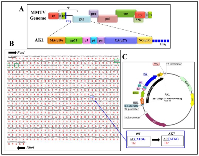
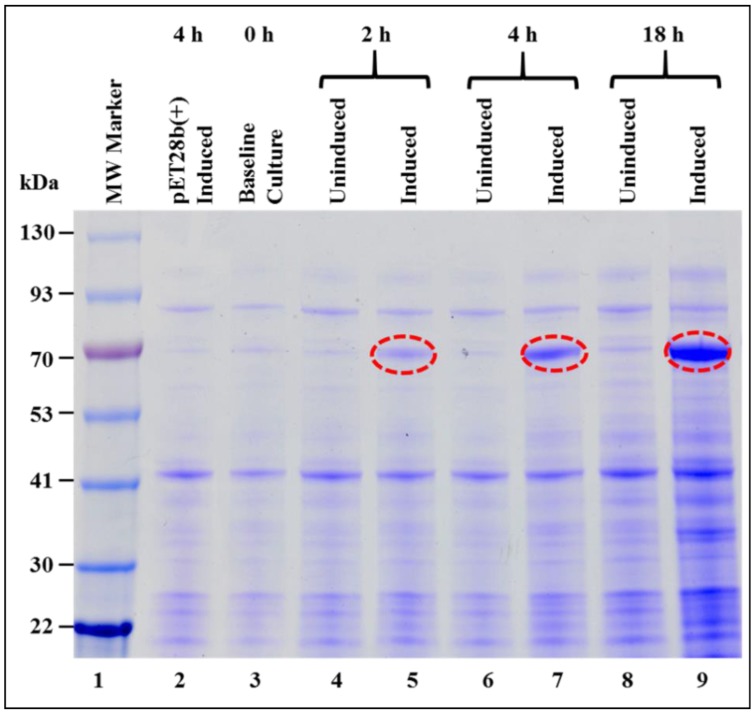
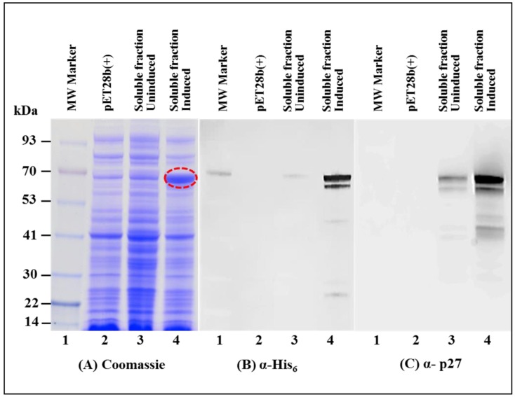
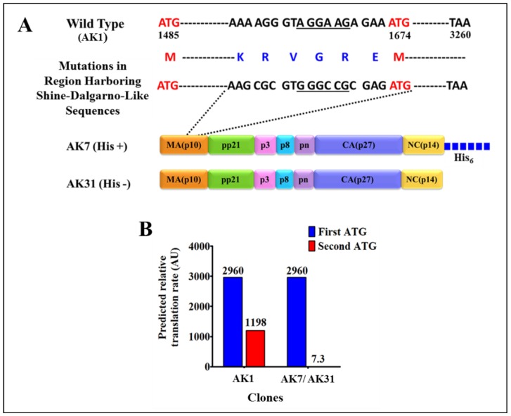
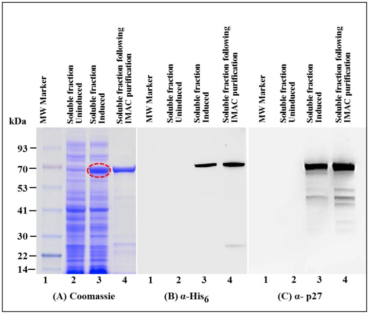
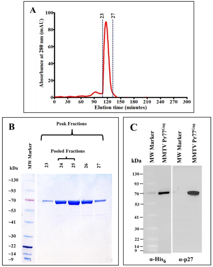
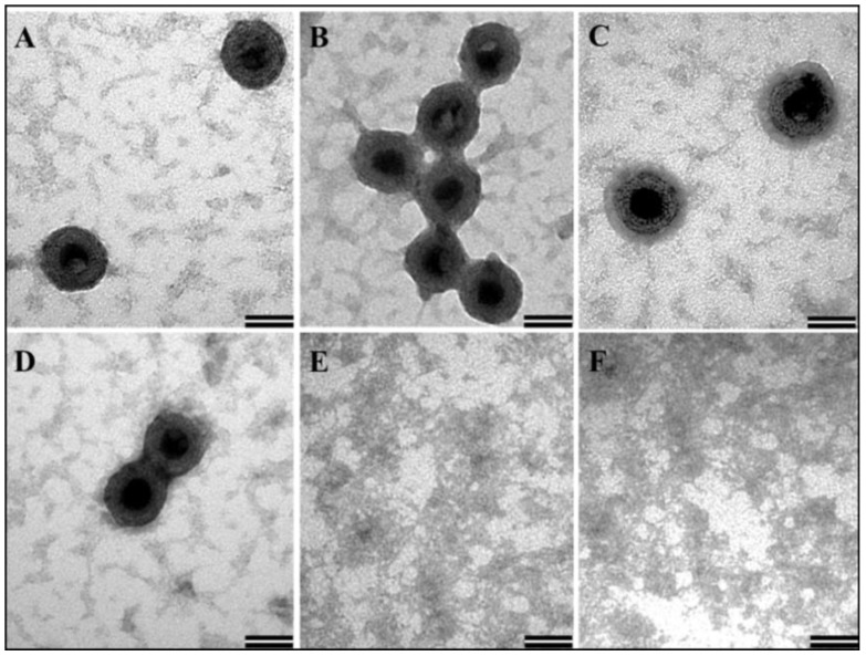
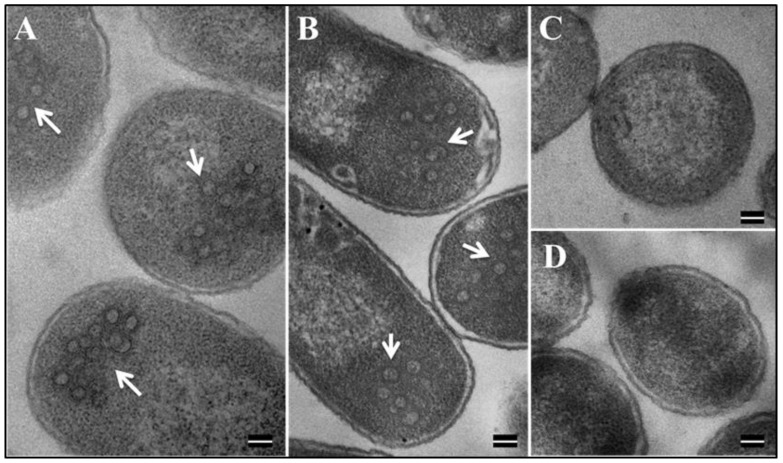
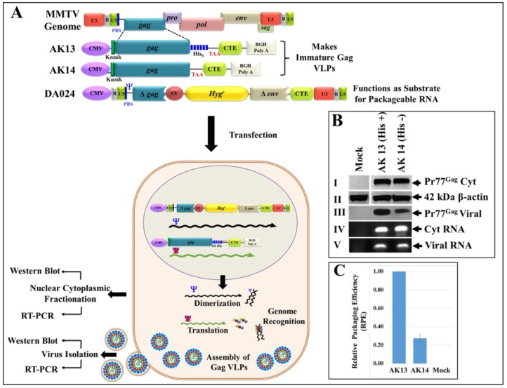
The mouse mammary tumor virus (MMTV) Pr77Gag polypeptide is an essential retroviral structural protein without which infectious viral particles cannot be formed. This process requires specific recognition and packaging of dimerized genomic RNA (gRNA) by Gag during virus assembly. Most of the previous work on retroviral assembly has used either the nucleocapsid portion of Gag, or other truncated Gag derivatives—not the natural substrate for virus assembly. In order to understand the molecular mechanism of MMTV gRNA packaging process, we expressed and purified full-length recombinant Pr77Gag-His₆-tag fusion protein from soluble fractions of bacterial cultures. We show that the purified Pr77Gag-His₆-tag protein retained the ability to assemble virus-like particles (VLPs) in vitro with morphologically similar immature intracellular particles. The recombinant proteins (with and without His₆-tag) could both be expressed in prokaryotic and eukaryotic cells and had the ability to form VLPs in vivo. Most importantly, the recombinant Pr77Gag-His₆-tag fusion proteins capable of making VLPs in eukaryotic cells were competent for packaging sub-genomic MMTV RNAs. The successful expression and purification of a biologically active, full-length MMTV Pr77Gag should lay down the foundation towards performing RNA–protein interaction(s), especially for structure-function studies and towards understanding molecular intricacies during MMTV gRNA packaging and assembly processes.

Keywords: Pr77Gag; RNA packaging; RNA–Gag interactions; RNA–protein interaction; mouse mammary tumor virus (MMTV); protein assembly; protein expression; protein purification; retrovirus.


Conflict of interest statement

The authors declare no conflict of interest.

Figures

Figure 1
Figure 1
Construction of the recombinant full-length Pr77Gag bacterial expression vector. (A) Domain organization of the mouse mammary tumor virus (MMTV) Gag precursor with His6-tag; (B) Nucleic acid and amino acid sequences of full-length MMTV gag gene. An internal NcoI site (boxed in blue color) was removed by introducing a silent mutation (shown in the inset) that preserved the threonine (Thr) amino acid. The Shine-Dalgarno-like sequence and second in-frame ATG are highlighted by green color; (C) Schematic representation of bacterial expression plasmid AK1 containing full-length MMTV Pr77Gag gene cloned into the NcoI and XhoI sites of the pET28b(+) vector.
Figure 2
Figure 2
Expression of recombinant Pr77Gag-His6-tag fusion protein in Escherichia coli (E. coli). Sodium dodecyl sulfate-polyacrylamide gel electrophoresis (SDS-PAGE) analysis showing full length Pr77Gag-His6-tag fusion protein expressed from AK1 in total bacterial cell lysates at 0, 2, 4, and 18 h post IPTG-induction and un-induced BL21(DE3) bacterial cells. The bacterial cells were grown at 37 °C overnight, but following IPTG induction, cultures were grown sub-optimally at 28 °C. MW: molecular weight.
Figure 3
Figure 3
Recombinant Pr77Gag-His6-tag fusion protein expressed in soluble fraction of E. coli. (A) SDS-PAGE analysis showing recombinant MMTV Pr77Gag-His6-tag fusion protein expression in the bacterial soluble fraction (lane 4) transformed with AK1; (B) western blot analysis of MMTV Pr77Gag expression by AK1 in soluble fraction analyzed with an α-His6 monoclonal antibody (lane 4); and (C) with an α-p27 monoclonal antibody (lane 4), respectively.
Figure 4
Figure 4
Silent mutations in the Shine-Dalgarno-like sequence and the predicted relative translation rates from the first and the second in-frame ATGs. (A) Illustration of the 18-nucleotide region mutated in MMTV gag gene to disrupt the Shine-Dalgarno-like sequence (underlined) 4 nts upstream of the second in-frame ATG (at nucleotide position 1674). These mutated sequences were cloned in both with and without His-tag clones AK7 and AK31, respectively; (B) Bar graphs showing the predicted translation rates from the legitimate first start codon and the second in-frame start codon in the wild type and in AK7(His+) and AK31(His−)-containing a mutated Shine-Dalgarno-like sequence.
Figure 5
Figure 5
Expression of recombinant Pr77Gag-His6-tag fusion protein from AK7 in soluble fractions of E. coli before and after immobilized metal affinity chromatography (IMAC) purification. (A) SDS-PAGE analysis showing recombinant MMTV Pr77Gag-His6-tag fusion protein expressed in the bacterial soluble fractions transformed with AK7 (lane 3), followed by IMAC purification (lane 4); (B) western blot analysis of MMTV Pr77Gag-His6-tag fusion protein analyzed with an α-His6 monoclonal antibody; and (C) with α-p27 monoclonal antibody, respectively.
Figure 6
Figure 6
Resolution of IMAC-purified recombinant Pr77Gag-His6-tag fusion protein by size exclusion chromatography and western blot analysis. (A) Absorbance versus elution time chromatogram of eluted fractions after size exclusion chromatography; (B) Coomassie Brilliant Blue-stained SDS-PAGE analysis of peak fractions 23 to 27, showing the resolution of purified recombinant MMTV Pr77Gag expressed from AK7; (C) western blot analysis of pooled peak fractions of purified MMTV Pr77Gag-His6-tag fusion protein analyzed with α-His6 and α-p27 monoclonal antibodies, respectively.
Figure 7
Figure 7
Transmission electron micrographs showing virus-like particles (VLPs) following in vitro assembly. (AD) In vitro assembled VLPs from purified recombinant Pr77Gag-His6-tag fusion expressed from AK7 in the presence of yeast tRNA; and (E,F) negative controls consisting of assembly buffer and yeast tRNA in the absence of any protein (Scale bar = 50 nm, 135,000× magnification).
Figure 8
Figure 8
Formation of VLPs by recombinant Pr77Gag-His6-tag fusion in E. coli BL21(DE3). Transmission electron micrographs showing VLPs assembled from (A) recombinant Pr77Gag-His6-tag fusion expressed in E. coli BL21(DE3) cells transformed with AK7 and (B) AK31 (without His6 tag); (C,D) un-induced BL21(DE3) cells transformed with AK7 and AK31, respectively (Scale bar = 100 nm; 60,000× magnification).
Figure 9
Figure 9
Schematic representation of the two-plasmid genetic complementation assay to demonstrate VLPs formation following Pr77Gag-His6-tag protein expression in eukaryotic cells and their ability to package MMTV sub-genomic RNA. (A) Upper panel; MMTV full-length Gag eukaryotic expression plasmids and MMTV sub-genomic transfer vector, DA024 [29]. (A) Lower panel; Graphical representation of the MMTV two-plasmid genetic complementation assay in which VLPs produced by recombinant MMTV Pr77Gag expression plasmids (AK13 and AK14) should package MMTV sub-genomic transfer vector (DA024) owing to the presence of the packaging sequences (Ψ). HEK 293T cells co-transfected with the two plasmids were subjected to nucleocytoplasmic fractionation. The cytoplasmic fractions and pelleted VLPs were analyzed for transfer vector RNA expression by RT-PCR; (B) western blots performed on cell lysates and ultracentrifuged transfected culture supernatants using α-MMTV p27 monoclonal antibody (panels I and III), and α-β-actin antibody (panel II), respectively. PCR amplification of cDNAs prepared from cytoplasmic (panel IV) and viral RNA (panel V) demonstrating RNA packaging using MMTV transfer vector (DA024)-specific primers (OTR671/OTR672) to amplify a 142 bp fragment. The RNA packaging experiment was performed more than three independent times followed by its analysis by RT-PCR and a representative blot of the packaged viral RNA is shown in panel V; (C) relative RNA packaging efficiency (RPE) by AK13 and AK14 of one of the representative experiments, as measured by quantitative real time PCR. Briefly, the real time experiments were conducted in triplicates (± standard deviation (SD)) and the relative quantification (RQ) values obtained for the packaged viral RNA in the Gag VLPs were normalized to the cytoplasmic expression of the transfer vector RNA (DA024) for the respective clones as described previously.

Similar articles

Cited by

References

    1. Bittner J.J. Some possible effects of nursing on the mammary gland tumor incidence in mice. Science. 1936;84:162. doi: 10.1126/science.84.2172.162. - DOI - PubMed
    1. Cardiff R.D., Kenney N. Mouse mammary tumor biology: A short history. Adv. Cancer Res. 2007;98:53–116. doi: 10.1016/S0065-230X(06)98003-8. - DOI - PubMed
    1. Duesberg P.H., Blair P.B. Isolation of the nucleic acid of mouse mammary tumor virus (MTV) Proc. Natl. Acad. Sci. USA. 1966;55:1490–1497. doi: 10.1073/pnas.55.6.1490. - DOI - PMC - PubMed
    1. Varmus H.E., Bishop J.M., Nowinski R.C., Sarker N.H. Mammary Tumour Virus Specific Nucleotide Sequences in Mouse DNA. Nat. New Biol. 1972;238:189–191. doi: 10.1038/newbio238189a0. - DOI - PubMed
    1. Dudley J.P., Golovkina T.V., Ross S.R. Lessons Learned from Mouse Mammary Tumor Virus in Animal Models. ILAR J. 2016;57:12–23. doi: 10.1093/ilar/ilv044. - DOI - PMC - PubMed

Publication types

MeSH terms

LinkOut - more resources