. 2018 Sep 1;42(5):694-717.
doi: 10.1093/femsre/fuy027.

Archaeal cell surface biogenesis

Affiliations

Archaeal cell surface biogenesis

Mechthild Pohlschroder et al. FEMS Microbiol Rev. .

Abstract

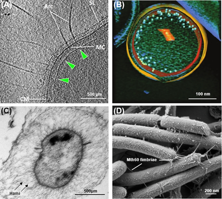
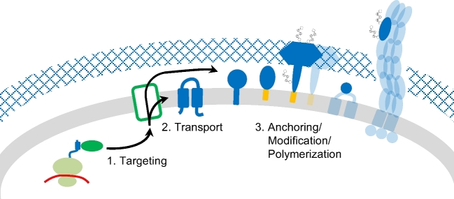
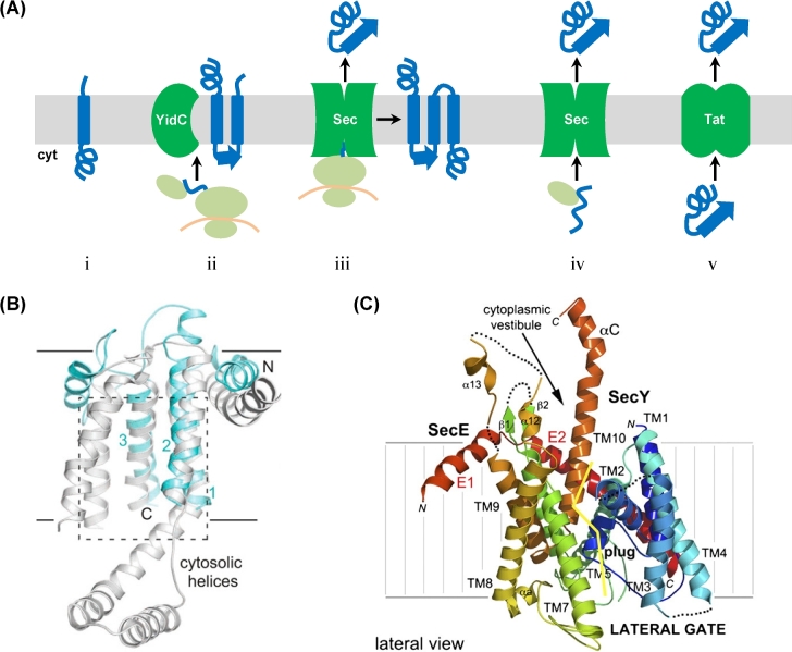
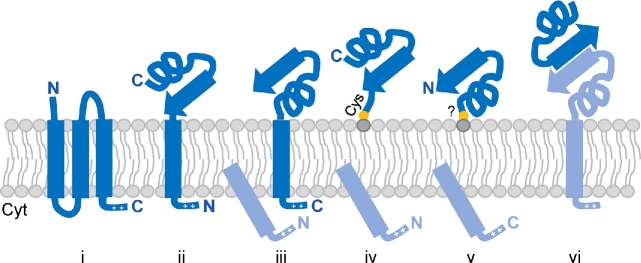
Cell surfaces are critical for diverse functions across all domains of life, from cell-cell communication and nutrient uptake to cell stability and surface attachment. While certain aspects of the mechanisms supporting the biosynthesis of the archaeal cell surface are unique, likely due to important differences in cell surface compositions between domains, others are shared with bacteria or eukaryotes or both. Based on recent studies completed on a phylogenetically diverse array of archaea, from a wide variety of habitats, here we discuss advances in the characterization of mechanisms underpinning archaeal cell surface biogenesis. These include those facilitating co- and post-translational protein targeting to the cell surface, transport into and across the archaeal lipid membrane, and protein anchoring strategies. We also discuss, in some detail, the assembly of specific cell surface structures, such as the archaeal S-layer and the type IV pili. We will highlight the importance of post-translational protein modifications, such as lipid attachment and glycosylation, in the biosynthesis as well as the regulation of the functions of these cell surface structures and present the differences and similarities in the biogenesis of type IV pili across prokaryotic domains.


Figures

Figure 1.
Figure 1.
Archaeal cell-surface components. (A) Electron cryotomography of Pyrococcus furiosus; displayed is a tomographic slice through a frozen-hydrated P. furiosus cell, showing archaella on the cell pole. Arc, archaella; SL, S-layer; CM, cell membrane; green arrowheads, polar cap. MC, (archaellar) motor complex. Image and modified legend adapted from Daum et al. (2017). (B) A 50 voxel-thick slice of a central section of an ultrasmall archaeal ARMAN cell with the segmented inner and outer membranes in orange and yellow, respectively. Ribosomes are represented with light blue spheres drawn to scale. Low mass densities in the volume-rendered cytoplasm are in green. Segmented orange label in the cytoplasm corresponds to the cross section of a tubular organelle structure. Image and modified legend adapted from Comolli et al. (2008). (C) Electron micrograph (ultrathin section) of Candidatus Altiarchaeum hamiconexum; the cell is surrounded by an EPS matrix and cell surface appendages (hami), which extend beyond the matrix. The cell has two membranes with a faint periplasm. FtsZ aggregates are located at the inner membrane. Image and modified legend adapted from Probst et al. (2014). (D) Scanning electron micrograph of M. thermautotrophicus containing Mth60 fimbriae grown on gold electron microscope grids. Image courtesy of Gerhard Wanner, Ludwig Maximilian University, Germany.
Figure 2.
Figure 2.
Schematic overview of common steps in the biosynthesis of archaeal cell surface proteins. During translation, hydrophobic domains of the nascent polypeptide chains are recognized, targeting the ribonucleic protein complex to the membrane (1). The cell surface proteins can be integrated into or transported across the membrane (2), followed by anchoring via transmembrane (TM) domains or covalent linkage to lipid anchors (3). Further post-translational modifications (PTMs) of cell surface proteins include the removal of the signal peptide as well as N- and O-glycosylation. Protein–protein interactions can lead to the binding of soluble proteins at the cell surface or to the polymerization into larger structures such as type IV pili or the S-layer. Proteins involved in distinct pathways associated with these processes (green), the cell surface proteins themselves (blue) as well as their various membrane anchors (orange) are not specified here but discussed in detail in this review.
Figure 3.
Figure 3.
Translocation of proteins into and across the membrane. (A) With the exception of spontaneous insertion of membrane proteins (i), four main routes for the translocation of cell surface protein into or across the membrane exist in archaea: co-translational YidC-dependent insertion (ii), co-translational insertion or translocation via the Sec complex (iii), Sec-dependent post-translational translocation (iv), and post-translational translocation in a folded confirmation by Tat (v). See the main text for more details. (B) Archaeal Mj0480 and bacterial YidC share key structural features. Structure-based alignment of M. jannaschii Mj0480 (light blue; 5C8J) and Bacillus halodurans BhYidC (gray; 3WO6) showing views from the plane of the membrane. The proteins superimpose with a root-mean-square deviation of 3.9 A° over 105 equivalent residues (out of 141 visible) (Borowska et al.2015). (C) Structure of P. furiosus Pfu-SecYE and the crystal packing. SecY is colored using a rainbow pattern. A ‘clam shell’ structure is formed by the 10 transmembrane (TM) helices with a lateral gate opening between transmembrane helices TM2 and TM3 (in the N-terminal half of the ‘clam shell’) and TM7 and TM8 (in the C-terminal half of the ‘clam shell’). The yellow line delineates the lateral gate on the SecY subunit. Structure and modified legend adapted from Egea and Stroud (2010).
Figure 4.
Figure 4.
Anchoring strategies of archaeal surface proteins. Proteins can be anchored via multiple TM domains (i), single N- or C-terminal TM domains (ii and iii, respectively); N- or C-terminal covalent lipid interactions (iv and v, respectively); or interactions with other surface-anchored proteins (vi). Cleaved signal peptides and interacting surface proteins are colored in light blue.
Figure 5.
Figure 5.
Post-translational modifications of S-layer glycoproteins. Schematic representations of S-layer glycoproteins from (A)H. volcanii P25062, based on Sumper et al. (1990), Kaminski et al. (2013), Parente et al. (2014), Kandiba et al. (2016), Abdul Halim et al. (2017); (B)H. salinarum B0R8E4, based on Mescher and Strominger (1976), Wieland (1988), Kikuchi, Sagami and Ogura (1999), Jarrell et al. (2014); (C)M. voltae Q50833, based on Voisin et al. (2005); and (D)S. acidocaldarius Q4J6E5, based on Peyfoon et al. (2010). Their signal peptides (last three amino acids before the cleavage site, defined as the peptide bond between positions –1 and +1, are indicated), N- and O-glycosylation as well as lipid modification are highlighted. The position of the PGF motif, conserved for ArtA substrates, is indicated but the processing site has not been resolved yet (*). Glycan compositions are given for confirmed glycosites; solid horizontal lines indicate that all subjacent glycosites were identified with the glycans given above the line. It should be noted that for H. volcanii and S. acidocaldarius shorter glycans of the corresponding N-glycosylation pathways were identified for several N-glycosites and that the extent and type of N-glycosylation can depend on the growth condition. Monosaccharides are depicted according to the Symbol Nomenclature for Glycans (Varki et al.2015). While this is a selection of well-characterized SLG N-glycosylation, a more comprehensive summary can be found in Jarrell et al. (2014).

References

    1. Abdul Halim MF, Karch KR, Zhou Y et al. . Permuting the PGF signature motif blocks both archaeosortase-dependent C-terminal cleavage and prenyl lipid attachment for the Haloferax volcanii S-layer glycoprotein. J Bacteriol 2016;198:808–15. - PMC - PubMed
    1. Abdul Halim MF, Pfeiffer F, Zou J et al. . Haloferax volcanii archaeosortase is required for motility, mating, and C-terminal processing of the S-layer glycoprotein. Mol Microbiol 2013;88:1164–75. - PubMed
    1. Abdul Halim MF, Rodriguez R, Stoltzfus JD et al. . Conserved residues are critical for Haloferax volcanii archaeosortase catalytic activity: Implications for convergent evolution of the catalytic mechanisms of non-homologous sortases from archaea and bacteria. Mol Microbiol 2018;108:276–87. - PubMed
    1. Abdul Halim MF, Stoltzfus JD, Schulze S et al. . ArtA-dependent processing of a tat substrate containing a conserved tripartite structure that is not localized at the C terminus. J Bacteriol 2017;199:00802–16. - PMC - PubMed
    1. Abu-Qarn M, Yurist-Doutsch S, Giordano A et al. . Haloferax volcanii AglB and AglD are involved in N-glycosylation of the S-layer glycoprotein and proper assembly of the surface layer. J Mol Biol 2007;374:1224–36. - PubMed

Publication types

MeSH terms