. 2018 Jun 18;14(6):e1007139.
doi: 10.1371/journal.ppat.1007139. eCollection 2018 Jun.

RD5-mediated lack of PE_PGRS and PPE-MPTR export in BCG vaccine strains results in strong reduction of antigenic repertoire but little impact on protection

Affiliations

RD5-mediated lack of PE_PGRS and PPE-MPTR export in BCG vaccine strains results in strong reduction of antigenic repertoire but little impact on protection

Louis S Ates et al. PLoS Pathog. .

Abstract

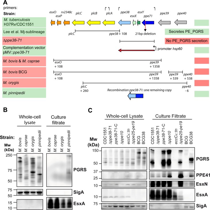
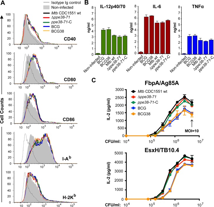
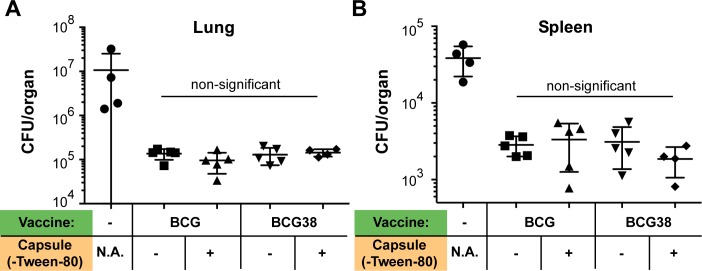
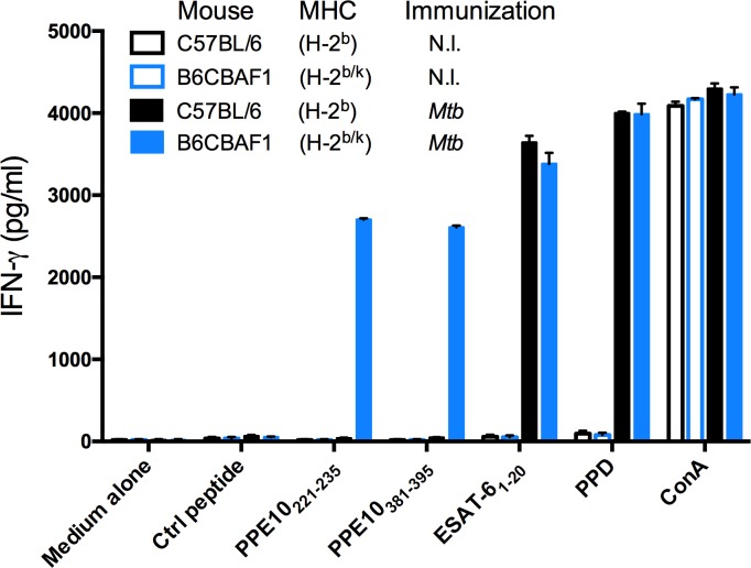
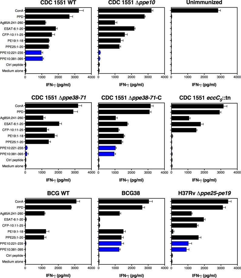
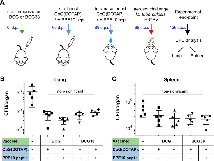
Tuberculosis is the deadliest infectious disease worldwide. Although the BCG vaccine is widely used, it does not efficiently protect against pulmonary tuberculosis and an improved tuberculosis vaccine is therefore urgently needed. Mycobacterium tuberculosis uses different ESX/Type VII secretion (T7S) systems to transport proteins important for virulence and host immune responses. We recently reported that secretion of T7S substrates belonging to the mycobacteria-specific Pro-Glu (PE) and Pro-Pro-Glu (PPE) proteins of the PGRS (polymorphic GC-rich sequences) and MPTR (major polymorphic tandem repeat) subfamilies required both a functional ESX-5 system and a functional PPE38/71 protein for secretion. Inactivation of ppe38/71 and the resulting loss of PE_PGRS/PPE-MPTR secretion were linked to increased virulence of M. tuberculosis strains. Here, we show that a predicted total of 89 PE_PGRS/PPE-MPTR surface proteins are not exported by certain animal-adapted strains of the M. tuberculosis complex including M. bovis. This Δppe38/71-associated secretion defect therefore also occurs in the M. bovis-derived tuberculosis vaccine BCG and could be partially restored by introduction of the M. tuberculosis ppe38-locus. Epitope mapping of the PPE-MPTR protein PPE10, further allowed us to monitor T-cell responses in splenocytes from BCG/M. tuberculosis immunized mice, confirming the dependence of PPE10-specific immune-induction on ESX-5/PPE38-mediated secretion. Restoration of PE_PGRS/PPE-MPTR secretion in recombinant BCG neither altered global antigenic presentation or activation of innate immune cells, nor protective efficacy in two different mouse vaccination-infection models. This unexpected finding stimulates a reassessment of the immunomodulatory properties of PE_PGRS/PPE-MPTR proteins, some of which are contained in vaccine formulations currently in clinical evaluation.


Conflict of interest statement

LM and RB are named inventors on a patent related to RD1, RD5 and RD8 regions of BCG. MAB is a named inventor on a separate patent related to genomic differences of the Mycobacterium tuberculosis complex. The other authors declare that no financial or competing interests exist.

Figures

Fig 1
Fig 1. RD5-like genetic deletions in the M. tuberculosis complex and their effect on PE_PGRS secretion.
A) The genetic organization of the RD5 locus in M. tuberculosis strains CDC1551 and H37Rv is depicted in colored arrows. Bars below the genes indicate the size and location of different RD5-like and ppe38-deletions examined in this work. Arrows above the genes indicate primers used in this study to verify the presence of RD5 associated genes, sequences can be found in S4 Table. Functional PE_PGRS secretion is indicated by shading of the strain name in green, while red shading represents strains in which PE_PGRS secretion is not functional (based on immunoblot analysis). Figure adapted from Mc Evoy et al. 2009 with permission [38]. B) Immunoblot secretion analysis of animal-adapted MTBC strains verifies that strains with RD5 deletions do not secrete PE_PGRS proteins. Samples were prepared as described in materials and methods section. C) Immunoblot secretion analysis reveals PE_PGRS secretion defect in BCG, comparable to the M. tuberculosis ppe38-71-deletion strain or a general ESX-5 secretion mutant (eccC5::tn). SigA was used as a loading and lysis control. Some lysis could be found in both BCG and BCG38, but was not markedly different between strains. Please note that these immunoblots correspond to the same pre-cultures as those that were used in the immunogenicity experiment depicted in Fig 5 and therefore also include the Δppe10 and Δppe25-pe19 isolates. Full western blots corresponding to panels depicted in B-C are depicted in S5 Fig and S7 Fig, respectively.
Fig 2
Fig 2. Secretion of PPE38 and PE_PGRS/PPE-MPTR proteins in BCG or M. tuberculosis does not alter phenotypic and functional maturation, or antigen presentation by innate immune cells.
A) BM-DCs (C57BL/6, H-2b) infected with the indicated mycobacterial strains were stained for surface expression of co-stimulation markers CD40, CD80 and CD86, or MHC components I-Ab and H-2Kb. Depicted are the cell counts (Y-axis) and fluorescent intensity (X-axis) as quantified by flow cytometric analyses. Quantification of mean fluorescent intensity and quantification of cell survival can be found in S1B Table) Culture supernatant of the experiment described in A was assessed for the presence of cytokines IL-12p40/70, IL-6 and TNF-α. No differences were detected between cells infected with the isogenic BCG or M. tuberculosis isolates. C) Antigenic presentation by infected DCs is not affected by disruption or restoration of PPE38–dependent protein secretion in M. tuberculosis or BCG. BM-DCs (BALB/c, H-2d) were infected with two-fold dilutions (data points in graph) of the indicated M. tuberculosis or BCG strains starting at MOI = 10 (indicated by black arrow). IL-2 production was quantified by ELISA after overnight co-culture with I-Ed-restricted T-cell hybridoma specific for FbpA (Ag85A101-120 (2A1), upper panel) or with I-Ad-restricted T-cell hybridoma specific for EsxH (TB10.474−88 (1G1), lower panel). Data are representative of biological duplicates.
Fig 3
Fig 3. Restoring PPE38-dependent protein secretion of BCG does not increase protection against M. tuberculosis in C57BL/6 mice.
Lung (A) or spleen (B) bacterial burdens of C57BL/6 mice infected with M. tuberculosis H37Rv via aerosol administration. Mice were vaccinated s.c. four weeks before the challenge, with 1 x 10⁶ CFU/mouse of either BCG or BCG38 (indicated in green). Both strains were prepared, either in standard culture conditions in medium containing 0.025% Tween-80 considered as no capsule (indicated with (-)), or in culture allowing capsule formation/retention in detergent free condition (indicated with (+)). Photographs of the assessed organs are depicted in S2A and S2B Fig. Each data point represents the CFU/organ of one single mouse counted and averaged from two technical duplicates. Error bars depict the standard deviation. Differences between different vaccination conditions were non-significant (p>0.05), but all vaccination conditions were statistically different from the unimmunized control group (p<0.01). For simplicity, this latter information is not depicted in the figure. Significance was calculated with Prism software using ordinary one-way ANOVA followed by Tukey’s test for multiple comparisons.
Fig 4
Fig 4. Epitope mapping of PPE10 identifies two novel immunogenic T-cell epitopes.
C57BL/6 H-2b (black) or C57BL/6 x CBA (H-2b/k) F1 mice (B6CBAF1, blue) were immunized s.c. with 1 x 10⁶ CFU/mouse of M. tuberculosis H37Rv (Mtb, filled bars), or were left non-immunized (N.I. empty bars). Three weeks post-immunization, splenocytes were stimulated with control peptides or a library of 15-mers spanning PPE10 excluding the PPE domain. T-cell mediated IFN-γ responses were quantified buy ELISA as a measure of immunogenicity. Two immunogenic PPE10-peptides were identified (PPE10221-235 & PPE10381-395) in B6CBAF1 mice. Error bars depict standard deviation over two technical replicates. This figure depicts only newly identified epitopes and controls. Full results of the pep-scan epitope mapping can be found in S2 Fig.
Fig 5
Fig 5. Ability of mycobacteria to induce T-cell responses against PPE-MPTR protein PPE10 is dependent on functional ESX-5- and PPE38-dependent secretion.
C57BL/6 x CBA F1 mice were immunized with the indicated mycobacterial strains. Three weeks post-immunization, splenocytes were stimulated with the indicated peptides and IFN-γ production was measured by ELISA. Responses to the newly identified PPE10-derived immunogenic peptides are depicted in blue. Error bars represent the standard deviation over two technical duplicates. The results are representative of two biological replicates performed on different timepoints.
Fig 6
Fig 6. Boosting PPE10 specific immune responses does not increase protection against M. tuberculosis.
A) Graphical representation of the prime-boost vaccination protocol. Mice were immunized with either BCG or BCG38 (Green). 60 days post-infection (d.p.i.) C57BL/6 x CBA F1 mice were injected s.c. with a booster consisting of adjuvant CpG(DOTAP), alone or in combination with a mix of PPE10221-235 and PPE10381-395 peptides (blue). The same formulation was intranasally administered four weeks later. Nine days after the intranasal boost, mice were exposed to M. tuberculosis H37Rv aerosol infection (220 CFU/lung 1 d.p.i). Bacterial lung (B) and Spleen (C) burdens were assessed by dilution and counting 4 weeks post-infection (experimental end-point) after being photographed for macroscopic investigation (S2C and S2D Fig). Each data point represents the CFU value of one organ from a single mouse, error bars depict the standard deviation. No significant differences between the vaccination conditions were detected by ordinary one-way ANOVA followed by Tukey’s test of multiple comparisons. All vaccination conditions resulted in a significant (p<0.01) reduction in lung burden compared to unimmunized controls (Ordinary one way ANOVA; Dunnett’s test of multiple comparisons against a single control). For simplicity, this latter information is not depicted in the figure. Reduction in spleen CFUs was not significant for any of the vaccination conditions. Statistical analyses were performed using PRISM software.

Similar articles

Cited by

References

    1. WHO. Global tuberculosis report 2017. WHO. World Health Organization; 2017; Available: http://www.who.int/tb/publications/global_report/en/
    1. Arnold A, Witney AA, Vergnano S, Roche A, Cosgrove CA, Houston A, et al. XDR-TB transmission in London: Case management and contact tracing investigation assisted by early whole genome sequencing. J Infect. 2016;73(3):210–8. doi: 10.1016/j.jinf.2016.04.037 - DOI - PubMed
    1. Dheda K, Limberis JD, Pietersen E, Phelan J, Esmail A, Lesosky M, et al. Outcomes, infectiousness, and transmission dynamics of patients with extensively drug-resistant tuberculosis and home-discharged patients with programmatically incurable tuberculosis: a prospective cohort study. Lancet Respir Med. 2017;5(4):269–281. doi: 10.1016/S2213-2600(16)30433-7 - DOI - PubMed
    1. Fletcher HA, Schrager L. TB vaccine development and the End TB Strategy: importance and current status. Trans R Soc Trop Med Hyg. 2016;110: 212–218. doi: 10.1093/trstmh/trw016 - DOI - PMC - PubMed
    1. Behr M., Small P. A historical and molecular phylogeny of BCG strains. Vaccine. 1999;17: 915–922. doi: 10.1016/S0264-410X(98)00277-1 - DOI - PubMed

Publication types