. 2018 Jun 18;13(6):e0198403.
doi: 10.1371/journal.pone.0198403. eCollection 2018.

Tongxinluo attenuates reperfusion injury in diabetic hearts by angiopoietin-like 4-mediated protection of endothelial barrier integrity via PPAR-α pathway

Affiliations

Tongxinluo attenuates reperfusion injury in diabetic hearts by angiopoietin-like 4-mediated protection of endothelial barrier integrity via PPAR-α pathway

Kang Qi et al. PLoS One. .

Abstract

Objective: Endothelial barrier function in the onset and Tongxinluo (TXL) protection of myocardial ischemia/reperfusion (I/R) injury, and TXL can induce the secretion of Angiopoietin-like 4 (Angptl4) in human cardiac microvascular endothelial cells during hypoxia/reoxygenation. We intend to demonstrate whether TXL can attenuate myocardial I/R injury in diabetes, characterized with microvascular endothelial barrier disruption, by induction of Angptl4-mediated protection of endothelial barrier integrity.

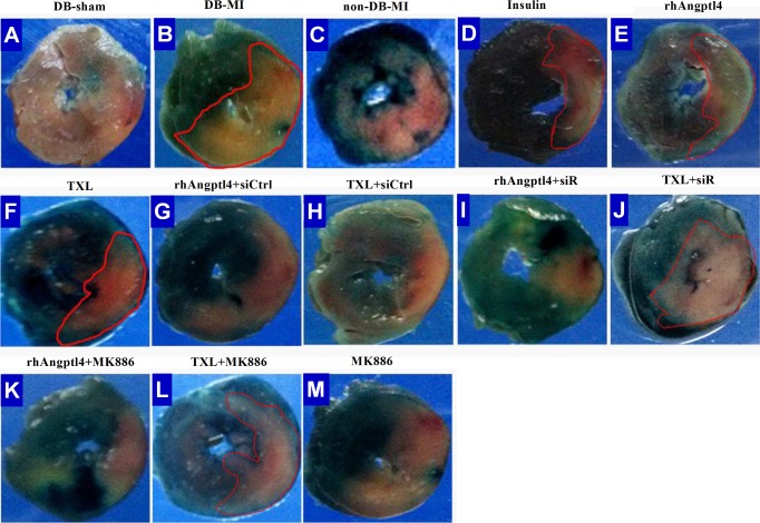
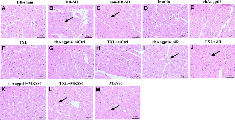
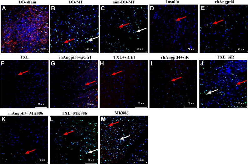
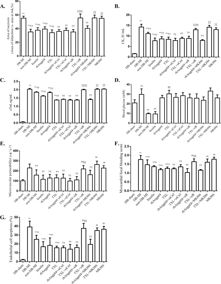
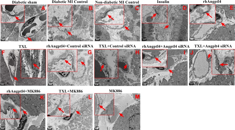
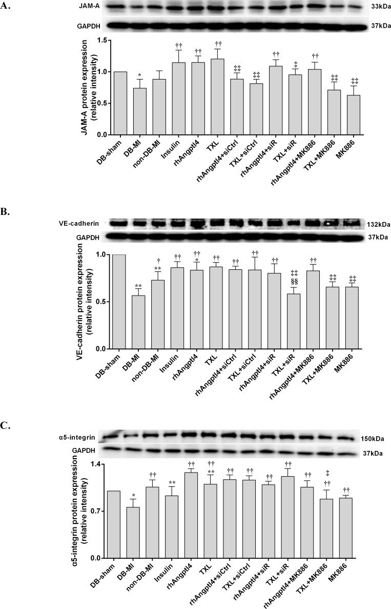
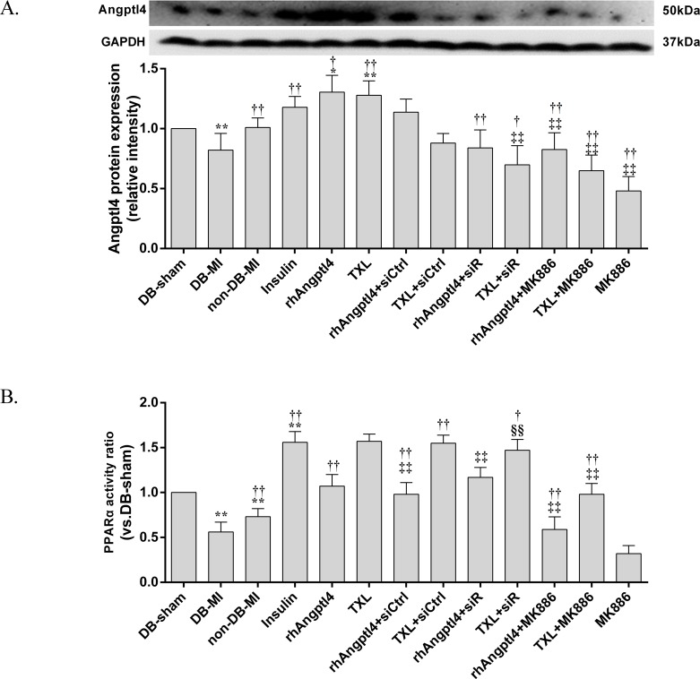
Methods and results: I/R injury was created by coronary ligation in ZDF diabetic and non-diabetic control rats. The animals were anesthetized and randomized to sham operation or I/R injury with or without the exposure to insulin, rhAngptl4, TXL, Angptl4 siRNA, and the PPAR-α inhibitor MK886. Tongxinluo, insulin and rhAngptl4 have the similar protective effect on diabetic hearts against I/R injury. In I/R-injured diabetic hearts, TXL treatment remarkably reduced the infarct size, and protected endothelial barrier integrity demonstrated by decreased endothelial cells apoptosis, microvascular permeability, and myocardial hemorrhage, fortified tight junction, and upregulated expression of JAM-A, integrin-α5, and VE-cadherin, and these effects of TXL were as effective as insulin and rhAngptl4. However, Angptl4 knock-down with siRNA interference and inhibition of PPAR-α with MK886 partially diminished these beneficial effects of TXL and rhAngptl4. TXL induced the expression of Angptl4 in I/R-injured diabetic hearts, and was canceled by Angptl4 siRNA and MK886. TXL treatment increased myocardial PPAR-α activity, and was abolished by MK886 but not by Angptl4 siRNA.

Conclusions: TXL protects diabetic hearts against I/R injury by activating Angptl4-mediated restoration of endothelial barrier integrity via the PPAR-α pathway.


Conflict of interest statement

The authors have declared that no competing interests exist.

Figures

Fig 1
Fig 1. Histopathologic assessments of the area at risk and necrosis in the infarcted hearts treated with or without TXL in the presence or absence of signal regulators.
The area at risk and necrosis was respectively examined by Evans blue and triphenyltetrazolium chloride (TTC) staining (n = 8 in each group). The health myocardium was stained blue by Evans blue, the area at risk (AAR) was not stained by Evans blue. TTC-unstained white myocardium was identified as the area of necrosis (AN). Abbreviations: DB-sham = Diabetic sham; DB-MI = Diabetic MI control; non-DB-MI = non-diabetic MI control; rhAngptl4 = recombinant human Angptl4; rhAngptl4+siCtrl = rhAngptl4+control siRNA; TXL+siCtrl = TXL+control siRNA; rhAngptl4+siR = rhAngptl4+Angptl4 siRNA; TXL+siR = TXL+Angptl4 siRNA.
Fig 2
Fig 2. Comparation of parameters of myocardial injury, blood glucose and endothelial barrier integrity in reperfused rats.
Area of necrosis, levels of CK activity and cTnI, blood glucose levels, microvascular permeability levels, myocardial focal bleeding score and endothelial cell apoptosis rate was compared among different groups. (A-C) Quantification of myocardial injury and necrosis; (D) Blood glucose levels; (E-G) Parameters of endothelial barrier integrity. Compared with the DB-sham group, *P<0.05, **P<0.01; Compared with the DB-MI group, P<0.05, ††P<0.01; Compared with the TXL group, P<0.05, ‡‡P<0.01; Compared with the rhAngptl4+siR group, §§P<0.01; Compared with the Insulin group, ¶¶P<0.01. N = 8 for each group. Abbreviations as in Fig 1.
Fig 3
Fig 3. Evaluation of intramyocardiac hemorrhage in the infarcted hearts treated with or without TXL.
Sections of the hearts were stained with hematoxylin-eosin (n = 8 in each group). DB-sham group has no obvious extravasation of red blood cells in the interstitial space (A). I/R injury induced apparent extravasation of red blood cells into the interstitial space in both DB-MI (B) and non-DB-MI (C) groups. Treatment with insulin (D), rhAngptl4 (E) or TXL (F) greatly decreased extravasation of red blood cells. Combination with angptl4 siRNA canceled the effects of rhAngptl4 (I) and TXL (J). However, combination with MK886 abolished the effect of TXL (L) but not rhAngptl4 (K). Images were taken under a Leica microscope with 40×objective. Black arrows indicate intra-myocardiac hemorrhage. Abbreviations as in Fig 1.
Fig 4
Fig 4. Identification of endothelial cell apoptosis in the I/R-injured hearts treated with or without TXL by confocal microscopy.
Endothelial cells were identified by red fluorescence (CD34), total cell number was detected by blue fluorescence (DAPI DNA staining), and apoptosis was detected by green fluorescence (TUNEL). Apoptotic endothelial cells were detected and counted by colocalized red and green (displayed as yellow). I/R injury induced significant ECs apoptosis in both MI control and diabetic MI rats (n = 8 in each group). Treatment with insulin, rhAngptl4 or TXL ameliorated ECs apoptosis compared with the diabetic MI controls. Whereas, co-treatment with Angptl4 siRNA partially blocked the beneficial effect of TXL. Administration of the PPARα inhibitor MK886 also reversed the inhibition effect of TXL on ECs apoptosis, but not reduce ECs apoptosis in the rhAngptl4-treated animals. Red arrows indicate endothelial cells and white arrows show apoptotic cells. Abbreviations as in Fig 1.
Fig 5
Fig 5. Ultrastructural assessment of the microvascular endothelial tight junction in I/R-injured myocardium.
Ultrastructural assessment of endothelial integrity was detected in the infarcted hearts in each group by transmission electron microscopy (×10000 and ×30000) (n = 3 in each group). Arrows indicate tight junctions (TJs). DB-sham group (A) showed a considerable overlap between adjacent ECs. In non-DB-MI (B) and DB-MI (C) groups, no obvious overlap and tight connection were found between adjacent ECs, and ECs appeared swollen with increased lysosomal-like inclusions. Treatment with insulin (D), rhAngptl4 (E) or TXL (F) almost reverted the I/R-induced injury of endothelial tight junctions. Combination with angptl4 siRNA or MK886 neutralized the protective effects of TXL (J, L) on endothelial integrity; whereas combination with angptl4 siRNA (I), but not MK886 (K), suppressed the effect of rhAngptl4. Abbreviations as in Fig 1.
Fig 6
Fig 6. Expression levels of JAM-A, VE-cadherin, and integrin-α5 in the I/R-injured hearts with or without TXL treatment.
I/R injury decreased the expression levels of JAM-A (A), VE-cadherin (B), and Integrin-α5 (C). Pre-treatment with insulin, rhAngptl4, or TXL up-regulated expression levels of JAM-A (A), VE-cadherin (B), and Integrin-α5 (C). Addition of Angptl4 siRNA canceled the effects of TXL-induced up-regulation of JAM-A (A) and VE-cadherin (B), but not Integrin-α5 (C). Co-treatment with MK886 abolished the TXL-upregulated expression of JAM-A (A), VE-cadherin (B), and Integrin-α5 (C). Compared with DB-sham group, *P<0.05, ** P<0.01; Compared with the DB-MI group, P<0.05, ††P<0.01; Compared with the TXL group, P<0.05, ‡‡P<0.01; Compared with the rhAngptl4+siR group, §§P<0.01. Abbreviations as in Fig 1.
Fig 7
Fig 7. Expression levels of Angptl4 and analysis of PPAR-α activity in the I/R-injured diabetic hearts treated with or without TXL in the presence or absence of signal regulators.
(A) Angptl4 expression was decreased by I/R injury, and pre-treatment with insulin, rhAngptl4, or TXL reverted this effect. Whereas, Angptl4 siRNA and MK886 abolished the TXL-induced upregulation of Angptl4. (B) I/R injury decreased the PPAR-α activity in the I/R-injured myocardium, and even worse in diabetic hearts. Pre-treatment of insulin, rhAngptl4 and TXL increased the PPAR-α activity. Addition of MK886 but not Angptl4 siRNA abolished the TXL-stimulated PPAR-α activation. Compared with the DB-sham group, *P<0.05, **P<0.01; Compared with the DB-MI group, P<0.05, ††P<0.01; Compared with the TXL group, P<0.05, ‡‡P<0.01; Compared with the rhAngptl4+siR group, §§P<0.01. Abbreviations as in Fig 1.

Similar articles

Cited by

References

    1. Hillis LD, Lange RA (2006) Myocardial infarction and the open-artery hypothesis. N Engl J Med 355: 2475–2477. doi: 10.1056/NEJMe068251 - DOI - PubMed
    1. Zhang L, Liu Y, Lu XT, Wu YL, Zhang C, Ji XP, et al. (2009) Traditional Chinese medication Tongxinluo dose-dependently enhances stability of vulnerable plaques: a comparison with a high-dose simvastatin therapy. Am J Physiol Heart Circ Physiol 297: H2004–2014. doi: 10.1152/ajpheart.00208.2009 - DOI - PubMed
    1. Li XD, Yang YJ, Geng YJ, Jin C, Hu FH, Zhao JL, et al. (2010) Tongxinluo reduces myocardial no-reflow and ischemia-reperfusion injury by stimulating the phosphorylation of eNOS via the PKA pathway. Am J Physiol Heart Circ Physiol 299: H1255–1261. doi: 10.1152/ajpheart.00459.2010 - DOI - PubMed
    1. Cheng YT, Yang YJ, Zhang HT, Qian HY, Zhao JL, Meng XM, et al. (2009) Pretreatment with Tongxinluo protects porcine myocardium from ischaemia/reperfusion injury through a nitric oxide related mechanism. Chin Med J (Engl) 122: 1529–1538. - PubMed
    1. Li XD, Yang YJ, Cheng YT, Dou KF, Tian Y, Meng XM (2013) Protein kinase A-mediated cardioprotection of Tongxinluo relates to the inhibition of myocardial inflammation, apoptosis, and edema in reperfused swine hearts. Chin Med J (Engl) 126: 1469–1479. - PubMed

Publication types

MeSH terms