Case Reports
. 2018 Jun 17;6(2):21.
doi: 10.3390/dj6020021.

The Rigid-Shield Technique: A New Contour and Clot Stabilizing Method for Ridge Preservation

Affiliations
Case Reports

The Rigid-Shield Technique: A New Contour and Clot Stabilizing Method for Ridge Preservation

Alessandro Mattiola et al. Dent J (Basel). .

Abstract

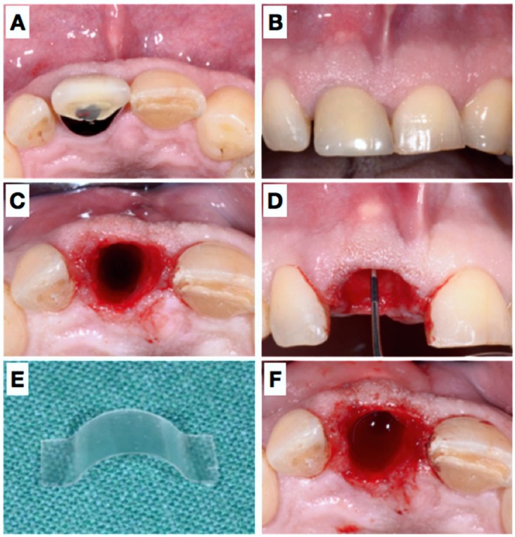
Tooth extraction causes vertical and horizontal alveolar bone loss and consequent remodeling. Several methods have been introduced in terms of so-called “ridge preservation” techniques, which mostly resemble guided bone regenerative (GBR) procedures using filler materials and membranes in order to stabilize the respective sites. This conceptual case report describes a novel approach using a degradable polylactic acid membrane covered with a collagen matrix, which aims to reshape the resorbed alveolar wall and thereby to stabilize the soft tissues during matrix formation and socket mineralization. Clinical re-entry, radiographic (CBCT) and histologic evaluation proved adequate for osteoneogenesis despite an unfavorable initial situation: An implant could be ideally placed, which was circumferentially covered by bone. This minimally invasive method could offer a new method to approach socket preservation without using filler materials and coverage of the socket entrance. However, more controlled research on this topic is needed.

Keywords: bone; dental implant; guided tissue regeneration; regeneration; ridge preservation; tooth extraction.


Conflict of interest statement

The authors declare no conflict of interest.

Figures

Figure 1
Figure 1
Pre-operative view of the tooth 11 still in situ (A,B) and after extraction (C). Panel (D) shows clinically the lack of the buccal plate elucidated with the periodontal probe causing local blood circulation impairment due to pressure at the respective apical gingiva. A resorbable rigid membrane was cut in shape (E) and placed under the resorbable collagen membrane (F), unfortunately not clearly visible in the image).
Figure 2
Figure 2
Post-operative images immediately after extraction and placement of the membranes (A) and after 3 days (B), 11 days (C), 4 weeks (D), 7 weeks (E) and 7 months (F). Please note the minute changes of the buccal contour and good coronal tissue closure.
Figure 3
Figure 3
Clinical situation before implant placement, 7 months after extraction (A,B). Panel (C) shows the situation after elevation of a flap and (D) elucidated the biopsy. Afterwards, an implant could be placed in an ideal position (E): A thin buccal bone coverage was achieved also at the buccal site. Nevertheless, a little GBR was performed using a filler material and a resorbable collagen membrane (F); wound closure (G,H).
Figure 4
Figure 4
After 6 months submerged healing of the implant (A,B), the mucosa was de-epithelialized (C) and a roll-flap was made to uncover the implant (D); a healing cap was placed, which held the buccally advance flap in situ. Situation after 3 weeks (E,F).
Figure 5
Figure 5
5 weeks later (A), the healing abutment was removed (B) and an impression was taken. Panels (C,D) show the situation after placement of the definitive crown placement (screw-retained) and the result two years later, clinically and radiographically (E,F).
Figure 6
Figure 6
Radiographic evaluation (CBCT) after 6 (A) and 16 (B) weeks. The arrows in panel (A) indicate the border of the buccal bone plate and the radiopaque membrane.
Figure 7
Figure 7
Paraffin (A) and acrylic resin (B) sections showing complete bone fill of the extraction socket 7 months after placement of a rigid, resorbable polymer membrane covering the missing buccal bone plate. The two histological sections show mature trabecular bone in the apical half with mature bone marrow, numerous blood vessels, and many embedded osteocytes, whereas mature compact bone prevails in the coronal-most region.

References

    1. Schmidlin P.R., Jung R.E., Schug J. Alveolarkammprävention nach Zahnextraktion-eine Literaturübersicht. Schweiz Monatsschr Zahnmed. 2004;114:328–336. - PubMed
    1. Tan W.L., Wong T.L.T., Wong M.C.M., Lang N.P. A systematic review of post-extractional alveolar hard and soft tissue dimensional changes in humans. Clin. Oral. Implants Res. 2012;23(Suppl. 5):1–21. doi: 10.1111/j.1600-0501.2011.02375.x. - DOI - PubMed
    1. Chappuis V., Engel O., Reyes M., Shahim K., Nolte L.P., Buser D. Ridge Alterations Post-extraction in the Esthetic site. J. Dent. Res. 2013;92(Suppl. 12):195S–201S. doi: 10.1177/0022034513506713. - DOI - PMC - PubMed
    1. Lisowski C.S. A comparative study of the resorption of alveolar ridge tissue under immediate dentures. Dent. Res. Grad. Study. 1944;45:11–13. - PubMed
    1. Von Wowern N., Winther S. Submergence of roots for alveolar ridge preservation. A failure (4-year follow-up study. Int. J. Oral. Surg. 1981;10:247–250. doi: 10.1016/S0300-9785(81)80066-X. - DOI - PubMed

Publication types