. 2018 Sep 15;24(18):4505-4517.
doi: 10.1158/1078-0432.CCR-18-0822. Epub 2018 Jun 18.

Zinc Metallochaperones Reactivate Mutant p53 Using an ON/OFF Switch Mechanism: A New Paradigm in Cancer Therapeutics

Affiliations

Zinc Metallochaperones Reactivate Mutant p53 Using an ON/OFF Switch Mechanism: A New Paradigm in Cancer Therapeutics

Xin Yu et al. Clin Cancer Res. .

Abstract

Purpose: Zinc metallochaperones (ZMC) are a new class of anticancer drugs that reactivate zinc-deficient mutant p53 by raising and buffering intracellular zinc levels sufficiently to restore zinc binding. In vitro pharmacodynamics of ZMCs indicate that p53-mutant activity is ON by 4-6 hours and is OFF by 24. We sought to understand the mechanism of this regulation and to translate these findings preclinically. We further sought to innovate the formulation of ZMCs to improve efficacy.Experimental Design: We performed in vitro mechanistic studies to determine the role of cellular zinc homeostatic mechanisms in the transient pharmacodynamics of ZMCs. We conducted preclinical pharmacokinetic, pharmacodynamic, and efficacy studies using a genetically engineered murine pancreatic cancer model (KPC) to translate these mechanistic findings and investigate a novel ZMC formulation.Results:In vitro, cellular zinc homeostatic mechanisms that restore zinc to its physiologic levels function as the OFF switch in ZMC pharmacodynamics. In vivo pharmacokinetic studies indicate that ZMCs have a short half-life (< 30 minutes), which is sufficient to significantly improve survival in mice expressing a zinc-deficient allele (p53R172H) while having no effect in mice expressing a non-zinc-deficient allele (p53R270H). We synthesized a novel formulation of the drug in complex with zinc and demonstrate this significantly improves survival over ZMC1.Conclusions: Cellular zinc homeostatic mechanisms function as an OFF switch in ZMC pharmacodynamics, indicating that a brief period of p53-mutant reactivation is sufficient for on-target efficacy. ZMCs synthesized in complex with zinc are an improved formulation. Clin Cancer Res; 24(18); 4505-17. ©2018 AACR.


Conflict of interest statement

Disclosure of Potential Conflicts of Interest: Darren Carpizo and S. David Kimball are affiliated with Z53 Therapeutics, Inc. as founder and scientific board member respectively.

Figures

Figure 1
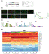
Figure 1. The pharmacodynamics of ZMC1 in cells over 24 hours mirrors the zinc kinetics
A, The p21 and p53 protein levels were examined using western blot in TOV112D cells (p53R175H) in a time course of treatment with 1 μM ZMC1for a 24 hour period. Actin is used as an internal loading control. The quantitation is shown on the right. B, Intracellular free zinc concentration was measured using FluoZin-3 fluorescent imaging. The TOV112D cells were treated with 1 μM ZMC1 over a 24 hour period. At 0, 1, 2, 4, 6, 8, 12, 16 and 24 hrs the cells were imaged with FluoZin-3. The intracellular free zinc concentration was measured according to the manufacturer's instructions. The scale bar = 25 μm. The quantitation of the zinc concentration (from Fig. 1B left panel) and the p21 protein levels (from Fig. 1A) are shown on the right. C, The expression of zinc homeostatic genes (ZnT1 and MT1A which function to decrease free zinc, and ZIP10 which increases intracellular free zinc) was measured by quantitative RT-PCR in TOV112D cells with treatment of 1 μM ZMC1 for over 24 hours. D, RNAseq analysis of TOV112D cells at baseline, 4 hours, 8 hours, and 24 hours after ZMC1 treatment. The subset of the 37 zinc homeostatic genes which were induced in response to ZMC1 treatment labeled with asterisks were shown in Supplemental Table 1.
Figure 2
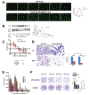
Figure 2. Cellular zinc ion homeostatic mechanisms function as the OFF switch in ZMC1 pharmacodynamics allowing a brief exposure to be efficacious
A, Intracellular free zinc concentration was measured using FluoZin-3 fluorescent imaging in TOV112D parental cells and MT2A/MT1A KO cells. The cells were treated with 1 μM ZMC1 over an 8 hour period. Scale bar = 50 μm. The quantitation of the zinc concentration is shown on the right. B, The p21 and p53 protein levels in TOV112D parental cells and MT2A/MT1A KO cells were measured by western blot. Actin is used as an internal loading control. The quantitation of p21 levels normalized with actin is shown on the right. The data of the parental cells (left panel) is from Figure 1A. C, Cell viability measurement of TOV112D parental cells and MT2A/MT1A KO cells upon treatment of the serial dilutions of ZMC1 for 72 hours using Calcein-AM assays. Statistical analysis was performed using Student’s t-test, comparing the viability of each drug concentration between the two cell lines. *, p value < 0.05. EC50s were shown below the graph. D, Cell growth measurement of TOV112D parental cells and MT2A/MT1A KO cells upon treatment of 1 μM ZMC1 for 24 hours. The cells were fixed and stained with Crystal violet. The viable cells were counted by using automated cell counting on the Keyence BZ-X software. The average cell count per field was normalized with the vehicle control samples. 4 fields per dish and 3 dishes per treatment or control were counted. **, P value < 0.0001. E, The TOV112D cells with various time of exposure of the serial dilutions of ZMC1 and then the drug-free medium was replaced for the incubation up to 72 hours. Cell viability measurement was performed using Guava ViaCount. Statistical analysis was performed using Student’s t-test, compared to 30 min treatment with 0.01 μM. *, p value = 0.006. **, p value < 0.0001. F, Long term effect of the different exposure time (15 min, 30 min, and 60 min) of 0.01 μM ZMC1 was evaluated by clonogenic assay. The cells were incubated in drug-free medium for 14 days to form colonies followed by Crystal violet staining. The quantitation is shown on the right. The efficiency of the p53 siRNA knockdown is measured by Western blot shown on the right.
Figure 3
Figure 3. ZMC1 Reactivates the p53R172H in the murine KPC pancreatic cancer model
A, Mutant p53 protein refolding was evaluated using conformation-specific immunofluorescent imaging with PAB240 antibody. The p53+/+ and p53R172H/+ cells were treated with 1 μM ZMC1 for 6 hours followed by immunofluorescent staining. The scale bar = 25 μm. B, The p21 protein levels were evaluated by western blot in K8484 cells upon treatment with 1 μM ZMC1 for up to 24 hours. GAPDH was used as the internal control. C, Sensitivity of K8484 (p53R172H/+), 270H (p53R270H/+) and K8483 (p53−/−) to ZMC1 was evaluated by cell growth inhibition assay (MTS assay). The cells were treated with serial dilutions of ZMC1 for 72 hours. D, Gene expression of 4 p53 regulated genes, p21, Puma, Noxa and Gdf15, after ZMC1 treatment was measured by qRT-PCR. E, Gene expression of 3 common zinc regulation genes (Znt1, Mt1, Zip10) after ZMC1 treatment was measured by qRT-PCR.
Figure 4
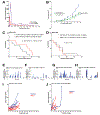
Figure 4. ZMC1 improves survival in the KPC model specifically in a zinc deficient allele
A, Kinetics of plasma concentrations of ZMC1 over 4 hours. B, Comparison of ZMC1 administered at 1 mg/kg IV bolus daily versus 1 mg/kg/24hr daily by continuous IV infusion (AZLET pump) in the K8484 subcutaneous model. Tumor size was measured every 2–3 days. The p-value of bolus IV vs pump IV was shown. C-D, Kaplan-Meier survival curves for KPC-172 (p53R172H) (C) and KPC-270 (p53R270H) (D) mice treated with vehicle control or ZMC1. The median survival time, number of mice in each group and the p value was shown. E-H, Relative individual tumor growth measured by ultrasound for KPC-172 and KPC-270 (Waterfall plot). The mice were treated with vehicle control or ZMC1 by IP with 5 mg/kg daily until the end point. Each bar color represents a measurement expressed as percentage change from baseline. E, KPC-172 mice treated with vehicle control. F, KPC-172 mice treated with ZMC1. G, KPC-270 mice treated with vehicle control. H KPC-270 mice treated with ZMC1. I-J, Individual tumor growth for KPC-172 (I) and KPC-270 (J).
Figure 5
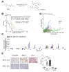
Figure 5. ZMC1 complexed with zinc (Zn-1), a novel formulation, exhibits greater efficacy and less toxicity
A, Synthesis of [Zn(ZMC1)2] (i.e. Zn-1). B, Kaplan-Meier survival curves for KPC-172 mice treated with Zn-1. The median survival time, number of mice in each group and the p value was shown. The data of vehicle control and ZMC1 treatment groups were from Fig. 4D. C, Relative individual tumor growth (waterfall plot) for KPC-172 mice treated with Zn-1 by IP with 5.6 mg/kg daily until the end point. Each bar color represents a measurement. D, Individual tumor growth for KPC-172 upon treatment of vehicle control, ZMC1 or Zn-1 (Spiderweb plot). The data of vehicle control and ZMC1 treatment groups were from Fig. 4J. E, Representative IHC staining with Cleaved caspase 3 for tumor tissues from KPC-172 and KPC-270 treated with vehicle, ZMC1 or Zn-1. The quantitation is shown on the right.
Figure 6
Figure 6. The ZMC Switch mechanism
A, At physiologic zinc levels (10−9 M), p53R175H is in its Apo (zinc free) state because the mutation weakens its binding at the native ligation site (p53R175H Zn Kd 2 × 10⁻⁶ (15)). This causes the protein to misfold and lose wild type transcriptional function. B, Upon treatment with a ZMC, intracellular zinc levels increase approximately 1000 fold (1.5 × 10⁻⁵ M), and this allows zinc to bind in the p53R175H native ligation site and the protein adopts a wild type conformation (ON switch). C, Once the protein undergoes a wild type conformation change, the p53R175H exhibits wild type transcriptional activity and an apoptotic mechanism is induced. D, Within hours the cell responds to this zinc surge by activating zinc homeostatic mechanisms (increasing expression of metallothioneins, zinc exporters, decreasing expression of zinc importers) that function to lower cellular zinc levels to physiologic range. Zinc is no longer at a sufficient concentration to remain bound in the p53R175H and the protein returns to its Apo state (OFF switch).

Similar articles

Cited by

References

    1. Haupt Y, Maya R, Kazaz A, Oren M. Mdm2 promotes the rapid degradation of p53. Nature 1997;387(6630):296-9. - PubMed
    1. Levine AJ, Oren M. The first 30 years of p53: growing ever more complex. Nature reviews Cancer 2009;9(10):749-58. - PMC - PubMed
    1. Joerger AC, Fersht AR. Structure-function-rescue: the diverse nature of common p53 cancer mutants. Oncogene 2007;26(15):2226-42. - PubMed
    1. Freed-Pastor WA, Prives C. Mutant p53: one name, many proteins. Genes & development 2012;26(12):1268-86. - PMC - PubMed
    1. Blanden AR, Yu X, Loh SN, Levine AJ, Carpizo DR. Reactivating mutant p53 using small molecules as zinc metallochaperones: awakening a sleeping giant in cancer. Drug Discov Today 2015;20(11):1391-7. - PMC - PubMed

Publication types