. 2018 Jul 6;38(4):BSR20180704.
doi: 10.1042/BSR20180704. Print 2018 Aug 31.

Long noncoding RNA MIAT regulates apoptosis and the apoptotic response to chemotherapeutic agents in breast cancer cell lines

Affiliations

Long noncoding RNA MIAT regulates apoptosis and the apoptotic response to chemotherapeutic agents in breast cancer cell lines

Zainab A Almnaseer et al. Biosci Rep. .

Abstract

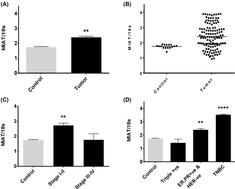
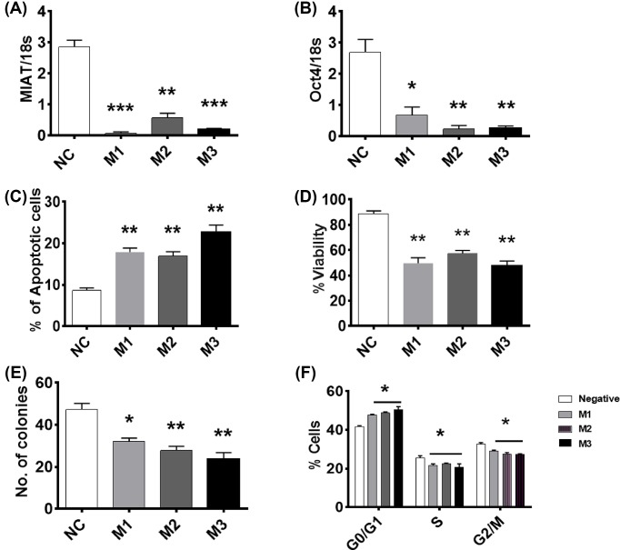
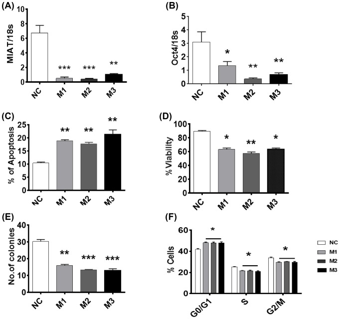
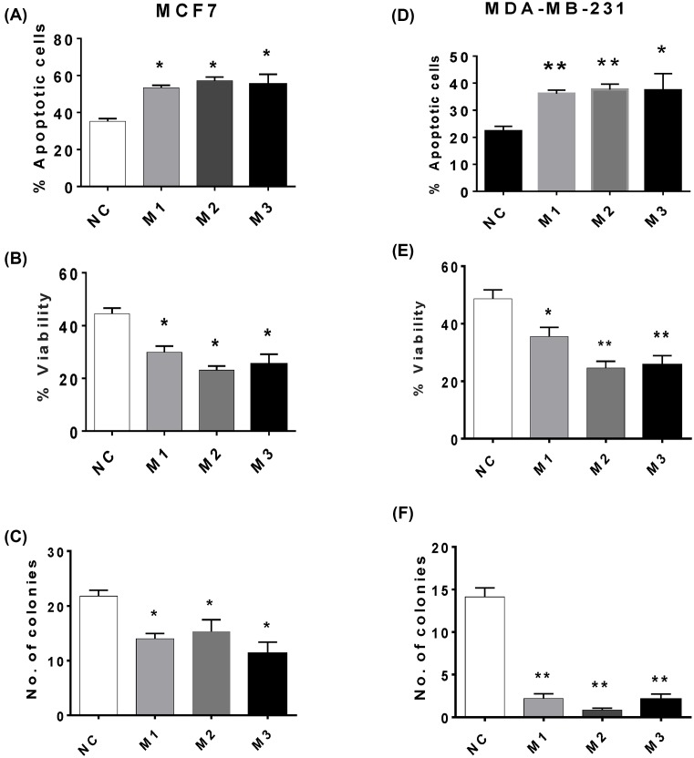
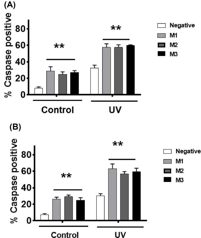
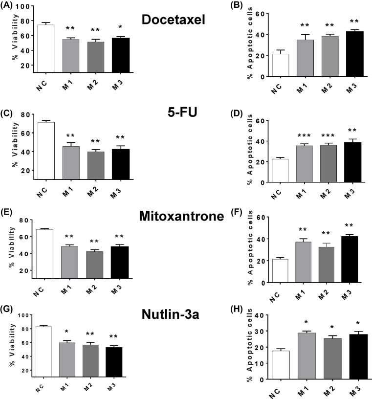
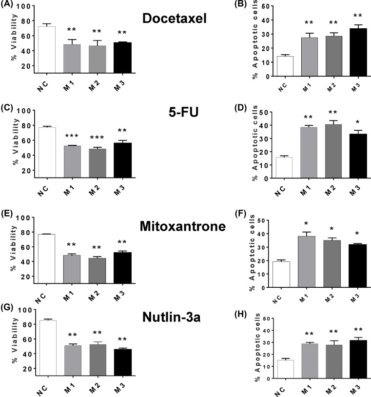
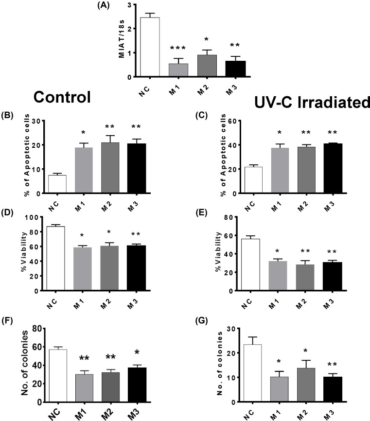
The long noncoding RNA myocardial infarction associated transcript (MIAT) is involved in a number of diseases, including myocardial infarction and diabetic retinopathy. Emerging evidence suggests that MIAT expression levels are increased in different type of cancers, including breast cancer. In the present study, we further evaluated the role of MIAT in breast cancer and investigated the consequences of its silencing on breast cancer response to chemotherapeutic agents. Expression levels of MIAT mRNA in breast cancer were determined using TissueScan™ Breast Cancer cDNA Arrays. Breast cancer cell lines were transfected with MIAT specific siRNAs, with silencing confirmed using RT-qPCR and the effects on breast cancer cell survival and response to different apoptotic stimuli determined. MIAT transcript levels were significantly elevated in breast cancer samples. Such increase was specific to the early stages of the disease, ER, PR +ve, HER -ve, and triple negative breast cancer samples. Silencing of MIAT induced growth arrest and increased basal apoptosis. Reduced levels of MIAT augmented the apoptotic response of breast cancer cells to a wide range of apoptotic stimuli. Our results also showed that MIAT down-regulation was associated with a decrease in OCT4 mRNA, suggesting the existence of a MIAT/OCT4 regulatory loop, similar to that observed in malignant mature B cells. Taken together with the recent demonstration of oncogene characteristics, our observations suggest that MIAT plays an important role in breast tumorigenesis. Strategies to decrease MIAT expression levels may improve sensitivity to therapy in breast cancer by enhancing the apoptotic responses to conventional chemotherapies.

Keywords: Apoptosis; Breast; Cancer; Chemotherapy; MIAT; OCT4.


Conflict of interest statement

The authors declare that there are no competing interests associated with the manuscript.

Figures

Figure 1
Figure 1. MIAT expression levels in breast cancer
The level of MIAT lncRNA was determined in 128 tumor samples and compared with 16 normal breast tissue samples. (A) MIAT expression showed significant increase in the full data sets of breast tumors compared with normal. (B) Corresponding scattergrams of these data are shown. (C) Analysis of the data revealed that there is a highly significant up-regulation of MIAT lncRNA in stage I–II disease. (D) Stratification of MIAT expression levels according to the molecular subtypes showed variation in MIAT expression. MIAT expression was not changed in the triple positive (ER, PR, and HER2 positive) samples, significantly increased in ER, PR +ve, HER –ve, and TNBC breast cancer subtypes (Mann–Whitney Rank Sum Test (A and B) and Kruskal–Wallis one-way ANOVA on ranks (C and D); **P<0.01 and ****P< 0.0001; n=16 (normal), 128 (breast adenocarcinoma specimens)).
Figure 2
Figure 2. Effect of MIAT silencing on the survival of MCF7 cells
MCF7 cells (n=4 cultures) were transfected with the indicated MIAT siRNA or negative control (NC) siRNA and harvested at 72 h post-transfection. Cells were replated for assessment of cell survival after a further 48 h. (A) At 72 h post-transfection, RT-qPCR analysis confirmed decreased cellular MIAT levels in cells treated with MIAT siRNAs. (B) Cells transfected with MIAT siRNAs expressed less OCT4 mRNA as confirmed with RT-qPCR. (C) Silencing of MIAT enhanced basal apoptosis. (D) Short-term viability was reduced 48 h post replating. (E) Colony formation in long-term clonogenic assays was significantly decreased. (F) Cell cycle analysis revealed that MIAT silencing caused an elevation in the percentage of cells in G0/G1 phase; *P<0.05, **P<0.01, and ***P<0.001 versus cells transfected with NC siRNA (one-way ANOVA and Bonferrroni’s MCT).
Figure 3
Figure 3. Effect of MIAT siRNAs on the survival of MDA-MB-231 cells
MDA-MB-231 cells were transfected with the indicated MIAT siRNA or negative control (NC) siRNA and harvested at 72 h post-transfection (n=4 cultures). (A) At 72 h post-transfection, RT-qPCR analysis confirmed decreased cellular MIAT levels. (B) mRNA levels of OCT4 were also reduced in MIAT transfected cells. (C) Apoptosis levels measured 48 h post replating showed that silencing of MIAT enhanced basal apoptosis. (D) Transfection of MIAT siRNAs caused a reduction in short-term viability. (E) Clonogenic ability was reduced in the cells transfected with MIAT siRNAs. (F) Cell cycle analysis revealed that MIAT silencing caused an elevation in the percentage of cells in G0/G1 phase; *P<0.05, **P<0.01, and ***P<0.001 versus cells transfected with NC siRNA (one-way ANOVA and Dunnett’s MCT).
Figure 4
Figure 4. Effect of MIAT siRNAs on UV-C induced cell death in MCF7 and MDA-MB-231 cells
Breast cancer cells (n=4 cultures) were transfected with the indicated MIAT siRNA or negative control (NC) siRNA and harvested at 72 h post transfection. Cells were exposed to UV-C irradiation and cell survival parameters were determined 48 h post-treatment. For all figure parts, MCF7 results are displayed in the left-hand panel and MDA-MB-231 cells in the right-hand panel. (A) and (D) Reduced levels of MIAT enhanced UV-C-induced apoptosis. (B) and (E) MIAT silencing increased UV-C-induced loss of cell survival. (C) and (F) MIAT silencing also promoted UV-induced loss of clonogenic activity; *P<0.05 and **P<0.01 versus cells transfected with NC siRNA (one-way ANOVA and Dunnett’s MCT).
Figure 5
Figure 5. MIAT silencing promotes caspase activation in breast cancer cells
Breast cancer cells were transfected with the indicated MIAT siRNA or negative control (NC) siRNA. At 72 h post-transfection, cells were trypsinized and treated ± UV-C light (20 J/m2). Cells were incubated for 24 h before the assessment of caspase activity. Percentage of cells containing activated caspases was determined using fluorescence microscopy. (A) MIAT silencing caused an enhanced caspase activity in MCF7 breast cancer cells. (B) Transfection of MIAT siRNAs caused an enhanced caspase activity in MDA-MB-231 breast cancer cells; **P<0.01 versus cells transfected with NC (one-way ANOVA and Bonferrroni’s MCT; n=4).
Figure 6
Figure 6. Effect of MIAT siRNAs on the survival of MCF7 cells after treatment with chemotherapeutic drugs
MCF7 cells (n=4 cultures) were transfected with MIAT siRNA or negative control (NC) siRNA and after 72 h, treated with either 5-FU (175 μM), docetaxel (10 μM), nutlin-3 (5 μM), mitoxantrone (50 μM) or vehicle (0.25% dimethyl sulfoxide). Following 48 h incubation, cell viability was determined by flow cytometry on harvested cells and apoptosis was determined by annexin V staining and flow cytometry. For all figure parts, cell viability is displayed in the left-hand panel and apoptosis in the right-hand panel. (A and B) MIAT silencing enhanced the loss of viability and apoptosis induced by docetaxel. (C and D) Similar effects were observed with 5-FU. (E and F) Cells transfected with MIAT siRNAs exhibit enhanced loss of cell viability and an increased in apoptosis level after mitoxantrone treatment. (G and H) Loss of viability and induction of apoptosis by nutlin-3a were enhanced in MIAT siRNA transfected cultures; *P<0.05, **P<0.01, and ***P<0.001 versus cells transfected with NC siRNA (one-way ANOVA and Bonferrroni’s MCT).
Figure 7
Figure 7. Effect of MIAT silencing on chemotherapeutic drug-induced death of MDA-MB-231 TNBC cell line
MDA-MB-231 cells (n=4 cultures) were transfected with the indicative MIAT siRNA or negative control (NC) siRNA. At 72 h post-transfection, cells were replated and treated with either 5-FU (175 μM), docetaxel (10 μM), nutlin-3 (5 μM), mitoxantrone (50 μM) or vehicle (0.25% dimethyl sulfoxide). After 48 h incubation, cell viability was determined by flow cytometry and apoptosis was determined by annexin V staining and flow cytometry. For all figure parts, cell viability is displayed in the left-hand panel and apoptosis in the right-hand panel. (A) and B) MIAT silencing enhanced the loss of viability and apoptosis caused by docetaxel. (B and C) Cells transfected with MIAT siRNAs exhibit enhanced loss of cell viability and an increased in apoptosis level after 5-FU treatment. (E and F) MIAT transfected cells showed decreased cell viability and enhanced apoptosis after mitoxantrone treatment. (G and H) Same results were obtained with nutlin; *P<0.05, **P<0.01, and ***P<0.001 versus cells transfected with NC siRNA (one-way ANOVA and Bonferrroni’s MCT).
Figure 8
Figure 8. Effect of MIAT knockdown on the basal survival and UV-C induced cell death in triple-negative Hs58T breast cancer cells
Hs58T cells (n=4 cultures) were transfected with the indicated MIAT siRNA or negative control (NC) siRNA. Cells were harvested at 72 h post-transfection and were treated ± UV-C irradiation and replated for a further 48 h before the assessment of cell survival. (A) RT-qPCR analysis confirmed decreased cellular MIAT levels at 72 h post-transfection in cells treated with MIAT siRNAs. (B) Basal apoptosis levels were increased in MIAT siRNA transfected cells. (C) Transfection with MIAT siRNAs enhanced UV-C-induced apoptosis. (D) Correspondingly, cell viability was also reduced in cells transfected with MIAT siRNAs. (E) Loss of viability caused by UV was more enhanced in the cells transfected with MIAT siRNAs. (F) MIAT silencing induces loss of clonogenic activity. (G) UV-C-induced loss of colony forming ability was more enhanced in MIAT siRNA transfected cells; *P<0.05, **P<0.01, and ***P<0.001 versus cells transfected with NC siRNA (one-way ANOVA and Bonferrroni’s MCT).

Similar articles

Cited by

References

    1. Mattick J.S. and Makunin I.V. (2006) Non-coding RNA. Hum. Mol. Genet. 15, R17–R29 10.1093/hmg/ddl046 - DOI - PubMed
    1. Wang K.C. and Chang H.Y. (2011) Molecular mechanisms of long noncoding RNAs. Mol. Cell 43, 904–914 10.1016/j.molcel.2011.08.018 - DOI - PMC - PubMed
    1. Quinn J.J. and Chang H.Y. (2016) Unique features of long non-coding RNA biogenesis and function. Nat. Rev. Genet. 17, 47–62 10.1038/nrg.2015.10 - DOI - PubMed
    1. Rasool M., Malik A., Zahid S., Ashraf M.A.B., Qazi M.H., Asif M. et al. (2016) Non-coding RNAs in cancer diagnosis and therapy. Non-Coding RNA Res. 1, 69–76 10.1016/j.ncrna.2016.11.001 - DOI - PMC - PubMed
    1. Dinger M.E., Amaral P.P., Mercer T.R., Pang K.C., Bruce S.J., Gardiner B.B. et al. (2008) Long noncoding RNAs in mouse embryonic stem cell pluripotency and differentiation. Genome Res. 18, 1433–1445 10.1101/gr.078378.108 - DOI - PMC - PubMed