. 2018 Sep 20;132(12):1318-1331.
doi: 10.1182/blood-2017-12-820308. Epub 2018 Jun 18.

Mutations in the SRP54 gene cause severe congenital neutropenia as well as Shwachman-Diamond-like syndrome

Affiliations

Mutations in the SRP54 gene cause severe congenital neutropenia as well as Shwachman-Diamond-like syndrome

Christine Bellanné-Chantelot et al. Blood. .

Abstract

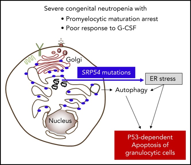
Congenital neutropenias (CNs) are rare heterogeneous genetic disorders, with about 25% of patients without known genetic defects. Using whole-exome sequencing, we identified a heterozygous mutation in the SRP54 gene, encoding the signal recognition particle (SRP) 54 GTPase protein, in 3 sporadic cases and 1 autosomal dominant family. We subsequently sequenced the SRP54 gene in 66 probands from the French CN registry. In total, we identified 23 mutated cases (16 sporadic, 7 familial) with 7 distinct germ line SRP54 mutations including a recurrent in-frame deletion (Thr117del) in 14 cases. In nearly all patients, neutropenia was chronic and profound with promyelocytic maturation arrest, occurring within the first months of life, and required long-term granulocyte colony-stimulating factor therapy with a poor response. Neutropenia was sometimes associated with a severe neurodevelopmental delay (n = 5) and/or an exocrine pancreatic insufficiency requiring enzyme supplementation (n = 3). The SRP54 protein is a key component of the ribonucleoprotein complex that mediates the co-translational targeting of secretory and membrane proteins to the endoplasmic reticulum (ER). We showed that SRP54 was specifically upregulated during the in vitro granulocytic differentiation, and that SRP54 mutations or knockdown led to a drastically reduced proliferation of granulocytic cells associated with an enhanced P53-dependent apoptosis. Bone marrow examination of SRP54-mutated patients revealed a major dysgranulopoiesis and features of cellular ER stress and autophagy that were confirmed using SRP54-mutated primary cells and SRP54 knockdown cells. In conclusion, we characterized a pathological pathway, which represents the second most common cause of CN with maturation arrest in the French CN registry.


Conflict of interest statement

Conflict-of-interest disclosure: The authors declare no competing financial interests.

Figures

None
Graphical abstract
Figure 1.
Figure 1.
WES of 4 pedigrees and description of SRP54 mutations. (A) Confirmation of SRP54 mutations by Sanger sequencing of DNA extracted from whole blood sample and fibroblasts. *Individuals analyzed by WES. (B) Exon-intron of the SRP54 gene locus and protein diagram with the positions of all the mutations identified in this study. (C) Ribbon representation of the three-dimensional structure of the human SRP54 (gold)/SRα (pink) dimer (pdb 5l3q) in 2 different orientations, with the positions of the 7 mutated amino acids (sphere and ball-and-stick representations on the left and right, respectively). The 2 molecules of GMPPNP (5-guanylyl-imidodiphosphate), a nonhydrolyzable GTP analog are shown. The positions of the 5 G elements are reported in green (G1) and red (G2-G5). Magnesium ions are shown as green balls. For a detailed description of the contacts and interactions made by the 7 mutated amino acids, see supplemental Data.
Figure 2.
Figure 2.
Cytological and ultrastructural characteristics of bone marrow granulocytic cells. (A-B) Cytology analysis after May-Grünwald Giemsa staining. Pictures represent granulocytic precursors (a-f) and neutrophils (g-i)) in different individuals P3, P10, P19, P20, and P23. (Original magnification ×100). (C) Comparative cytological bone marrow analysis of 16 individuals with SRP54 mutations and 16 patients with ELANE mutations Significant difference was observed between SRP54 and ELANE patients using nonparametric Kruskal-Wallis test (P = .02). (D-E) Analysis by TEM of the ultrastructural aspect of bone marrow cells from the granulocytic lineage in patient P1. Enlarged ER was observed around the nucleus (N) and thorough the cytoplasm (Cyt) in promyelocytes (D). The black arrow points to nascent autophagosome. Right panels (E) show pre-apoptotic neutrophil granulocytes, a major characteristic of the sample.
Figure 3.
Figure 3.
Increased expression of SRP54 during granulocyte differentiation. CD34+ cells from healthy donors (controls) or patients were cultured for 21 days in serum free-medium with SCF, IL-3, and G-CSF. (A) May-Grünwald Giemsa was performed to determine the purity and stages of differentiation at days (D) 7, 14, and 21. The expression levels of (B) SRP54, (C) CSF3R, and (D) SRP72 were quantified by quantitative reverse transcription polymerase chain reaction (qRT-PCR) related to PPIA and HPRT at days (D) 0, 7, 14, 21. Results are the mean ± SEM of 4 to 8 control samples performed in triplicate. Student t test: *P < .05; **P < .01; ***P < .001. (E) The expression level of SRP54 was quantified by qRT-PCR related to PPIA and HPRT in CD34+ progenitors, CD36+GPA+ erythroblasts, and CD41+CD42+ megakaryocytes. Results are the mean ± SEM of 4 to 8 control samples performed in triplicate. (F) The expression level of SRP54 was quantified by qRT-PCR related to PPIA and HPRT in granulocytic precursors at days 7 and 14 in both controls (n = 5) and patients (n = 5). Results are the mean ± SEM performed in triplicate. (G) The expression level of SRP54 protein was evaluated by western blot analysis on day 10 granulocytic precursors in controls (n = 2) and patients (n = 2), using specific antibodies.
Figure 4.
Figure 4.
SRP54 mutations induce a great impairment of granulocytic differentiation. CD34+ cells from healthy donors (controls) or patients were cultured for 14-21 days in serum free-medium with SCF, IL-3, and G-CSF. (A) The fold increase in proliferation in log scale was assessed individually by Trypan blue exclusion, using controls (= 4) and patients (n = 5). (B) Fold increase in proliferation in patients and controls at days (D) 7, 10, and 14. Mean ± SEM; ***P < .001. (C) Apoptotic granulocytic cells were analyzed by flow cytometry using Annexin V+ assay in controls (n = 2) and patients (n = 2). The MDM2 antagonist, Nutlin 3a (20 µM for 24 hours), was used as positive control at D9. (D) The results represent the mean of 3 independent experiments at days 7, 9, and 13 with controls (n = 2) and patients (n = 2). Mean ± SEM; *P < .05; ***P < .001. (E) P53 target genes (P21, BAX, NOXA) expression levels were checked by qRT-PCR related to PPIA and HPRT at days (D) 7, 10, and 14 with controls (n = 3-4) and patients (n = 2-4). Mean ± SEM; *P < .05. (F) Granulocytic cells were sorted as CD15+CD11b+CD36 cells between days 10 and 14, using CD36 to specifically eliminate monocytes. P53 target genes (P21, BAX, NOXA) expression levels were checked by qRT-PCR related to PPIA and HPRT with controls (n = 4) and patients (n = 3). Mean ± SEM; *P < .05; **P < .01.
Figure 5.
Figure 5.
SRP54 mutant induce ER-stress and autophagy. mRNA expression levels of (A) ATF4, CHOP, and spliced XBP1 for ER stress and (B) ULK1 for autophagy were examined by qRT-PCR related to PPIA and HPRT at days (D) 7, 10, and 14 of culture of control (n = 3-4) and patient (n = 2-4) cells. Mean ± SEM; unpaired t test, 2-tailed on controls vs patients combing all days, *P < .05. mRNA expression levels of (C) CHOP and (D) ULK1 were also checked after sorting CD36CD15+CD11b and CD15+CD11b+CD36 granulocytic cells between days 10 and 14. Controls (n = 3) and patients (n = 3); mean ± SEM; unpaired t test, 2-tailed on controls vs patients; *P < .05; **P < .01. (E) Using primary fibroblasts, autophagy was evaluated by the lipidation of LC3 (LC3-II) by western blot analysis in controls (n = 2) and SRP54-mutated patients (n = 8). β-Actin was used as a loading control. Fold induction of LC3-II/β-Actin was quantified using Image J software.
Figure 6.
Figure 6.
SRP54 knockdown induces a defect in proliferation and increased ER stress and autophagy. (A-D) HL-60 cell lines stably expressing either shRNA targeting SRP54 (1 or 2) or a SCR were generated after transduction with lentiviral particles and after sorting on GFP+ after 72 hours. Nontransduced (NT, not transduced) HL-60 cell line was used as control. (A) SRP54 mRNA expression level was checked by qRT-PCR related to PPIA (n = 4). Mean ± SEM; unpaired t test, 2-tailed; *P < .05; **P < .01. (B) Fold proliferation was assessed after Trypan blue exclusion for 3 days (n = 4). Mean ± SEM; unpaired t test, 2-tailed; *P < .05. (C) Spliced XBP1 mRNA expression level was checked by qRT-PCR related to PPIA, CFL1, and H2AZ (n = 4). Mean of SCR or shRNA_1 and 2 ± SEM; Unpaired t test, 2-tailed; *P < .05. (D) P-eIF2a, P-PERK, P-ULK1 were evaluated by western blot analysis using specific antibodies. β-Actin was used as loading control. (E-G) Normal CD34+ progenitors were transduced with 2 lentiviruses expressing either shRNA targeting SRP54 (ShSRP54_2) or SCR. CD34+GFP+ cells were sorted 72 hours later and were cultured for 10 days in serum-free medium with SCF, IL-3, and G-CSF. (E) Granulocytic cells were counted by Trypan blue exclusion (n = 3) or sorted the same day on the CD36CD15+CD11b+ phenotype for analysis of (F) SRP54 gene expression level (n = 4) and (G) spliced XBP1 (n = 3) mRNA expression level by qRT-PCR related to PPIA and HPRT. Mean ± SEM; unpaired t test, 2-tailed; **P < .01; ***P < .001.

Comment in

Similar articles

Cited by

References

    1. Donadieu J, Beaupain B, Fenneteau O, Bellanné-Chantelot C. Congenital neutropenia in the era of genomics: classification, diagnosis, and natural history. Br J Haematol. 2017;179(4):557-574. - PubMed
    1. Donadieu J, Beaupain B, Mahlaoui N, Bellanné-Chantelot C. Epidemiology of congenital neutropenia. Hematol Oncol Clin North Am. 2013;27(1):1-17, vii. - PubMed
    1. Glaubach T, Minella AC, Corey SJ. Cellular stress pathways in pediatric bone marrow failure syndromes: many roads lead to neutropenia. Pediatr Res. 2014;75(1-2):189-195. - PubMed
    1. Skokowa J, Dale DC, Touw IP, Zeidler C, Welte K. Severe congenital neutropenias. Nat Rev Dis Primers. 2017;3:17032. - PMC - PubMed
    1. Grenda DS, Murakami M, Ghatak J, et al. . Mutations of the ELA2 gene found in patients with severe congenital neutropenia induce the unfolded protein response and cellular apoptosis. Blood. 2007;110(13):4179-4187. - PMC - PubMed

Publication types

MeSH terms

Substances

Supplementary concepts