

Bradykinin Stimulates Renal Na+ and K+ Excretion by Inhibiting the K+ Channel (Kir4.1) in the Distal Convoluted Tubule
- PMID: 29915013
- PMCID: PMC6043363
- DOI: 10.1161/HYPERTENSIONAHA.118.11070
Bradykinin Stimulates Renal Na+ and K+ Excretion by Inhibiting the K+ Channel (Kir4.1) in the Distal Convoluted Tubule
Abstract
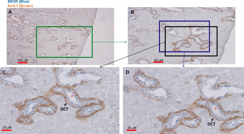
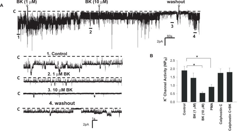
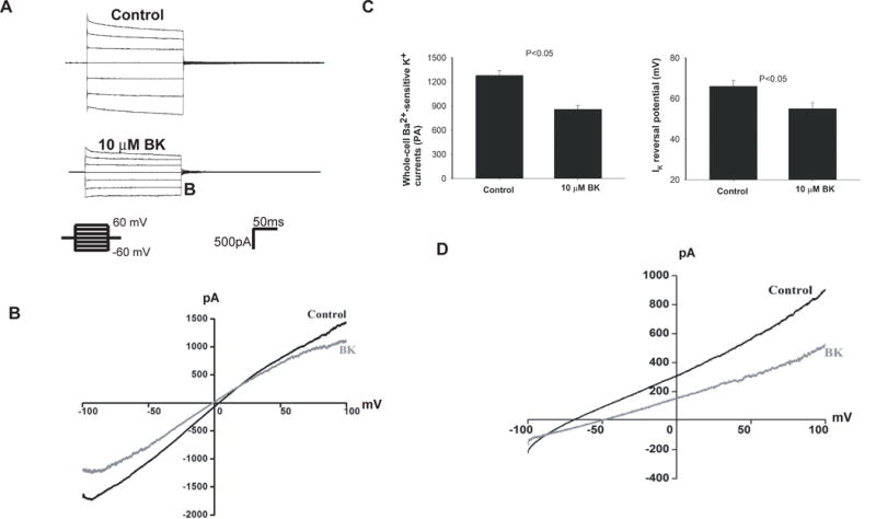
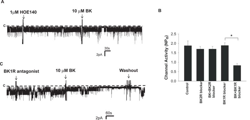
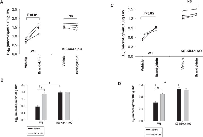
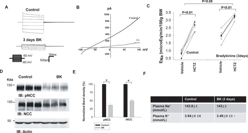
Stimulation of BK2R (bradykinin [BK] B2 receptor) has been shown to increase renal Na+ excretion. The aim of the present study is to explore the role of BK2R in regulating Kir4.1 and NCC (NaCl cotransporter) in the distal convoluted tubule (DCT). Immunohistochemical studies demonstrated that BK2R was highly expressed in both apical and lateral membrane of Kir4.1-positive tubules, such as DCT. Patch-clamp experiments demonstrated that BK inhibited the basolateral 40-pS K+ channel (a Kir4.1/5.1 heterotetramer) in the DCT, and this effect was blocked by BK2R antagonist but not by BK1R (BK B1 receptor) antagonist. Whole-cell recordings also demonstrated that BK decreased the basolateral K+ conductance of the DCT and depolarized the membrane. Renal clearance experiments showed that BK increased urinary Na+ and K+ excretion. However, the BK-induced natriuretic effect was completely abolished in KS-Kir4.1 KO (kidney-specific conditional Kir4.1 knockout) mice, suggesting that Kir4.1 activity is required for BK-induced natriuresis. The continuous infusion of BK with osmotic pump for 3 days decreased the basolateral K+ conductance and the negativity of the DCT membrane. Western blot showed that infusion of BK decreased the expression of total NCC and phosphorylated NCC. Renal clearance experiments demonstrated that thiazide-induced natriuresis was blunted in the mice receiving BK infusion, suggesting that BK inhibited NCC function. Consequently, mice receiving BK infusion for 3 days were hypokalemic. We conclude that stimulation of BK2R inhibits NCC activity, increases urinary K+ excretion, and causes mice hypokalemia and that Kir4.1 is required for BK2R-mediated stimulation of urinary Na+ and K+ excretion.
Keywords: bradykinin B2, receptor; diuretics; hypertension; ion transport; sodium-chloride symporter.
© 2018 American Heart Association, Inc.
Conflict of interest statement
None.
Figures
Similar articles
-
Deletion of renal Nedd4-2 abolishes the effect of high K+ intake on Kir4.1/Kir5.1 and NCC activity in the distal convoluted tubule.Am J Physiol Renal Physiol. 2021 Jul 1;321(1):F1-F11. doi: 10.1152/ajprenal.00072.2021. Epub 2021 May 24. Am J Physiol Renal Physiol. 2021. PMID: 34029145 Free PMC article.
-
AT2R (Angiotensin II Type 2 Receptor)-Mediated Regulation of NCC (Na-Cl Cotransporter) and Renal K Excretion Depends on the K Channel, Kir4.1.Hypertension. 2018 Apr;71(4):622-630. doi: 10.1161/HYPERTENSIONAHA.117.10471. Epub 2018 Feb 26. Hypertension. 2018. PMID: 29483225 Free PMC article.
-
Potassium intake modulates the thiazide-sensitive sodium-chloride cotransporter (NCC) activity via the Kir4.1 potassium channel.Kidney Int. 2018 Apr;93(4):893-902. doi: 10.1016/j.kint.2017.10.023. Epub 2018 Jan 6. Kidney Int. 2018. PMID: 29310825 Free PMC article.
-
Basolateral Kir4.1 activity in the distal convoluted tubule regulates K secretion by determining NaCl cotransporter activity.Curr Opin Nephrol Hypertens. 2016 Sep;25(5):429-35. doi: 10.1097/MNH.0000000000000248. Curr Opin Nephrol Hypertens. 2016. PMID: 27306796 Free PMC article. Review.
-
Inwardly rectifying K+ channels 4.1 and 5.1 (Kir4.1/Kir5.1) in the renal distal nephron.Am J Physiol Cell Physiol. 2022 Aug 1;323(2):C277-C288. doi: 10.1152/ajpcell.00096.2022. Epub 2022 Jun 27. Am J Physiol Cell Physiol. 2022. PMID: 35759440 Free PMC article. Review.
Cited by
-
Dietary Potassium Downregulates Angiotensin-I Converting Enzyme, Renin, and Angiotensin Converting Enzyme 2.Front Pharmacol. 2020 Jun 18;11:920. doi: 10.3389/fphar.2020.00920. eCollection 2020. Front Pharmacol. 2020. PMID: 32625100 Free PMC article.
-
Inhibition of AT2R and Bradykinin Type II Receptor (BK2R) Compromises High K+ Intake-Induced Renal K+ Excretion.Hypertension. 2020 Feb;75(2):439-448. doi: 10.1161/HYPERTENSIONAHA.119.13852. Epub 2019 Dec 23. Hypertension. 2020. PMID: 31865783 Free PMC article.
-
Modifying Dietary Sodium and Potassium Intake: An End to the 'Salt Wars'?Hypertension. 2024 Mar;81(3):415-425. doi: 10.1161/HYPERTENSIONAHA.123.19487. Epub 2023 Oct 12. Hypertension. 2024. PMID: 37823260 Free PMC article. Review.
-
A mechanistic model and therapeutic interventions for COVID-19 involving a RAS-mediated bradykinin storm.Elife. 2020 Jul 7;9:e59177. doi: 10.7554/eLife.59177. Elife. 2020. PMID: 32633718 Free PMC article.
-
Imbalance in Renal Vasoactive Enzymes Induced by Mild Hypoxia: Angiotensin-Converting Enzyme Increases While Neutral Endopeptidase Decreases.Front Physiol. 2018 Dec 11;9:1791. doi: 10.3389/fphys.2018.01791. eCollection 2018. Front Physiol. 2018. PMID: 30618804 Free PMC article.
References
-
- Vio CP, Figueroa CD. Evidence for a stimulatory effect of high potassium diet on renal kallikrein. Kidney Internat. 1987;31(6):1327–1334. - PubMed
-
- Jin L, Chao L, Chao J. Potassium supplement upregulates the expression of renal kallikrein and bradykinin B2 receptor in SHR. Am J Physiol Renal Physiol. 1999;276:F476–F484. - PubMed
-
- Suzuki T, Katori M, Fujita T, Kumagai Y, Majima M. Involvement of the renal kallikrein-kinin system in K(+)-induced diuresis and natriuresis in anesthetized rats. Eur J Pharmacol. 2000;399:223–227. - PubMed
-
- Wang D, Yoshida H, Song Q, Chao L, Chao J. Enhanced renal function in bradykinin B2 receptor transgenic mice. American Journal of Physiology-Renal Physiology. 2000;278(3):F484–F491. - PubMed
Publication types
MeSH terms
Substances
Grants and funding
LinkOut - more resources
Full Text Sources
Other Literature Sources
Research Materials
