. 2018 Jul 3;115(27):E6135-E6144.
doi: 10.1073/pnas.1805159115. Epub 2018 Jun 18.

Hyperstimulation of CaSR in human MSCs by biomimetic apatite inhibits endochondral ossification via temporal down-regulation of PTH1R

Affiliations

Hyperstimulation of CaSR in human MSCs by biomimetic apatite inhibits endochondral ossification via temporal down-regulation of PTH1R

Melika Sarem et al. Proc Natl Acad Sci U S A. .

Abstract

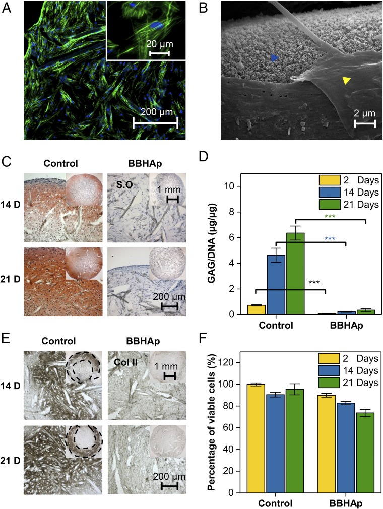
In adult bone injuries, periosteum-derived mesenchymal stem/stromal cells (MSCs) form bone via endochondral ossification (EO), whereas those from bone marrow (BM)/endosteum form bone primarily through intramembranous ossification (IMO). We hypothesized that this phenomenon is influenced by the proximity of MSCs residing in the BM to the trabecular bone microenvironment. Herein, we investigated the impact of the bone mineral phase on human BM-derived MSCs' choice of ossification pathway, using a biomimetic bone-like hydroxyapatite (BBHAp) interface. BBHAp induced hyperstimulation of extracellular calcium-sensing receptor (CaSR) and temporal down-regulation of parathyroid hormone 1 receptor (PTH1R), leading to inhibition of chondrogenic differentiation of MSCs even in the presence of chondroinductive factors, such as transforming growth factor-β1 (TGF-β1). Interestingly rescuing PTH1R expression using human PTH fragment (1-34) partially restored chondrogenesis in the BBHAp environment. In vivo studies in an ectopic site revealed that the BBHAp interface inhibits EO and strictly promotes IMO. Furthermore, CaSR knockdown (CaSR KD) disrupted the bone-forming potential of MSCs irrespective of the absence or presence of the BBHAp interface. Our findings confirm the expression of CaSR in human BM-derived MSCs and unravel a prominent role for the interplay between CaSR and PTH1R in regulating MSC fate and the choice of pathway for bone formation.

Keywords: Wnt signaling; bone remodeling; calcium phosphate; regenerative medicine; stem cell niche.


Conflict of interest statement

The authors declare no conflict of interest.

Figures

Fig. 1.
Fig. 1.
Characterization of BBHAp. (A) SEM revealing a nanoscale homogeneous coating of apatite along the PET fibers (Inset). (B) Transmission electron microscopy confirming the presence of apatite with all of the characteristic diffraction rings of biogenic apatite (Inset). (C) Raman spectroscopy of BBHAp on PET fibers showing clear similarities between BBHAp and human cancellous bone.
Fig. 2.
Fig. 2.
In vitro characterization of human MSC-seeded scaffolds in conditions promoting chondrogenic differentiation. (A) MSCs were able to adhere to the BBHAp interface. F-actin is shown in green, and nuclei are shown in blue. (B) SEM confirmed that the apatite mineral phase is retained in MSC-laden BBHAp scaffolds even after 21 d of in vitro culture. The yellow arrowhead points to a matrix produced by MSCs, and the blue arrowhead points to the BBHAp phase. (C) Tissue produced by MSCs in control constructs (PET) was rich in GAG as visualized by Safranin-O (S.O) staining, while tissue within BBHAp constructs showed no staining for GAGs. (D) Quantification of GAG/DNA in control and BBHAp scaffolds. (E) BBHAp inhibits the expression of collagen type II (Col II) by MSCs as visualized by immunohistochemistry. (F) Cell viability quantified by trypan blue exclusion assay on cells dissociated from scaffolds at different time points. Data are normalized to the number of viable cells retrieved from the control condition at day 2 of in vitro culture.
Fig. 3.
Fig. 3.
Molecular effects of hyperstimulation of extracellular CaSR on human MSCs. (A) Western blot analysis of total cell protein lysate at days 14 and 21 of in vitro culture. BBHAp notably induced the expression of CaSR, Cav-1, β-catenin (β-Cat), and pERK1/2, all with known roles in skeletal development. GAPDH was used as a loading control. Ctrl, control. (B) Immunofluorescent staining confirming the higher expression of CaSR and Cav-1 at day 14 of in vitro culture. (C) Flow cytometry analysis of MSCs constitutively expressing mCherry and EGFP+ under β-catenin promoter retrieved from BBHAp constructs, indicating an increase in β-catenin signaling in comparison to controls (*P < 0.05; ***P < 0.001). (Inset) Representative image of cells residing on fibers coated with BBHAp, coexpressing mCherry and EGFP. (Scale bar: 10 μm.) (D) Heat map showing 2D clustering of the 100 differentially regulated genes with a role in MSC adhesion, proliferation, and differentiation at day 2 of in vitro culture using log fold change (logFC) = 2 and P < 0.05. Expression intensity is represented by red and green, for high and low intensities, respectively. The genes of interest are indicated with arrows. (E) Hierarchical cluster analysis and heat map of the top 100 differentially regulated genes between conditions at two different time points. (F) K-mean clustering gene network analysis of genes shown in E plus CaSR and CTNBB1. The genes with no connection are not shown in the network. Blue cluster shows genes involved in the EO signaling pathway, while the genes in the green cluster belong to peptide ligand-binding SuperPath or immunoregulatory interactions between a lymphoid cell SuperPath and a nonlymphoid cell SuperPath. The network reveals that CaSR is connected to genes modulating the EO pathway (blue clusters) via the Wnt signaling cascade and GNG11 plays a central role in signal transduction. (G) Cfu assays for cfu-c, cfu-o, and cfu-f cells retrieved from the control and BBHAp scaffolds. MSCs once retrieved from the control and BBHAp environments are equally competent in forming colonies.
Fig. 4.
Fig. 4.
Effect of stimulation of PTH1R using PTH (1–34) on human MSC-seeded scaffolds under conditions promoting chondrogenic differentiation. (A) Western blot analysis of total cell protein lysate following stimulation of PTH1R for 2 d showing that PTH (1–34) rescued the expression of PTH1R significantly in the BBHAp group. GAPDH was used as a loading control (Ctrl). (B) Safranin-O staining of cryosectioned slides from conditions with and without PTH (1–34) supplementation at 14 d (Top) and 21 d (Middle). Tissues within BBHAp constructs stimulated with PTH (BBHAp+PTH) showed expression of GAGs at day 21 of in vitro culture in the periphery region (red-orange regions) as assessed by positive staining by Safranin-O, while tissue within BBHAp constructs showed no staining for GAGs. Both control and control scaffolds stimulated with PTH (Control+PTH) were rich in GAG. (Bottom) Higher magnification images of cells residing within the dashed black boxes (Middle) are shown in this row.
Fig. 5.
Fig. 5.
Bone-like mineral phase inhibits EO in vivo. Safranin-O (S.O) (A) and H&E staining (B) of control and BBHAp constructs after 2, 4, and 8 wk following ectopic implantation in nude mice. Control samples showed GAG-rich cartilaginous matrix, which remodels into bone through EO after 8 wk. While the cartilaginous matric was remarkably absent in the BBHAp constructs at all time points, the bony ossicles were formed after 8 wk via IMO. The magnified area is denoted with a dashed black rectangle in the images. (C) In situ hybridization for visualization of human nuclei using an Alu probe. The region within the section that was probed using Alu is denoted by the dashed black rectangles in B. The presence of Alu+ nuclei (arrows) confirmed that the formation of bone in both conditions involves the participation of transplanted human MSCs. Explanted BBHAp constructs after 4 wk lack expression of characteristics of bone formation via EO, such as collagen type II (Col II) (D), MMP-9 (E), and TRAP (F). Coll II and MMP-9 are visualized as brown-colored regions positive for horseradish peroxidase activity. TRAP-positive areas in the control section are indicated by an arrow. (G) Three-dimensional reconstructed μCT images after 8 wk of in vivo implantation confirms the presence of mineralized tissue in both conditions. (H) Degree of maturation in mineralized tissue is visualized by an X-ray attenuation heat map. (I) Bone mineral density in both conditions calculated based on X-ray attenuation. ***P < 0.001. (J) SEM confirming the presence of BBHAp coating even after 8 wk of implantation. Magnified image of the region within the rectangular box on the Top is shown in the Bottom. (K) Fluorescent microscopy images of the area represented by the dashed circle in B (8-wk time point), which corresponds to bone deposits after staining for mouse CD45+ cells, showing the presence of immune cells in close proximity to ectopic bone. Magnified image of the region within the rectangular box on the Top is shown in the Bottom. (L) Visual representation of a proposed mechanism showing the counteractive effects of the PTH1R and IGF1 signaling pathways on hyperstimulation of CaSR between days 2 and 21 in vitro preceding the formation of bone via IMO in BBHAp constructs upon implantation.
Fig. 6.
Fig. 6.
Effect of CaSR KD on the fate and bone-forming capacity of MSCs. (A) MSCs transduced with GIPZ lentiviral shRNA targeting CaSR showing TurboGFP expression 2 d after initial transduction and before puromycin selection. Arrow points to a TurboGFP positive cell. (B) Western blot analysis of total cell lysate for CaSR and Cav-1 in wild type (WT), nonsilencing control (NS), and CaSR KD MSCs after stimulation with 24 mM calcium chloride. GAPDH was used as a loading control. (C) Quantification of Western blot showing an ∼75% decrease in CaSR expression and a slight increase in Cav-1 expression in CaSR KD cells (values are normalized to WT). (D) Frequency of cell cycle per time unit was calculated for NS and KD cells and normalized to WT cells. (Inset) Suppression of growth rate in KD cells. Safranin-O (S.O) staining (E) and immunohistochemistry for collagen type II (Col II) (F) of KD cells seeded in control and BBHAp constructs after 3 wk of in vitro culture revealed that CaSR KD inhibits MSC chondrogenic differentiation independent of the substrate. (G) Representative fluorescent microscopy image of scaffold seeded with CaSR KD cells before implantation clearly showing the expression of TurboGFP. (HJ) In vivo implantation of CaSR KD constructs in an ectopic site of the nude mice after 4 and 8 wk revealed that the potential of MSCs for bone formation via EO and IMO is completely hampered by CaSR KD, indicating that CaSR signaling in MSCs is essential for bone formation. Both control and BBHAp constructs lack expression of GAG after 4 and 8 wk (H), and, furthermore, no characteristic of bone or cartilage tissue is observed in H&E-stained sections (I). (J) Negative TRAP staining indicates an absence of cells of osteoclastic lineage.

References

    1. Long F, Ornitz DM. Development of the endochondral skeleton. Cold Spring Harb Perspect Biol. 2013;5:a008334. - PMC - PubMed
    1. Colnot C. Skeletal cell fate decisions within periosteum and bone marrow during bone regeneration. J Bone Miner Res. 2009;24:274–282. - PMC - PubMed
    1. Karaplis AC. Embryonic development of bone and regulation of intramembranous and endochondral bone formation. Princ Bone Biol. 2008;1:53–84.
    1. Kronenberg HM. Developmental regulation of the growth plate. Nature. 2003;423:332–336. - PubMed
    1. Silver IA, Murrills RJ, Etherington DJ. Microelectrode studies on the acid microenvironment beneath adherent macrophages and osteoclasts. Exp Cell Res. 1988;175:266–276. - PubMed

Publication types

MeSH terms

LinkOut - more resources