. 2018 Aug 1;84(16):e00144-18.
doi: 10.1128/AEM.00144-18. Print 2018 Aug 15.

Francisella marina sp. nov., Etiologic Agent of Systemic Disease in Cultured Spotted Rose Snapper (Lutjanus guttatus) in Central America

Affiliations

Francisella marina sp. nov., Etiologic Agent of Systemic Disease in Cultured Spotted Rose Snapper (Lutjanus guttatus) in Central America

Esteban Soto et al. Appl Environ Microbiol. .

Abstract

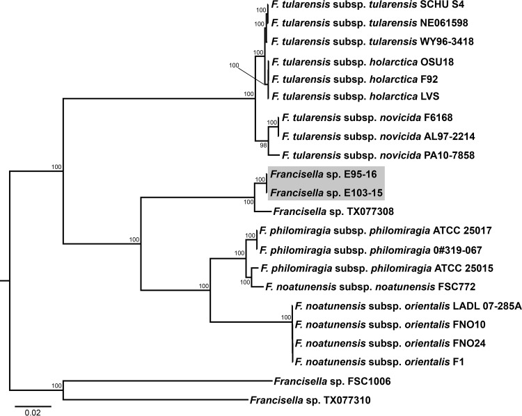
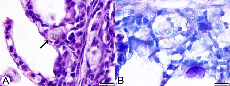
Historically, piscine francisellosis in various warm-, temperate-, and cold-water fish hosts has been attributed to Francisella noatunensis From 2015 to 2016, an undescribed Francisella sp. was recovered during mortality events in cultured spotted rose snapper (Lutjanus guttatus) off the Pacific coast of Central America. Despite high mortality and emaciation, limited gross findings were observed in affected fish. Histological examination revealed multifocal granulomatous lesions, with the presence of numerous small, pleomorphic coccobacilli, predominantly in the peritoneum, spleen, kidneys, liver, pancreas, heart, and intestine. Sequencing of an ∼1,400-bp fragment of the 16S rRNA gene demonstrated these isolates to be most similar (99.9% identity) to Francisella sp. isolate TX077308 cultured from seawater in the Gulf of Mexico, while sharing <99% similarity to other Fransicella spp. Biochemical analysis, multilocus sequence comparisons of select housekeeping genes, repetitive extragenic palindromic PCR fingerprinting, matrix-assisted laser desorption ionization-time of flight mass spectrometry, and fatty acid methyl ester analysis revealed marked differences between these isolates and other described members of the genus. Koch's postulates were fulfilled by experimental intracoelomic injection and immersion trials using Nile (Oreochromis niloticus) and blue (Oreochromis aureus) tilapia. Based on observed phenotypic and genotypic differences from recognized Francisella spp., the name Francisellamarina sp. nov. (NRRL B-65518) is proposed to accommodate these novel strains.IMPORTANCE Finfish aquaculture is the fastest growing global food production sector. Infectious disease, particularly emergent pathogens, pose a significant threat to established and nascent aquaculture industries worldwide. Herein, we characterize a novel pathogen isolated from mortality events in cultured spotted rose snapper in Central America. The bacteria recovered from these outbreaks were genetically and phenotypically dissimilar from other known Francisella spp. from fish, representing a previously unrecognized member of the genus Francisella, for which the name Francisella marina sp. nov. is proposed.

Keywords: Francisella; aquaculture; fish pathogens; snapper.


Figures

FIG 1
FIG 1
Histologic lesions in a spotted rose snapper (Lutjanus guttatus) naturally infected with a novel Francisella sp. (A) Low-magnification image of liver with a large focus of necrosis and granulomatous inflammatory infiltrates (arrows). H&E stain. Bar, 200 μm. (B) The spleen (lower left) is enlarged by inflammatory infiltrates dominated by macrophages. An arrow indicates an area of submucosal inflammation. The pale-staining cells are macrophages with cytoplasmic vacuoles containing bacteria. H&E stain. Bar, 50 μm. (C) Higher-magnification image of liver with a small focal area of necrosis and granulomatous inflammation. Arrowheads indicate vacuolated macrophages with intracellular bacteria. H&E stain. Bar, 50 μm. (D) Giemsa-stained section of liver with an irregular, pale-staining focus composed of macrophages with small intracytoplasmic bacteria typical of Francisella spp. Arrowheads indicate vacuolated macrophages with intracellular bacteria. Bar, 50 μm.
FIG 2
FIG 2
Repetitive extragenic palindromic PCR (Rep-PCR) analysis of unknown Francisella sp. isolates from cultured spotted rose snapper and archived F. noatunensis subsp. orientalis isolates. Amplification was performed using the ERIC I-ERIC II primer set. Lanes L, HyperLadder, 50 bp; lanes 1 to 10, F. noatunensis subsp. orientalis isolates 1 to 10, respectively (Table 7); lane 11, E95-16; lane 12, E103-15; N, no-template control.
FIG 3
FIG 3
Bayesian inference tree for Francisella spp. based on 1,357 bp of the 16S rRNA gene sequence. Numbers adjacent to branches represent posterior probability values (values of <0.50 are not shown). Francisella sp. isolates recovered from mortality events in snapper mariculture in Central America are highlighted in gray. GenBank accession numbers for reference genomes used in this study are listed in Tables 2 and 3.
FIG 4
FIG 4
Bayesian inference tree for Francisella spp. based on concatenated 16S rRNA > dnaK > gyrB > mutS > pgm > prfB > rpoB > sodB sequences. Numbers adjacent to branches represent posterior probability values (values of <0.50 are not shown). Francisella sp. isolates recovered from mortality events in snapper mariculture in Central America are highlighted in gray. GenBank accession numbers for reference genomes used in this study are listed in Tables 2 and 3.
FIG 5
FIG 5
Dendrogram constructed from MALDI-TOF MS main spectrum profiles of 19 Francisella sp. strains, including five Francisella noatunensis subsp. orientalis (Fno) strains (Table 7) and two novel Francisella marina isolates recovered from cultured snapper (Lutjanus guttatus) (E103-15 and E95-16).
FIG 6
FIG 6
Dendrogram of fatty acid profiles of five Francisella noatunensis subsp. orientalis (Fno) isolates and two Francisella marina isolates (E103-15 and E95-16) (Table 7).
FIG 7
FIG 7
Hematoxylin and eosin (H&E)-stained tissue sections from Nile and blue tilapia challenged with Francisella marina isolate E95-16 by intracoelomic injection. Lesions were similar in both species and most commonly affected the head kidney and spleen. (A) Head kidney with a small lesion composed of vacuolated macrophages. The arrow indicates a cluster of pleomorphic intracellular bacteria. Bar, 10 μm. (B) More advanced head kidney lesion with a central region containing apoptotic cells and necrotic debris. Bar, 50 μm. (C) Head kidney lesion dominated by typical vacuolated macrophages. A small granuloma with central caseation surrounded by epithelioid cells is visible in the upper right. Granuloma formation was rare and interpreted as a final stage in lesion progression. Bar, 20 μm. (D) Low-magnification image of head kidney with extensive, frequently coalescing lesions. Bar, 500 μm. (E) Splenic lesions began with expansion of periarteriolar sheaths by vacuolated macrophages (arrows). Bar, 50 μm. (F) Liver with a small perivascular cluster of macrophages containing intracellular bacteria. Bar, 10 μm.
FIG 8
FIG 8
Gill lesions in blue tilapia challenged with Francisella marina isolate E95-16 by intracoelomic injection. (A) Gill changes were inconspicuous, often involving only single macrophages laden with bacteria (arrow) within capillary lumens at the bases of lamellae. H&E stain. Bar, 10 μm. (B) In all tissue locations, the small bacteria were best visualized using Giemsa stains. Bar, 10 μm.

Similar articles

Cited by

References

    1. Food Agriculture Organization, United Nations. 2011. The state of world fisheries and aquaculture 2010. Food Agriculture Organization, United Nations, Rome, Italy.
    1. Cressey D. 2009. Aquaculture: future fish. Nature 458:398–400. doi: 10.1038/458398a. - DOI - PubMed
    1. Krkosek M. 2010. Host density thresholds and disease control for fisheries and aquaculture. Aquac Environ Interact 1:21–32. doi: 10.3354/aei0004. - DOI
    1. Colquhoun DJ, Duodu S. 2011. Francisella infections in farmed and wild aquatic organisms. Vet Res 42:47. doi: 10.1186/1297-9716-42-47. - DOI - PMC - PubMed
    1. Petersen JM, Carlson J, Yockey B, Pillai S, Kuske C, Garbalena G, Pottumarthy S, Chalcraft L. 2009. Direct isolation of Francisella spp. from environmental samples. Lett Appl Microbiol 48:663–667. doi: 10.1111/j.1472-765X.2009.02589.x. - DOI - PubMed