Review
. 2018 Jul 2;150(7):933-947.
doi: 10.1085/jgp.201711957. Epub 2018 Jun 18.

Known structures and unknown mechanisms of TMEM16 scramblases and channels

Affiliations
Review

Known structures and unknown mechanisms of TMEM16 scramblases and channels

Maria E Falzone et al. J Gen Physiol. .

Abstract

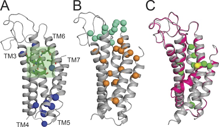
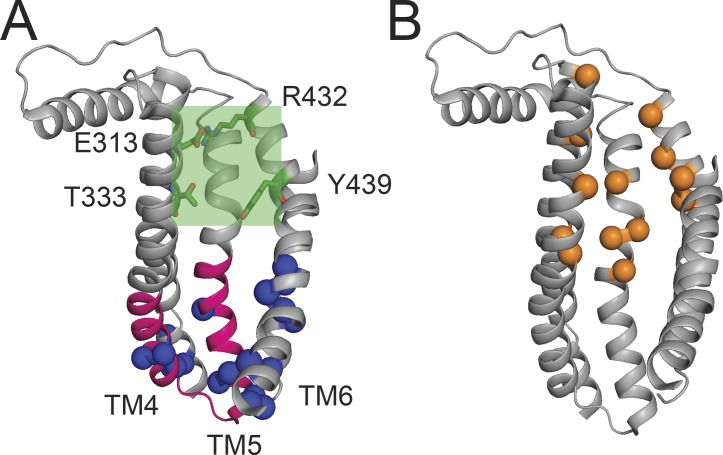
The TMEM16 family of membrane proteins is composed of both Ca2+-gated Cl- channels and Ca2+-dependent phospholipid scramblases. The functional diversity of TMEM16s underlies their involvement in numerous signal transduction pathways that connect changes in cytosolic Ca2+ levels to cellular signaling networks. Indeed, defects in the function of several TMEM16s cause a variety of genetic disorders, highlighting their fundamental pathophysiological importance. Here, we review how our mechanistic understanding of TMEM16 function has been shaped by recent functional and structural work. Remarkably, the recent determination of near-atomic-resolution structures of TMEM16 proteins of both functional persuasions has revealed how relatively minimal rearrangements in the substrate translocation pathway are sufficient to precipitate the dramatic functional differences that characterize the family. These structures, when interpreted in the light of extensive functional analysis, point to an unusual mechanism for Ca2+-dependent activation of TMEM16 proteins in which substrate permeation is regulated by a combination of conformational rearrangements and electrostatics. These breakthroughs pave the way to elucidate the mechanistic bases of ion and lipid transport by the TMEM16 proteins and unravel the molecular links between these transport activities and their function in human pathophysiology.


Figures

Figure 1.
Figure 1.
Phylogenetic organization of the TMEM16 family. The unrooted phylogenetic tree was derived from the original alignment from the Pfam database (PF04547) and constructed by using FastTree with the Jones-Taylor-Thornton model and CAT approximation for branch lengths (Price et al., 2010). Red, dual-function scramblases/channels; blue, CaCCs; green, scramblases with no reported channel activity; black, homologues with unknown function; *, homologues whose function has been demonstrated in vitro (afTMEM16, nhTMEM16, mTMEM16F, and hTMEM16A). For clarity, only the 10 human TMEM16 homologues are shown (TMEM16A–K), Subdued is a CaCC from D. melanogaster, AXS is a functionally uncharacterized homologue from D. melanogaster, and ANOH-1 is a scramblase from C. elegans.
Figure 2.
Figure 2.
TMEM16 architecture. (A and B) The structures the mTMEM16A Cl channel (A) and the nhTMEM16 scramblase (B) with two bound Ca2+ ions are viewed from the plane of the membrane. For clarity, one monomer is shown in gray. The cytosolic domain is in orange, the permeation pathway is in green, the extracellular domain in TMEM16A is in red, Ca2+ ions are in pink, and the remainder of the protein is in blue. (C and D) Close-up view of the Ca2+-binding site of TMEM16A (C) and nhTMEM16 (D). Conserved acidic residues are shown as yellow sticks, and polar residues are shown as green sticks. (E) Sequence conservation of the Ca2+-binding sites. Alignment of the regions shown in C and D of select TMEM16 family members. Coordinating acidic residues are highlighted in yellow, and polar residues in green. PDB accession nos.: 5OYB (mTMEM16A) and 4WIS (nhTMEM16).
Figure 3.
Figure 3.
The mTMEM16A pore. (A) Close-up view of the mTMEM16A pore. Basic residues lining the pore are shown as blue spheres, the neck constriction region is shown as a green shaded area, and key residues are shown as green sticks (Paulino et al., 2017a). (B) Residues important for ion permeation and selectivity: positions within the pore are shown as orange spheres; positions outside the pore that play a role in permeation are shown in cyan (Yu et al., 2012; Dang et al., 2017; Paulino et al., 2017a,b). (C) Structural alignment of the Ca2+-bound structure of mTMEM16A (gray) with the zero-Ca2+ structure (pink). TM6 bends into the pore around G644, shown as a yellow sphere. Green spheres are residues important for Ca2+ gating (Dang et al., 2017; Paulino et al., 2017a).
Figure 4.
Figure 4.
The lipid pathway of nhTMEM16. (A) Ribbon representation of the lipid pathway of nhTMEM16. Charged residues forming the intracellular lipid binding site are shown as blue spheres, the “scramblase domain” is shown in pink, and the narrowest region of the pathway is shown as shaded green box. Residues important in controlling this narrow region are shown as green sticks. (B) Residues important for lipid permeation (orange spheres; Bethel and Grabe, 2016; Jiang et al., 2017; Lee et al., 2018).
Figure 5.
Figure 5.
Structural differences between the TMME16 ion and lipid pores. (A) Structural alignment of nhTMEM16 (gray) and TMEM16A (pink) pathways. (B) Credit card model of lipid scrambling. (C) Alignment of the permeation pathways of the high-resolution structures of nhTMEM16 and TMEM16A highlighting the different tilts of TM4. The TM3 and TM5 from each structure are shown as gray ribbons, TM6 are shown as black ribbons, and TM4 are shown in color. Colors of TM4 are as follows: nhTMEM16 is red, mTMEM16A +Ca2+ is orange, mTMEM16A −Ca2+ is yellow, mTMEM16A in nanodiscs is green, and mTMEM16A in Lauryl Maltose Neopentyl Glycol is blue. (D) Cartoon depicting the ‘dynamic switch’ model for the dual-function activity of the TMEM16 scramblases. See text for details.

References

    1. Almaça J., Tian Y., Aldehni F., Ousingsawat J., Kongsuphol P., Rock J.R., Harfe B.D., Schreiber R., and Kunzelmann K.. 2009. TMEM16 proteins produce volume-regulated chloride currents that are reduced in mice lacking TMEM16A. J. Biol. Chem. 284:28571–28578. 10.1074/jbc.M109.010074 - DOI - PMC - PubMed
    1. Arreola J., Melvin J.E., and Begenisich T.. 1995. Inhibition of Ca(2+)-dependent Cl- channels from secretory epithelial cells by low internal pH. J. Membr. Biol. 147:95–104. 10.1007/BF00235400 - DOI - PubMed
    1. Aryal P., Abd-Wahab F., Bucci G., Sansom M.S.P., and Tucker S.J.. 2014. A hydrophobic barrier deep within the inner pore of the TWIK-1 K2P potassium channel. Nat. Commun. 5:4377 10.1038/ncomms5377 - DOI - PMC - PubMed
    1. Baig A.A., Haining E.J., Geuss E., Beck S., Swieringa F., Wanitchakool P., Schuhmann M.K., Stegner D., Kunzelmann K., Kleinschnitz C., et al. . 2016. TMEM16F-mediated platelet membrane phospholipid scrambling is critical for hemostasis and thrombosis but not thromboinflammation in mice-brief report. Arterioscler. Thromb. Vasc. Biol. 36:2152–2157. 10.1161/ATVBAHA.116.307727 - DOI - PubMed
    1. Balasubramanian K., and Schroit A.J.. 2003. Aminophospholipid asymmetry: A matter of life and death. Annu. Rev. Physiol. 65:701–734. 10.1146/annurev.physiol.65.092101.142459 - DOI - PubMed

Publication types