. 2018 Jun 18;8(1):9241.
doi: 10.1038/s41598-018-27581-0.

BMP and WNT signalling cooperate through LEF1 in the neuronal specification of adult hippocampal neural stem and progenitor cells

Affiliations

BMP and WNT signalling cooperate through LEF1 in the neuronal specification of adult hippocampal neural stem and progenitor cells

Tomás Armenteros et al. Sci Rep. .

Abstract

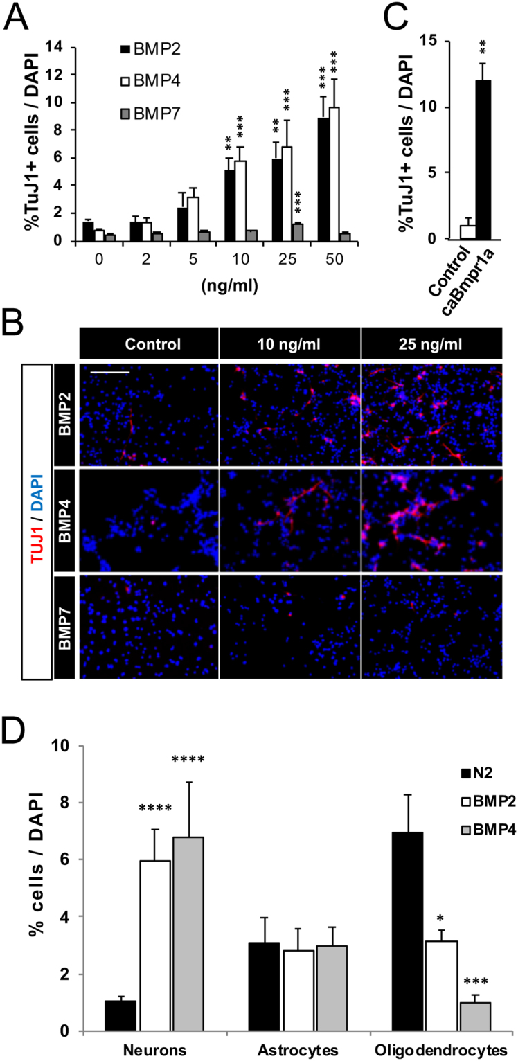
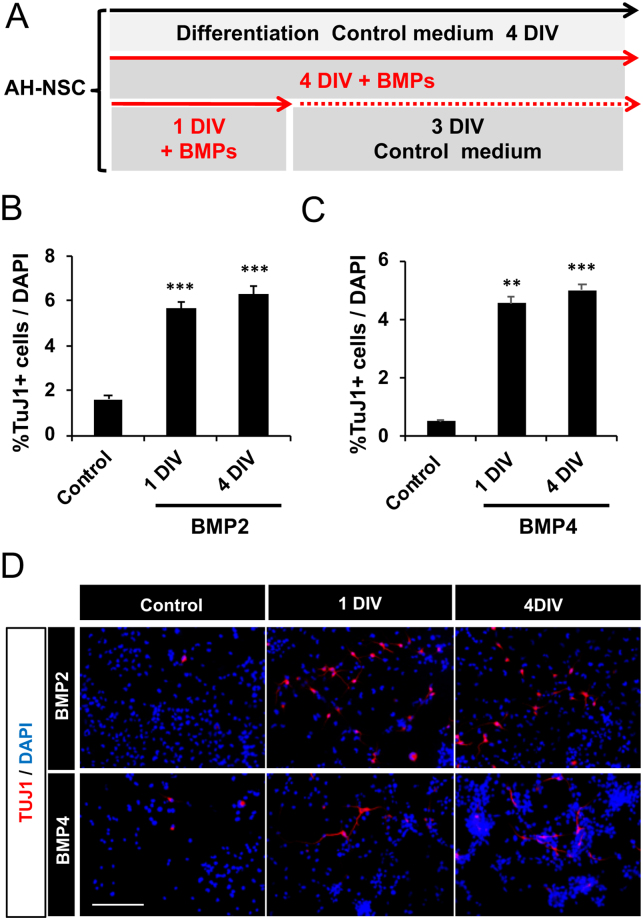
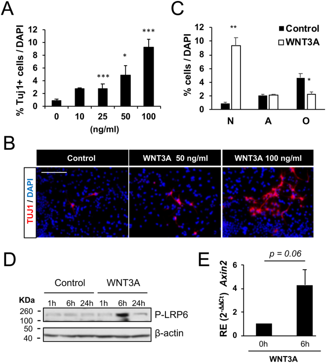
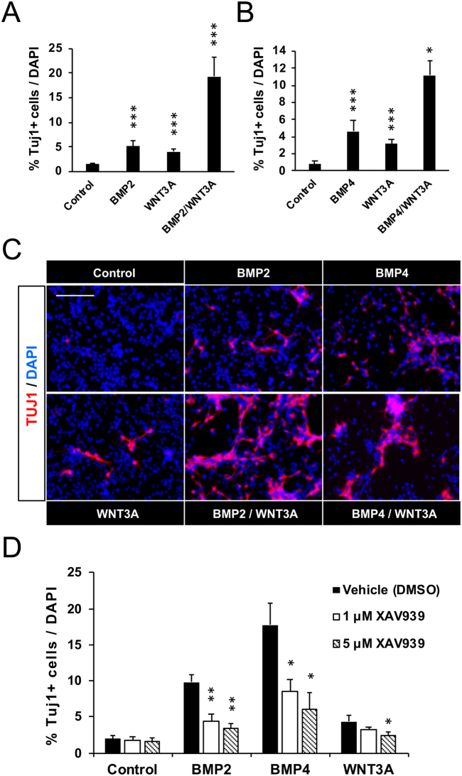
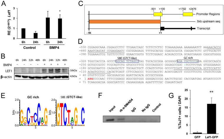
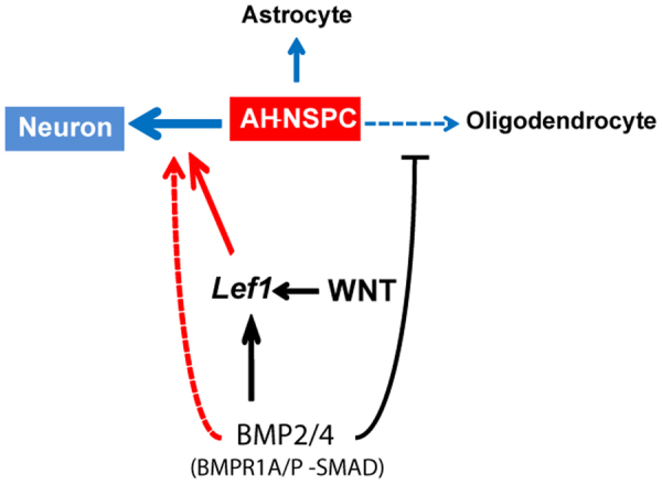
Neuronal production from neural stem cells persists during adulthood in the subgranular zone of the hippocampal dentate gyrus. Extracellular signals provided by the hippocampal microenvironment regulate the neuronal fate commitment of the stem cell progeny. To date, the identity of those signals and their crosstalk has been only partially resolved. Here we show that adult rat hippocampal neural stem and progenitor cells (AH-NSPCs) express receptors for bone morphogenetic proteins (BMPs) and that the BMP/P-Smad pathway is active in AH-NSPCs undergoing differentiation towards the neuronal lineage. In vitro, exposure to the BMP2 and BMP4 ligands is sufficient to increase neurogenesis from AH-NSPCs in a WNT dependent manner while decreasing oligodendrogenesis. Moreover, BMP2/4 and WNT3A, a key regulator of adult hippocampal neurogenesis, cooperate to further enhance neuronal production. Our data point to a mechanistic convergence of the BMP and WNT pathways at the level of the T-cell factor/lymphoid enhancer factor gene Lef1. Altogether, we provide evidence that BMP signalling is an important regulator for the neuronal fate specification of AH-NSPCs cultures and we show that it significantly cooperates with the previously described master regulator of adult hippocampal neurogenesis, the WNT signalling pathway.


Conflict of interest statement

The authors declare no competing interests.

Figures

Figure 1
Figure 1
Expression of BMP ligands and BMP receptors in the adult hippocampal dentate gyrus and in AH-NSPCs undergoing differentiation. (A) Phylogenetic Tree showing distances between the mouse BMPs (left). The tree was generated using the alignment viewer ‘AliView’. The preferred type1/2 receptors bound by the different BMP ligands are also shown (right). (BC) Differentiation of AH-NSPCs in control medium (N2) or N2 supplemented with Retinoic Acid (RA, 1 µM) and Forskolin (FSK, 5 µM) during 4 days in vitro. The percentage of neurons (N), astrocytes (A) and oligodendrocytes (O) was analysed by immunostaining against Tubulin βIII (Tuj1), Glial Fibrillary Acidic Protein (GFAP) and Myelin Basic Protein (MBP), respectively. The RA + FSK treatment favoured neuronal differentiation (average ± sem, n = 3, two-tailed T-test: ***P < 0,001). (DK) Relative gene expression (RE) patterns during the time course of AH-NSPC differentiation for Tubb3 (D), Nes (E), Bmpr1a (F), Bmpr1b (G), Acvr1 (H), Bmpr2 (I), Acvr2a (J) and Acvr2b (K). Sdha was used as the housekeeping gene and expression levels were referred to those of proliferating AH-NSPCs grown in fibroblast growth factor 2 (FGF). Data correspond to average ± sem of n = 3 independent experiments analysed by the 2−ΔΔCt method (ANOVA: *P < 0.05; **P < 0.01; ***P < 0.001). Scale bar in B, 25 µm.
Figure 2
Figure 2
BMP2, BMP4 and caBMPR1A increase neurogenesis in AH-NSPC differentiation assays. (A) Percentage of Tubulin βIII (Tuj1) positive cells out of the total number of cells differentiated from AH-NSPCs in the presence of increasing concentrations of BMP2, BMP4 or BMP7. Data correspond to the average ± sem of n = 3. (B) Immunofluorescence images showing the levels of Tuj1 positive cells (red) and DAPI stain (blue) at 0, 10 and 25 ng/ml of BMP2, BMP4 and BMP7. As illustrated, BMP2 and BMP4 (but not BMP7) markedly induce neurogenesis in vitro in a dose-dependent manner. (C) Percentage of Tuj1 positive cells out of the total number of cells differentiated from AH-NSPCs transduced with a retroviral vector overexpressing a constitutive active form of BMPR1A (caBmpr1a) or with an empty vector as a control. (D) Percentage of neurons (N, Tubulin βIII positive cells), astrocytes (A, Glial Fibrillary Acidic Protein, GFAP positive cells) and oligodendrocytes (O, Myelin Basic Protein, MBP positive cells) in the absence (N2) or presence of 25 ng/ml of BMP2 or BMP4. The cell lineage analysis shows an increase in neurons and a decrease in oligodendrocytes in the presence of BMPs. Scale bar on B, 100 µm. *P < 0.05; **P < 0.01; ***P < 0.001, ****P < 0.0001 by ANOVA.
Figure 3
Figure 3
BMP2 and BMP4 influence the neuronal fate choice decision of the AH-NSPC progeny at an early timepoint. (A) Diagram describing the procedure. AH-NSPCs were stimulated transiently during 1 DIV or continuously during 4 DIV with 10 ng/ml of BMP2 or BMP4. Cells were fixed at 4 DIV and neurogenesis was measured by immunostaining against Tubulin βIII (Tuj1). (B,C) Percentage of Tuj1 positive cells after stimulating for 1 or 4 DIV with BMP2 (B) and BMP4 (C). Data correspond to the average ± sem of n = 3 independent experiments. (D) Immunofluorescence images showing the levels of Tuj1 positive cells (red) and DAPI stain (blue) at 4DIV in cultures stimulated transiently (1 DIV) or continuously (4 DIV) with BMP2 or BMP4. Scale bar in D, 100 µm. **P < 0.01; ***P < 0.001 by two-tailed T-test.
Figure 4
Figure 4
BMP2 and BMP4 induce neurogenesis through the activation of the P-SMAD canonical pathway in AH-NSPCs. (A,B) Immunofluorescence images of AH-NSPCs showing the increase in P-SMAD1/5/8 levels and the nuclear translocation of the phosphorylated proteins upon BMP2 stimulation (A) or BMP4 stimulation (B) for 6 hours. (C) Western immunoblot of whole cell lysates from AH-NSPCs treated with BMP2 or BMP4, separated by SDS-PAGE and blotted sequentially with antibodies against P-SMAD1/5/8, total SMAD1/5/8 and β-actin as loading control. Full-length blots are included in Supplementary Fig. S5. (D) Relative gene expression (RE) patterns for the canonical BMP pathway target gene Id1 upon BMP2 or BMP4 stimulation at the indicated time points. Sdha was used as the housekeeping gene and expression levels were referred to untreated AH-NSPCs (Control). Data correspond to average ± sem of n = 3 independent experiments analysed by the 2−ΔΔCt method (two-tailed T-test: *P < 0.05; **P < 0.01). (E) Percentage of Tubulin βIII (Tuj1) positive cells out of the total number of cells differentiated from AH-NSPCs during 4 days in vitro in the presence of Retinoic Acid (RA, 1 µM) and Forskolin (FSK, 5 µM), BMP2 (25 ng/ml) or BMP4 (25 ng/ml) and P38MAPK inhibitor SB203580 or DMSO as a control. Data correspond to the average ± sem of n = 3. Inhibition of the non-canonical BMP signalling employing SB203580 had no effect on the increase in neurogenesis. Scale bar in A and B, 100 µm.
Figure 5
Figure 5
WNT3A increases neurogenesis in AH-NSPC differentiation assays. (A) Percentage of Tubulin βIII (Tuj1) positive cells out of the total number of cells differentiated from AH-NSPCs in the presence of increasing concentrations of WNT3A. Data correspond to the average ± sem of n = 3 (*P < 0.05; ***P < 0.001 by ANOVA). (B) Immunofluorescence images showing the levels of Tuj1 positive cells (red) and DAPI stain (blue) at 0, 50 and 100 ng/ml of WNT3A. As illustrated, WNT3A induces neurogenesis in vitro in a dose-dependent manner. (C) Percentage of neurons (N, Tubulin βIII positive cells), astrocytes (A, Glial Fibrillary Acidic Protein, GFAP positive cells) and oligodendrocytes (O, Myelin Basic Protein, MBP positive cells) in the absence (N2) or presence of 100 ng/ml of WNT3A. The cell lineage analysis shows an increase in neurons and a decrease in oligodendrocytes in the presence of WNT3A (*P < 0.05; **P < 0.01 by ANOVA). (D) Western immunoblot of whole cell lysates from AH-NSPCs treated with WNT3A (100 ng/ml), separated by SDS-PAGE and blotted sequentially with antibodies against P-LRP6 and β-actin as loading control. Full-length blots are included in Supplementary Fig. S7. (E) Relative gene expression (RE) pattern for the WNT pathway target gene Axin2 upon WNT3A (100 ng/ml) stimulation at 6 h. Sdha was used as the housekeeping gene and expression levels were referred to untreated AH-NSPCs (0 h). Data correspond to average ± sem of n = 3 independent experiments analysed by the 2−ΔΔCt method (two-tailed T-test: P = 0.06). Scale bar in B, 100 µm.
Figure 6
Figure 6
BMP2/4 require endogenous WNT signalling and synergize with exogenous WNT3A to increase neurogenesis in AH-NSPC differentiation assays. (A) Percentage of Tubulin βIII (Tuj1) positive cells out of the total number of cells differentiated from AH-NSPCs during 4 days in vitro in the presence of BMP2 (25 ng/ml), WNT3A (25 ng/ml) or a combination of both BMP2 and WNT3A. Data correspond to the average ± sem of n ≥ 5 (2-way ANOVA: ***P < 0.001). (B) Percentage of Tubulin βIII (Tuj1) positive cells out of the total number of cells differentiated from AH-NSPCs during 4 days in vitro in the presence of BMP4 (25 ng/ml), WNT3A (25 ng/ml) or a combination of both BMP4 and WNT3A. Data correspond to the average ± sem of n ≥ 6. (2-way ANOVA: *P < 0.05; ***P < 0.001). (C) Immunofluorescence images showing the levels of Tuj1 positive cells (red) and DAPI stain (blue) at the indicated conditions. As illustrated, the number of neurons in the combined BMP2/4 + WNT3A treatment is higher than in the BMP2/4 or WNT3A independent treatments, evidencing a synergistic effect in neurogenesis of the BMP and WNT pathways. (D) Percentage of Tubulin βIII (Tuj1) positive cells out of the total number of cells differentiated from AH-NSPCs during 4 days in vitro in the presence of BMP2 (50 ng/ml), BMP4 (50 ng/ml) or WNT3A (50 ng/ml) and XAV939 (a compound that blocks Wnt/b-catenin activity through the stabilization of Axin via tankyrase inhibition) or DMSO as a control. Data correspond to the average ± sem of n = 3 (two-tailed T-test: *P < 0.05; **P < 0.01). Scale bar in B, 100 µm.
Figure 7
Figure 7
Lef1 is a direct target of BMP4 signalling in AH-NSPCs. (A) Relative gene expression (RE) pattern for the Lef1 gene upon BMP4 (50 ng/ml) stimulation at the indicated time points. Sdha was used as the housekeeping gene and expression levels were referred to untreated AH-NSPCs (Control). (B) Western immunoblot of whole cell lysates from AH-NSPCs treated with BMP4 at the indicated time points, separated by SDS-PAGE and blotted sequentially with antibodies against LEF1 and β-actin as loading control. Full-length blots are included in Supplementary Fig. S8. (C) Lef1 promoter region analysis using Genomatix Promoter Inspector software. Image shows the retrieved 5 kb sequence (orange) upstream of the Lef1 coding sequence (black) and two validated promoter regions that result from the promoter analysis (yellow). (D,E) Nucleotide sequence upstream of the Lef1 translational start site (ATG, red). Two SMAD transcription factor binding sites were predicted: the first one, positioned at −274/−264, corresponds to a SMAD Binding Element (SBE) (GTCT-like), and the second one, positioned at −233/−223, corresponds to a GC rich element. (F) ChIP assay of BMP4-treated AH-NSPCs using a SMAD4 rabbit antibody. For the PCR amplification of the precipitated material, the underlined primers in (D) were employed. Rabbit IgG (IgG) was used as a control. Full-length gel is included in Supplementary Fig. S9. (G) Percentage of Tubulin βIII (Tuj1) positive cells out of the total number of cells differentiated from AH-NSPCs transduced with a lentiviral vector overexpressing Lef1-GFP or GFP as a control. Cells were cultured during 4 days in vitro. *P < 0.05; **P < 0.01 by two-tailed T-test.
Figure 8
Figure 8
BMP2/4 increase neurogenesis from adult hippocampal NSPCs and synergize with WNT3A. Schematic of our proposed model summarizing the main results. BMP2/4 induce neurogenesis through the activation of the P-SMAD canonical pathway downstream of the BMPR1A type 1 receptor. The pro-neurogenic effect of BMP signalling is partly dependent on endogenous WNT signalling, and the mechanism relies on the up-regulation of the Lef1 gene, a direct SMAD target. WNT-independent effects of the BMPs are not excluded (dashed line, red). BMP2/4 also decrease the number of oligodendrocytes.

Similar articles

Cited by

References

    1. Lim, D. A. & Alvarez-Buylla, A. The Adult Ventricular-Subventricular Zone (V-SVZ) and Olfactory Bulb (OB) Neurogenesis. Cold Spring Harb Perspect Biol8 (2016). - PMC - PubMed
    1. Aimone JB, et al. Regulation and function of adult neurogenesis: from genes to cognition. Physiol Rev. 2014;94:991–1026. doi: 10.1152/physrev.00004.2014. - DOI - PMC - PubMed
    1. Choe Y, Pleasure SJ, Mira H. Control of Adult Neurogenesis by Short-Range Morphogenic-Signaling Molecules. Cold Spring Harb Perspect Biol. 2015;8:a018887. doi: 10.1101/cshperspect.a018887. - DOI - PMC - PubMed
    1. Ciani L, Salinas PC. WNTs in the vertebrate nervous system: from patterning to neuronal connectivity. Nat Rev Neurosci. 2005;6:351–362. doi: 10.1038/nrn1665. - DOI - PubMed
    1. Inestrosa NC, Arenas E. Emerging roles of Wnts in the adult nervous system. Nat Rev Neurosci. 2010;11:77–86. doi: 10.1038/nrn2755. - DOI - PubMed

Publication types

MeSH terms

LinkOut - more resources