. 2018 Jun 18;9(7):715.
doi: 10.1038/s41419-018-0743-2.

Natural cyclopeptide RA-V inhibits the NF-κB signaling pathway by targeting TAK1

Affiliations

Natural cyclopeptide RA-V inhibits the NF-κB signaling pathway by targeting TAK1

Zhe Wang et al. Cell Death Dis. .

Abstract

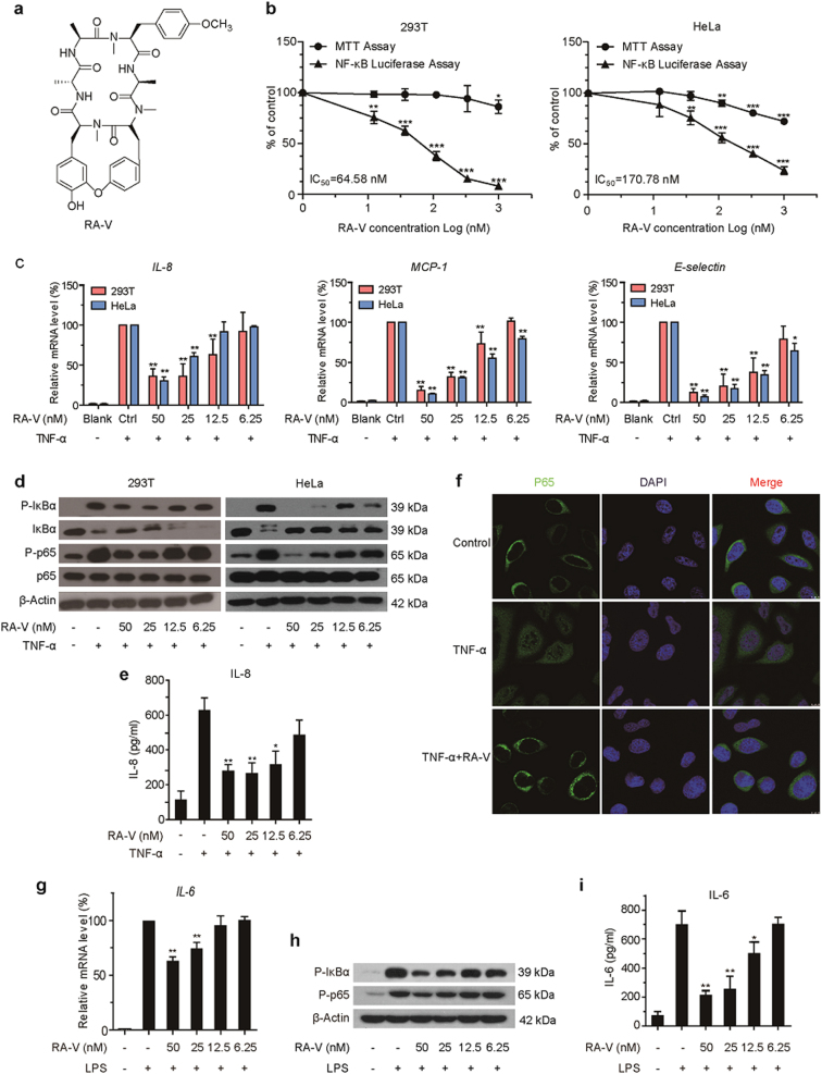
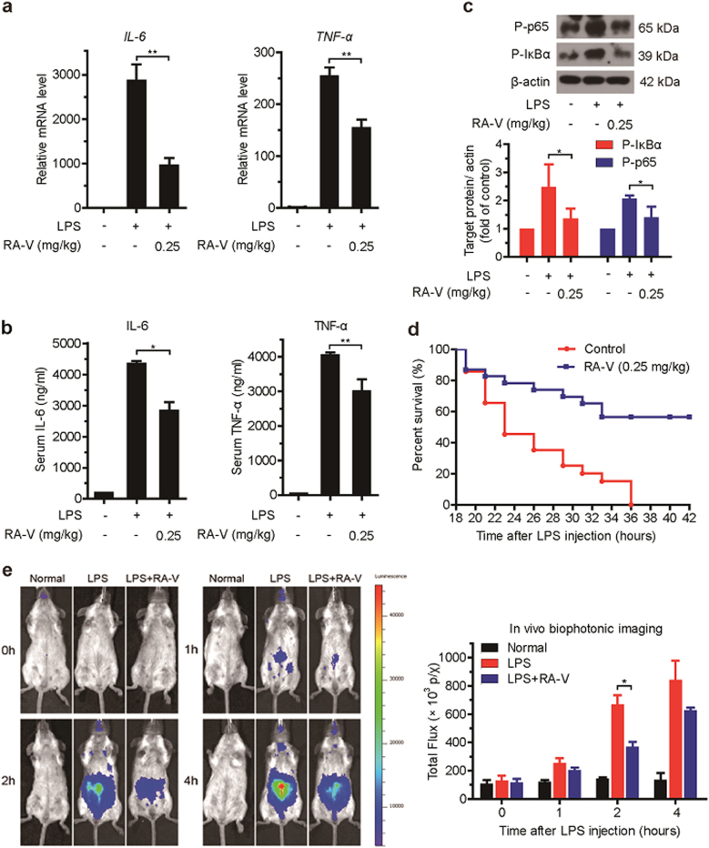
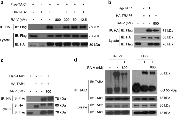
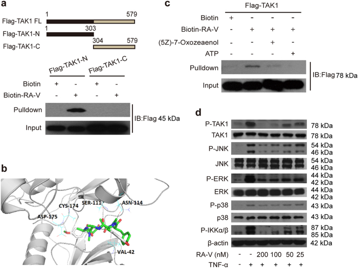
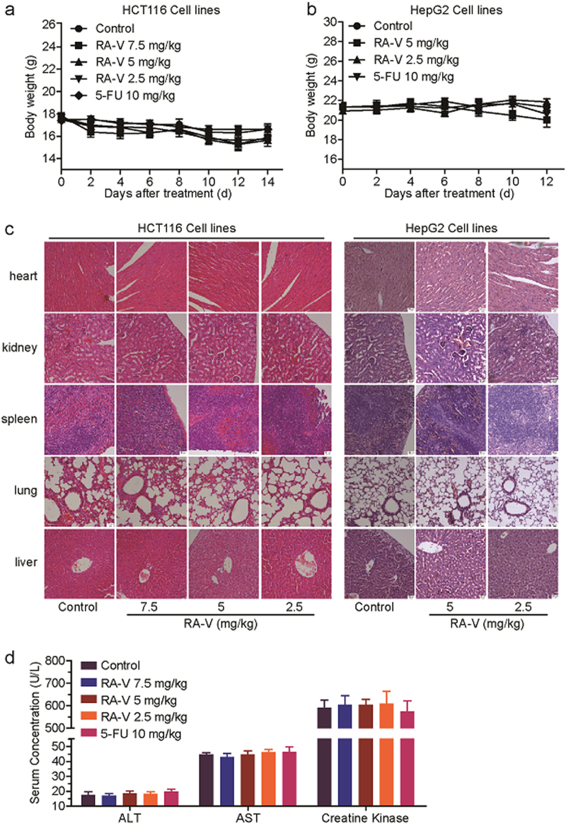
Rubiaceae-type cyclopeptides (RAs) are a type of plant cyclopeptides from the Rubia that have garnered significant attention owing to their unique bicyclic structures and amazing antitumour activities. Our recent work has shown that RAs suppress inflammation and angiogenesis and induce apoptosis. However, the underlying mechanism and targets remained unknown. Nuclear factor κB (NF-κB) signaling pathway plays a critical role in these biological processes, prompting us to investigate whether and how RAs affect this pathway. By screening compound libraries using NF-κB-dependent luciferase reporter, we observed that RA-V is the best NF-κB inhibitor. Further experiments demonstrated that RA-V interrupted the TAK1-TAB2 interaction and targeted TAK1 in this pathway. Moreover, RA-V prevented endotoxin shock and inhibited NF-κB activation and tumor growth in vivo. These findings clarify the mechanism of RA-V on NF-κB pathway and might account for the majority of known bioactivities of RA-V, which will help RA-V develop as new antiinflammatory and antitumour therapies.


Conflict of interest statement

The authors declare that they have no conflict of interest.

Figures

Fig. 1
Fig. 1. RA-V inhibits TNF-α- and LPS-induced activation of NF-κB.
a Chemical structure of RA-V. b RA-V inhibited the NF-κB signaling pathway in a dose-dependent manner. HEK293T or HeLa cells were transfected with the 5 × κB-luciferase and pTK-Renilla reporters. Twenty-four hours after transfection, the cells were incubated with various concentrations of RA-V for 6 h, and then treated with 10 ng/mL TNF-α for 2 h before the luciferase activity assay and MTT assay. c RA-V reduced TNF-α-induced expression of NF-κB target genes. HEK293T or HeLa cells were treated with various concentrations of RA-V for 24 h and stimulated with 10 ng/mL TNF-α for 2 h. The expression of the NF-κB target genes, IL-8, MCP-1, and E-selectin was measured by quantitative RT-PCR and normalized to GADPH expression. d RA-V inhibited TNF-α-induced p65 phosphorylation, IκBα phosphorylation and IκBα degradation. HEK293T or HeLa cells were incubated with various concentrations of RA-V for 24 h and treated with 10 ng/mL TNF-α for 10 min. The cell lysates were prepared and subjected to western blot analysis with the indicated antibodies. e RA-V inhibited TNF-α-induced IL-8 production. HeLa cells were treated with various concentrations of RA-V for 12 h before treatment with 10 ng/mL TNF-α for 2 h. The culture supernatant was collected and subjected to ELISA analysis. f RA-V inhibited the TNF-α-induced nuclear translocation of p65. HeLa cells were incubated with 200 nM RA-V for 6 h, treated with 10 ng/mL TNF-α for 15 min, and then subjected to immunocytochemical analysis. g RA-V reduced LPS-induced IL-6 expression. RAW264.7 cells were treated with various concentrations of RA-V for 24 h and stimulated with 1 μg/mL LPS for 3 h. The expression of IL-6 was measured. h RA-V inhibited LPS-induced p65 phosphorylation, IκBα phosphorylation. RAW264.7 cells were incubated with various concentrations of RA-V for 24 h and treated with 1 μg/mL LPS for 3 h. The protein expression was measured. i RA-V inhibited LPS-induced IL-6 production. RAW264.7 cells were treated with various concentrations of RA-V for 12 h before treatment with 1 μg/mL LPS for 3 h. The expression of IL-6 was measured. The data in b, c, e, g and i are presented as the means ± S.D. from three independent experiments. *, p< 0.05; **, p< 0.01; ***, p< 0.001
Fig. 2
Fig. 2. RA-V prevents endotoxic shock and inhibits NF-κB activation in vivo.
ac RA-V prevents endotoxic shock in vivo. Mice (n = 6 per group) were intravenously injected with RA-V micelles (0.25 mg/kg body weight) or control micelles 52, 28, and 4 h before an intraperitoneal injection of LPS (10 mg/kg). One hour later, the relative levels of IL-6 and TNF-α mRNAs in the liver were assessed by quantitative RT-PCR (a), the serum IL-6 and TNF-α levels were quantified by ELISA (b), and the expression of p65 and IκBα phosphorylation in the liver were measured by immunoblot analysis c. d RA-V improved animal survival. Mice (n = 20 per group) were intravenously injected with RA-V micelles (0.25 mg/kg body weight) or control micelles 52, 28, and 4 h before intraperitoneal injection of LPS (20 mg/kg). Animal survival was recorded in 2–3 hours intervals. e RA-V inhibits NF-κB activation in vivo. The NF-κB-luc transgenic mice (n = 4 per group) were intravenously injected with RA-V micelles (0.25 mg/kg body weight) or control micelles 52, 28, and 4 h before intraperitoneal injection with LPS (10 mg/kg). NF-κB-luc transgenic mice were photographed at different time points and representative images are shown. The bioluminescent signals from all of the mice were detected and quantified. The data are presented as the means ± S.D. *, p< 0.05; **, p< 0.01
Fig. 3
Fig. 3. RA-V represses NF-κB activation upstream of the IKK protein complex.
a RA-V inhibited the TRAF2- and MyD88-induced expression of the NF-κB reporter in a dose-dependent manner. The indicated plasmids were transfected into HEK293T cells together with the 5 × κB-luciferase and pTK-Renilla reporters. Twenty-four hours after transfection, the cells were incubated with various concentrations of RA-V for 48 h before luciferase assays were performed. b RA-V inhibited TRAF2- and MyD88-induced p65 phosphorylation and IκBα phosphorylation in a dose-dependent manner. HEK293T cells were transfected with TRAF2, MyD88, IKKβ, or p65 for 24 h and then treated with various concentrations of RA-V for 48 h. The cell lysates were immunoblotted with the indicated antibodies. c RA-V likely exerts its inhibitory activity on the NF-κB pathway by acting on the TRAF6-TAK1–TAB1/2 complex. Using drugCIPHER, ARRB1 is a potential target of RA-V, as ARRB1 ranks 44th of 13388 genome-wide candidates. TRAF6-TAK1–TAB1/2 complex is a key link that connects ARRB1 and the NF-κB signaling pathway by searching the STRING database. The data in a and b are presented as the means ± S.D. from three independent experiments. *, p< 0.05; **, p< 0.01
Fig. 4
Fig. 4. RA-V blocks the interaction between TAK1 and TAB2.
a RA-V blocked the exogenous TAK1–TAB2 interaction. HEK293T cells were transfected with Flag-TAK1 and HA-TAB2 for 24 h and then incubated with various concentrations of RA-V for 6 h. The cell lysates were immunoprecipitated with HA antibody and then immunoblotted with the indicated antibodies. b, c RA-V does not disrupt TAK1–TAB1 or TAK1–TRAF6 interactions. HEK293T cells were transfected with Flag-TAK1 and HA-TRAF6 or HA-TAB1 for 24 h and then incubated with RA-V (800 nM) for 6 h. The cell lysates were immunoprecipitated with HA antibody, and immunoblots were performed with the indicated antibodies. d RA-V blocked the endogenous TAK1–TAB2 interaction. HEK293T (left) or RAW264.7 (right) cells were incubated with 800 nM RA-V for 6 h and stimulated with 10 ng/mL TNF-α for 2 h or 1 μg/mL LPS for 3 h, respectively. The cell lysates were immunoprecipitated with TAK1 antibody or control IgG and then immunoblotted with the indicated antibodies. Each experiment was repeated at least three times
Fig. 5
Fig. 5. RA-V targets the TAK1 protein.
a Chemical structure of CB12. b CB12 binds the exogenous TAK1. HEK293T cells were transfected with TAK1 for 24 h, then the cell lysates were incubated with biotin or CB12 followed by streptavidin agarose pull-down. The immunoprecipitates were incubated with a 10-fold excess of RA-V or biotin and western blotted with Flag antibody. c CB12 binds the endogenous TAK1. HEK293T cell lysates were incubated with CB12 or biotin and precipitated with streptavidin agarose, followed by western blotting with TAK1 antibody. d CB12 did not bind other key proteins of the NF-κB signaling pathway. HEK293T cells were transfected with the indicated plasmids for 24 h. The cell lysates were pulled down with streptavidin agarose and then immunoblotted with HA or Flag antibody. Each experiment was repeated at least three times
Fig. 6
Fig. 6. RA-V binds the kinase domain of TAK1.
a CB12 directly binds to the TAK1 kinase domain. HEK293T cells were transfected with TAK1-N or TAK1-C for 24 h. The cells lysates were incubated with CB12 or biotin and precipitated with streptavidin-coated sepharose and were analyzed by immunoblotting with Flag antibody. b The model for RA-V binding to the crystal structure of the TAK1–TAB1 fusion protein (PDB 4GS6). The blue dotted lines represent the hydrogen bonds between RA-V (green) and TAK1–TAB1. c RA-V probably binds the ATP-binding pocket of TAK1. HEK293T cells were transfected with TAK1 for 24 h, and then the cell lysates were incubated with biotin or CB12 before precipitation with streptavidin agarose. The immunoprecipitates were treated with ATP or (5Z)-7-oxozeaenol and western blotted with Flag antibody. d RA-V inhibits TAK1, JNK, ERK, p38, and IKKα/β phosphorylation. HeLa cells were incubated with various concentrations of RA-V for 12 h and then treated with 10 ng/mL TNF-α for 10 min. The cell lysates were prepared and western blotted with the indicated antibodies. Each experiment was repeated at least three times
Fig. 7
Fig. 7. RA-V inhibits tumor growth and the expression of NF-κB target genes in a nude xenograft model.
ad Inhibition of the growth of HCT116 and HepG2 xenograft tumors by RA-V. Female athymic nude BALB/c mice bearing HCT116 (n = 8) or HepG2 (n = 7) xenograft tumors were intravenously injected with various concentrations of RA-V micelles or control micelles every other day. 5-FU (10 mg/kg) group as positive control. Effects of RA-V on the growth curves of subcutaneoue xenografts of HCT116 (a) and HepG2 (b) and effects on the tumor weight in the HCT116 (c) and HepG2 (d) models. ef Tumors removed were photographed. gh The expression of the NF-κB target genes, IL-8, MCP-1, and CXCL-1, in HCT116 (g) and HepG2 (h) tumor tissue from vehicle- and various concentrations of RA-V-treated group was determined by quantitative RT-PCR and normalized to GADPH expression. *, p< 0.05; **, p< 0.01; ***, p< 0.001
Fig. 8
Fig. 8. Analysis of potential side effects for treatment of RA-V.
a–b The change curves of body weights of BALB/c bearing HCT116 (a) or HepG2 (b) xenograft tumors (n = 8 or 7). c Representative hematoxylin–eosin staining of heart, kidney, spleen, lung, and liver from vehicle- and various concentrations of RA-V-treated group. d The evaluation of serum ALT, AST, and creatine kinase for vehicle- and various concentrations of the RA-V-treated group

Similar articles

Cited by

References

    1. Koehn FE, Carter GT. The evolving role of natural products in drug discovery. Nat. Rev. Drug Discov. 2005;4:206–220. doi: 10.1038/nrd1657. - DOI - PubMed
    1. Newman DJ, Cragg GM. Natural products as sources of new drugs over the 30 years from 1981 to 2014. J. Nat. Prod. 2016;79:629–661. doi: 10.1021/acs.jnatprod.5b01055. - DOI - PubMed
    1. Tan NH, Zhou J. Plant cyclopeptides. Chem. Rev. 2006;106:840–895. doi: 10.1021/cr040699h. - DOI - PubMed
    1. Zhao SM, et al. Antitumor cyclic hexapeptides from Rubia plants: history, chemistry, and mechanism (2005-2011) Chimia. 2011;65:952–956. doi: 10.2533/chimia.2011.952. - DOI - PubMed
    1. Xu K, et al. Quinone derivatives from the genus Rubia and their bioactivities. Chem. Biodivers. 2014;11:341–363. doi: 10.1002/cbdv.201200173. - DOI - PubMed

Publication types

LinkOut - more resources