. 2018 Jun 18;8(1):9281.
doi: 10.1038/s41598-018-27615-7.

Liver X receptors regulate hepatic F4/80 + CD11b+ Kupffer cells/macrophages and innate immune responses in mice

Affiliations

Liver X receptors regulate hepatic F4/80 + CD11b+ Kupffer cells/macrophages and innate immune responses in mice

Kaori Endo-Umeda et al. Sci Rep. .

Abstract

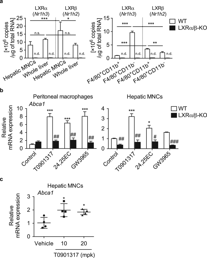
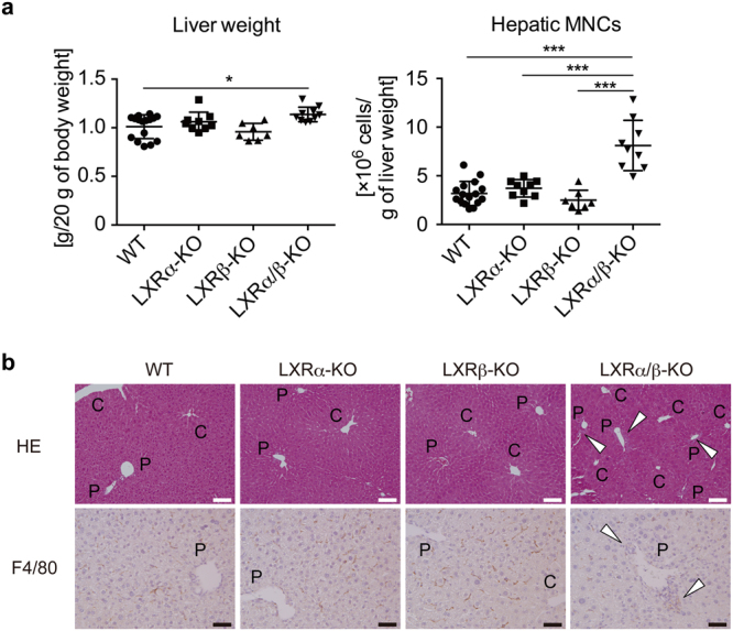
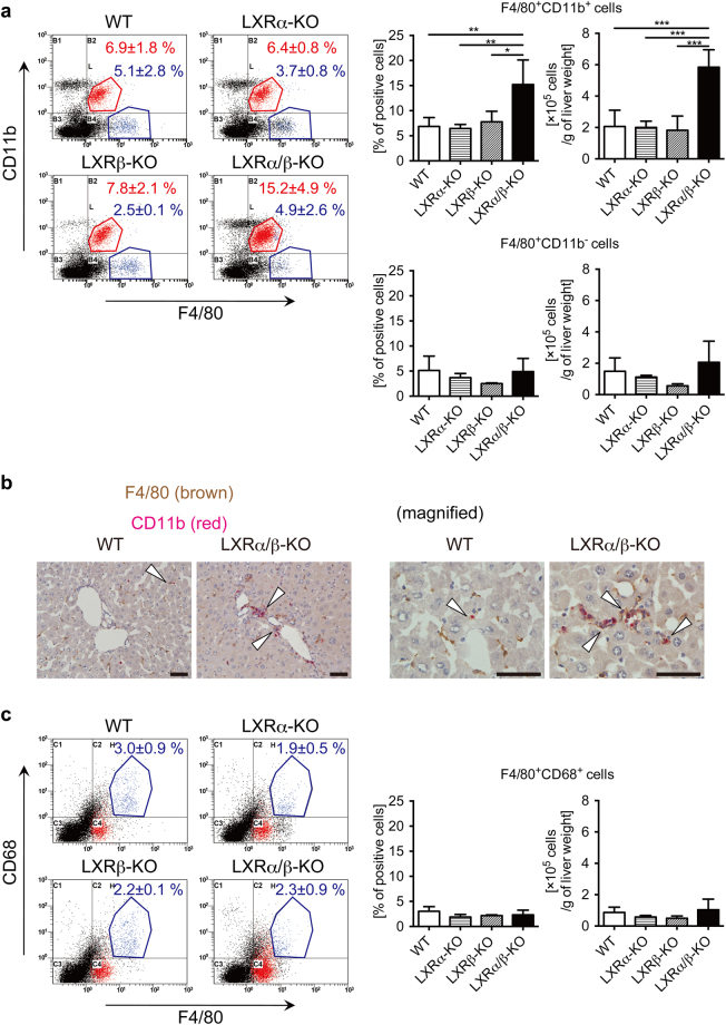
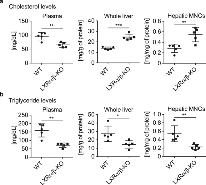
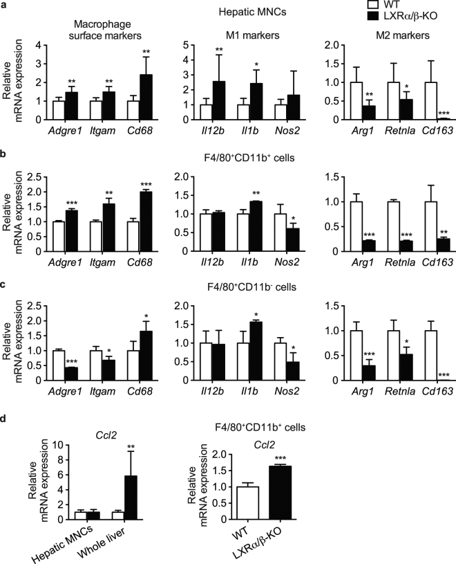
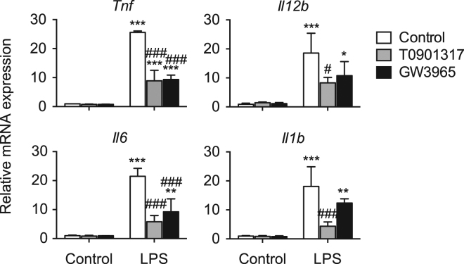
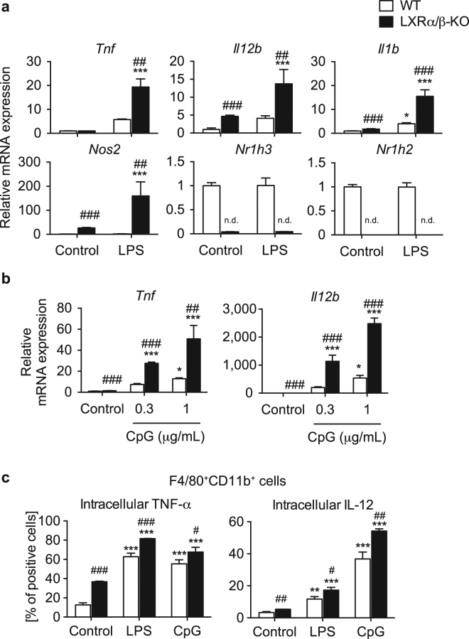
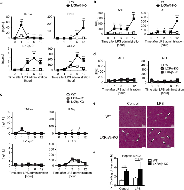
The liver X receptors (LXRs), LXRα and LXRβ, are nuclear receptors that regulate lipid homeostasis. LXRs also regulate inflammatory responses in cultured macrophages. However, the role of LXRs in hepatic immune cells remains poorly characterized. We investigated the role of LXRs in regulation of inflammatory responses of hepatic mononuclear cells (MNCs) in mice. Both LXRα and LXRβ were expressed in mouse hepatic MNCs and F4/80+ Kupffer cells/macrophages. LXRα/β-knockout (KO) mice had an increased number of hepatic MNCs and elevated expression of macrophage surface markers and inflammatory cytokines compared to wild-type (WT) mice. Among MNCs, F4/80+CD11b+ cells, not F4/80+CD11b- or F4/80+CD68+ cells, were increased in LXRα/β-KO mice more than WT mice. Isolated hepatic MNCs and F4/80+CD11b+ cells of LXRα/β-KO mice showed enhanced production of inflammatory cytokines after stimulation by lipopolysaccharide or CpG-DNA compared to WT cells, and LXR ligand treatment suppressed lipopolysaccharide-induced cytokine expression in hepatic MNCs. Lipopolysaccharide administration also stimulated inflammatory cytokine production in LXRα/β-KO mice more effectively than WT mice. Thus, LXR deletion enhances recruitment of F4/80+CD11b+ Kupffer cells/macrophages and acute immune responses in the liver. LXRs regulate the Kupffer cell/macrophage population and innate immune and inflammatory responses in mouse liver.


Conflict of interest statement

The authors declare no competing interests.

Figures

Figure 1
Figure 1
Expression and function of LXRs in hepatic MNCs. (a) Hepatic MNCs were isolated from the liver of WT mice, and mRNA copy numbers of LXRα (Nr1h3) and LXRβ (Nr1h2) were compared to those in whole liver samples (n = 8 for each group). LXRα (Nr1h3) and LXRβ (Nr1h2) mRNA levels were also evaluated in isolated F4/80+CD11b+ and F4/80+CD11b cells (n = 4 for each group). n.d., not detected. *P < 0.05; **P < 0.01; ***P < 0.001; n.s., not significant (one-way ANOVA followed by Tukey’s multiple comparisons for 4 groups of WT samples). (b) Thioglycolate elucidated-peritoneal macrophages and hepatic MNCs isolated were isolated from WT and LXRα/β-KO mice and were treated with vehicle control (ethanol), T0901317 (1 μM), 24,25(S)-epoxycholesterol (24,25EC) (10 μM), or GW3965 (1 μM) for 18 hours. Abca1 mRNA levels were quantified (n = 3 for each group). *P < 0.05; ***P < 0.001 compared to Control (one-way ANOVA followed by Tukey’s multiple comparisons); #P < 0.05; ##P < 0.01, ###P < 0.001 compared to WT (Student’s t test). (c) WT mice were fed a diet containing vehicle control (corn oil), or T0901317 (10 or 20 mg/kg/day (mpk)) for 7 days. Abca1 mRNA levels in hepatic MNCs were examined (n = 4 for each group). *P < 0.05 compared to control (one-way ANOVA followed by Tukey’s multiple comparisons). mRNA values were normalized with Ppib (a) or Gapdh (b,c) mRNA levels.
Figure 2
Figure 2
Increased hepatic MNCs in LXR-deficient mice. (a) Liver weight and hepatic MNC number in WT, LXRα-KO, LXRβ-KO, and LXRα/β-KO mice. WT, n = 16; LXRα-KO, n = 9; LXRβ-KO, n = 7; LXRα/β-KO, n = 10. *P < 0.05; ***P < 0.001 (one-way ANOVA followed by Tukey’s multiple comparisons). (b) Hematoxylin and eosin staining and F4/80 immunostaining of the liver samples from WT, LXRα-KO, LXRβ-KO, and LXRα/β-KO mice. The open triangles indicate accumulated MNCs. C and P indicate central vein and portal vein, respectively. C, central vein; P, portal vein. Scale bar = 100 μm (white) or 50 μm (black).
Figure 3
Figure 3
Increased population of F4/80+CD11b+ Kupffer cells/macrophages in the liver of LXRα/β-KO mice. (a) Representative flow cytometry for F4/80 and CD11b staining. Percentages and numbers of F4/80+CD11b+ cells and F4/80+CD11b cells were shown in right graphs. (b) Double immunostaining of F4/80 (brown) and CD11b (red) in the liver of WT and LXRα/β-KO mice. The open triangles indicate F4/80+CD11b+ cells. Scale bar = 50 μm. (c) Representative flow cytometry for F4/80 and CD68 staining. Percentages and numbers of F4/80+CD68+ cells were shown in right graphs. Hepatic MNCs were isolated from WT, LXRα-KO, LXRβ-KO, and LXRα/β-KO mice, stained with FITC-conjugated anti-F4/80, PE-Cy5-conjugated anti-CD11b, biotin-conjugated anti-CD68 and PE-streptavidin, and examined with flow cytometric analysis (a,c: WT, n = 6; LXRα-KO, n = 5; LXRβ-KO, n = 3; LXRα/β-KO, n = 4). *P < 0.05; **P < 0.01; ***P < 0.001 (one-way ANOVA followed by Tukey’s multiple comparisons).
Figure 4
Figure 4
Dysregulated lipid metabolism in whole liver and hepatic MNCs of LXRα/β-KO mice. Measurements of cholesterol levels (a) and triglyceride levels (b) in plasma, whole liver, and hepatic MNCs from WT and LXRα/β-KO mice (n = 5 for each group). *P < 0.05; **P < 0.01; ***P < 0.001 (Student’s t test).
Figure 5
Figure 5
Elevated expression of M1 macrophage markers in hepatic MNCs and F4/80+ Kupffer cells/macrophages of LXRα/β-KO mice. mRNA expression of macrophage surface markers (Adgre1, Itgam, Cd68), M1 and M2 macrophage markers (M1: Il12b, Il1b, Nos2; M2: Arg1, Retnla, Cd163) in hepatic MNCs (a), F4/80+CD11b+ cells (b), and F4/80+CD11b cells (c) were evaluated (MNCs, n = 8; F4/80+CD11b+ cells, n = 4; F4/80+CD11b cells, n = 4 for each genotype). mRNA expression of monocyte chemoattractant protein-1 (Ccl2) (d) in hepatic MNCs, whole liver and F4/80+CD11b+ cells from WT and LXRα/β-KO mice were also examined (MNCs, n = 8; whole liver, n = 8; F4/80+CD11b+ cells, n = 4 for each genotype). mRNA levels were quantified and normalized with Gapdh or Ppib mRNA levels. *P < 0.05; **P < 0.01; ***P < 0.001 compared to WT (Student’s t test).
Figure 6
Figure 6
LXR ligand activation suppresses LPS-induced pro-inflammatory cytokine expression in hepatic MNCs. Hepatic MNCs were isolated from WT mice and treated with vehicle control, T0901317 (1 μM) or GW3965 (1 μM) for 18 hours, and then stimulated without or with LPS (1 ng/mL) for 3 hours. mRNA levels of Tnf, Il12b, Il6 and Il1b were quantified and normalized with Gapdh mRNA levels (n = 3 for each group). *P < 0.05; **P < 0.01; ***P < 0.001 compared to LPS-unstimulated control; #P < 0.05; ###P < 0.001 compared to LPS-stimulated control (one-way ANOVA followed by Tukey’s multiple comparisons).
Figure 7
Figure 7
LXR-deficient hepatic MNCs enhance inflammatory gene expression and LXR deletion increases TNF-α and IL-12 levels in F4/80+CD11b+ cells. Hepatic MNCs were isolated from WT and LXRα/β-KO mice and stimulated without or with LPS (1 ng/mL) (a) or CpG-DNA (0.3 or 1 μg/mL) (b) for 3 hours. mRNA levels of Tnf, Il12b, Il1b and Nos2 were quantified and normalized with Gapdh mRNA levels (n = 4 for each group). n.d., not detected. (c) Intracellular protein expression of TNF-α and IL-12 in F4/80+CD11b+ cells. Hepatic MNCs isolated from WT and LXRα/β-KO mice were stimulated without or with LPS (1 μg/mL) or CpG-DNA (10 μg/mL) for 4 hours, and the percentages of TNF-α positive cells and IL-12 positive cells in F4/80+CD11b+ cells were evaluated by flow cytometric analysis. *P < 0.05; **P < 0.01; ***P < 0.001 compared to control (one-way ANOVA followed by Tukey’s multiple comparisons); #P < 0.05; ##P < 0.01; ###P < 0.001 compared to WT (Student’s t test).
Figure 8
Figure 8
LXR deletion exacerbates LPS-induced acute hepatic inflammation in mice. LPS (2.5 mg/kg) was administrated to WT, LXRα-KO, LXRβ-KO and LXRα/β-KO mice via intravenous injection (a, b: WT, n = 9; LXRα/β-KO, n = 3; (c,d): WT, n = 4; LXRα-KO, n = 4; LXRβ-KO, n = 5; (f): WT, n = 4; LXRα/β-KO, n = 4). (a) Plasma TNF-α, IFN-γ, IL-12p70 and CCL2 levels were measured by ELISA. (b) Plasma aspartate aminotransferase (AST) and alanine aminotransferase (ALT) levels. Two-way ANOVA analysis shows that values between WT and LXRα/β-KO in (a) and (b) were statistically significant (two-way ANOVA). *P < 0.05; **P < 0.01; ***P < 0.001 compared to WT at the same time point (Student’s t test). (c) Plasma TNF-α, IFN-γ, IL-12p70 and CCL2 levels were measured by ELISA. (d) Plasma AST and ALT levels. *P < 0.05 (WT versus LXRα-KO); #P < 0.05 (WT versus LXRβ-KO); + P < 0.05; ++ P < 0.01 (LXRα-KO versus LXRβ-KO) (Student’s t test). (e) Liver histology of WT or LXRα/β-KO mice after treatment of control (PBS) or LPS (2.5 mg/kg) for 12 hours. Scale bar = 100 μm. White arrows indicate infiltrated immune cells in periportal area. (f) The number of hepatic MNCs per g of liver weights in control (PBS) or LPS-treated WT or LXRα/β-KO mice (12 hours). ***P < 0.001 (Student’s t test).

Similar articles

Cited by

References

    1. Seki S, Nakashima H, Nakashima M, Kinoshita M. Antitumor immunity produced by the liver Kupffer cells, NK cells, NKT cells, and CD8 CD122 T cells. Clin. Dev. Immunol. 2011;2011:868345. doi: 10.1155/2011/868345. - DOI - PMC - PubMed
    1. Gao B, Jeong WI, Tian Z. Liver: An organ with predominant innate immunity. Hepatology. 2008;47:729–736. doi: 10.1002/hep.22034. - DOI - PubMed
    1. Kinoshita M, et al. Characterization of two F4/80-positive Kupffer cell subsets by their function and phenotype in mice. J. Hepatol. 2010;53:903–910. doi: 10.1016/j.jhep.2010.04.037. - DOI - PubMed
    1. Gomez Perdiguero E, et al. Tissue-resident macrophages originate from yolk-sac-derived erythro-myeloid progenitors. Nature. 2015;518:547–551. doi: 10.1038/nature13989. - DOI - PMC - PubMed
    1. Hashimoto D, et al. Tissue-resident macrophages self-maintain locally throughout adult life with minimal contribution from circulating monocytes. Immunity. 2013;38:792–804. doi: 10.1016/j.immuni.2013.04.004. - DOI - PMC - PubMed

Publication types

MeSH terms

LinkOut - more resources