

Formation of self-assembled gold nanoparticle supercrystals with facet-dependent surface plasmonic coupling
- PMID: 29915321
- PMCID: PMC6006263
- DOI: 10.1038/s41467-018-04801-9
Formation of self-assembled gold nanoparticle supercrystals with facet-dependent surface plasmonic coupling
Abstract
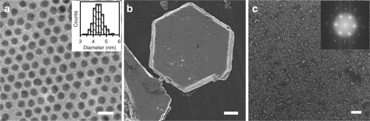
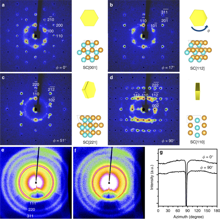
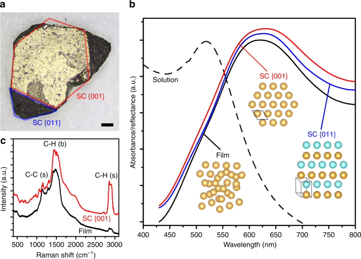
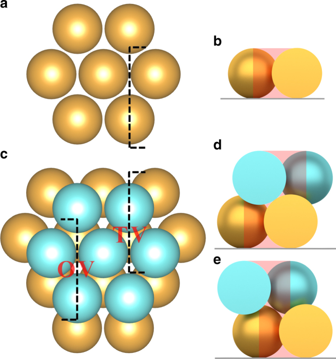
Metallic nanoparticles, such as gold and silver nanoparticles, can self-assemble into highly ordered arrays known as supercrystals for potential applications in areas such as optics, electronics, and sensor platforms. Here we report the formation of self-assembled 3D faceted gold nanoparticle supercrystals with controlled nanoparticle packing and unique facet-dependent optical property by using a binary solvent diffusion method. The nanoparticle packing structures from specific facets of the supercrystals are characterized by small/wide-angle X-ray scattering for detailed reconstruction of nanoparticle translation and shape orientation from mesometric to atomic levels within the supercrystals. We discover that the binary diffusion results in hexagonal close packed supercrystals whose size and quality are determined by initial nanoparticle concentration and diffusion speed. The supercrystal solids display unique facet-dependent surface plasmonic and surface-enhanced Raman characteristics. The ease of the growth of large supercrystal solids facilitates essential correlation between structure and property of nanoparticle solids for practical integrations.
Conflict of interest statement
The authors declare no competing interests.
Figures
Similar articles
-
Plasmonic Supercrystals.Acc Chem Res. 2019 Jul 16;52(7):1855-1864. doi: 10.1021/acs.accounts.9b00213. Epub 2019 Jun 25. Acc Chem Res. 2019. PMID: 31243968
-
A Shape-Induced Orientation Phase within 3D Nanocrystal Solids.Adv Mater. 2018 Aug;30(32):e1802078. doi: 10.1002/adma.201802078. Epub 2018 Jun 26. Adv Mater. 2018. PMID: 29944182
-
Controlled growth and shape-directed self-assembly of gold nanoarrows.Sci Adv. 2017 Oct 27;3(10):e1701183. doi: 10.1126/sciadv.1701183. eCollection 2017 Oct. Sci Adv. 2017. PMID: 29098180 Free PMC article.
-
Fabrication of Plasmonic Supercrystals and Their SERS Enhancing Properties.ACS Omega. 2020 Sep 30;5(40):25485-25492. doi: 10.1021/acsomega.0c03412. eCollection 2020 Oct 13. ACS Omega. 2020. PMID: 33073075 Free PMC article. Review.
-
Foldectures: 3D Molecular Architectures from Self-Assembly of Peptide Foldamers.Acc Chem Res. 2017 Apr 18;50(4):832-841. doi: 10.1021/acs.accounts.6b00545. Epub 2017 Feb 13. Acc Chem Res. 2017. PMID: 28191927 Review.
Cited by
-
Hierarchical supercrystalline nanocomposites through the self-assembly of organically-modified ceramic nanoparticles.Sci Rep. 2019 Mar 5;9(1):3435. doi: 10.1038/s41598-019-39934-4. Sci Rep. 2019. PMID: 30837545 Free PMC article.
-
Recent progress in multifunctional, reconfigurable, integrated liquid metal-based stretchable sensors and standalone systems.Prog Mater Sci. 2024 Apr;142:101228. doi: 10.1016/j.pmatsci.2023.101228. Epub 2023 Dec 14. Prog Mater Sci. 2024. PMID: 38745676 Free PMC article.
-
Dissipative Self-Assembly of Patchy Particles under Nonequilibrium Drive: A Computational Study.J Chem Theory Comput. 2024 Oct 22;20(20):8844-8861. doi: 10.1021/acs.jctc.4c00856. Epub 2024 Oct 4. J Chem Theory Comput. 2024. PMID: 39365844 Free PMC article.
-
Supercrystallography-Based Decoding of Structure and Driving Force of Nanocrystal Assembly.Materials (Basel). 2019 Nov 17;12(22):3771. doi: 10.3390/ma12223771. Materials (Basel). 2019. PMID: 31744175 Free PMC article. Review.
-
Surfactant-Assisted Cooperative Self-Assembly of Nanoparticles into Active Nanostructures.iScience. 2019 Jan 25;11:272-293. doi: 10.1016/j.isci.2018.12.025. Epub 2018 Dec 27. iScience. 2019. PMID: 30639850 Free PMC article. Review.
References
-
- Murray CB, Kagan CR, Bawendi MG. Synthesis and characterization of monodisperse nanocrystals and close-packed nanocrystal assemblies. Annu Rev. Mater. Sci. 2000;30:545–610. doi: 10.1146/annurev.matsci.30.1.545. - DOI
Publication types
LinkOut - more resources
Full Text Sources
Other Literature Sources
