

Inhibition of Semicarbazide-sensitive Amine Oxidase Reduces Atherosclerosis in Cholesterol-fed New Zealand White Rabbits
- PMID: 29915377
- PMCID: PMC6006253
- DOI: 10.1038/s41598-018-27551-6
Inhibition of Semicarbazide-sensitive Amine Oxidase Reduces Atherosclerosis in Cholesterol-fed New Zealand White Rabbits
Abstract
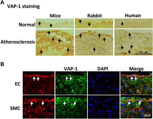
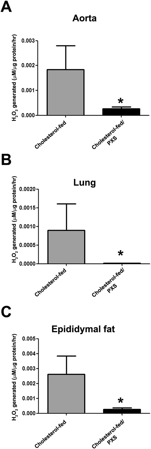
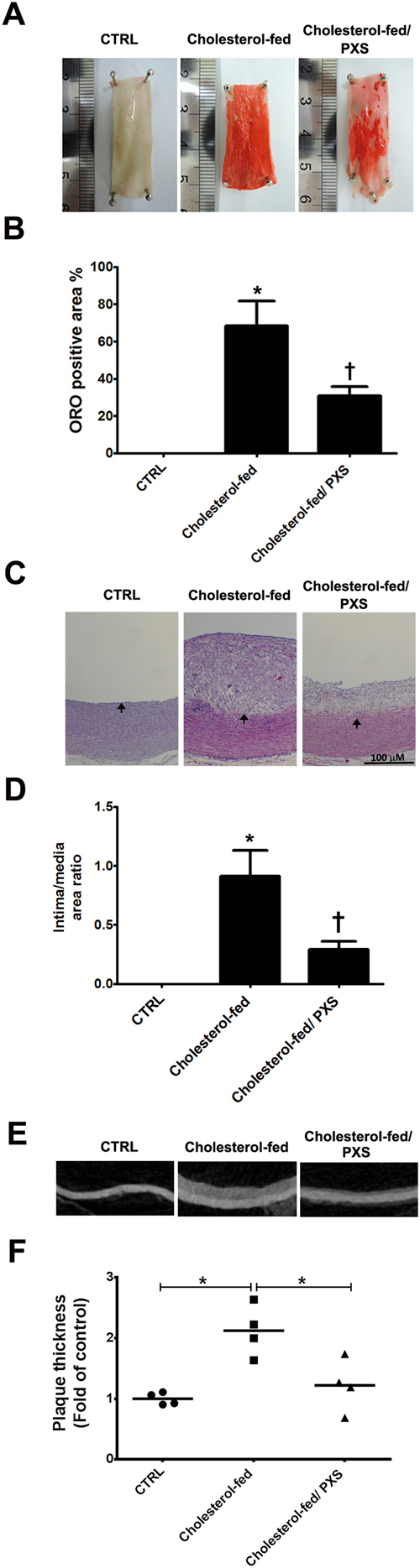
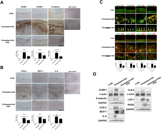
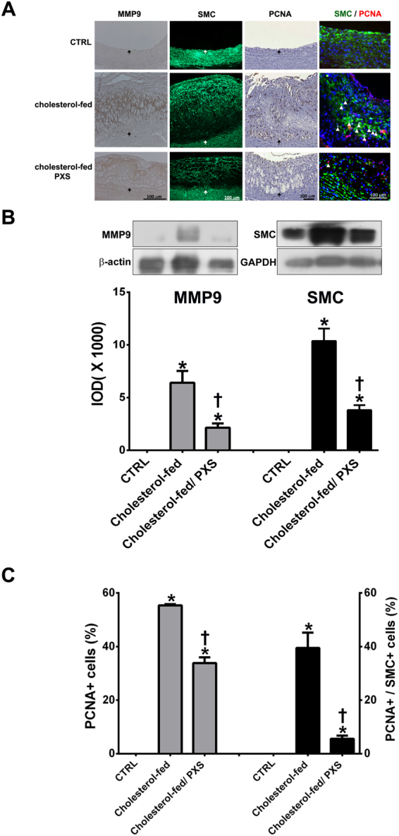
Inflammation, oxidative stress, and the formation of advanced glycated end-products (AGEs) are important components of atherosclerosis. Vascular adhesion protein-1 (VAP-1) participates in inflammation. Its enzymatic activity, semicarbazide-sensitive amine oxidase (SSAO), can catalyze oxidative deamination reactions to produce hydrogen peroxide and aldehydes, leading to the subsequent generation of AGEs. This study aimed to investigate the effect of VAP-1/SSAO inhibition on atherosclerosis. In our study, immunohistochemical staining showed that atherosclerotic plaques displayed higher VAP-1 expression than normal arterial walls in apolipoprotein E-deficient mice, cholesterol-fed New Zealand White rabbits and humans. In cholesterol-fed rabbits, VAP-1 was expressed on endothelial cells and smooth muscle cells in the thickened intima of the aorta. Treatment with PXS-4728A, a selective VAP-1/SSAO inhibitor, in cholesterol-fed rabbits significantly decreased SSAO-specific hydrogen peroxide generation in the aorta and reduced atherosclerotic plaques. VAP-1/SSAO inhibition also lowered blood low-density lipoprotein cholesterol, reduced the expression of adhesion molecules and inflammatory cytokines, suppressed recruitment and activation of macrophages, and decreased migration and proliferation of SMC. In conclusion, VAP-1/SSAO inhibition reduces atherosclerosis and may act through suppression of several important mechanisms for atherosclerosis.
Conflict of interest statement
The authors declare no competing interests.
Figures
Similar articles
-
Inhibition of semicarbazide-sensitive amine oxidase reduces atherosclerosis in apolipoprotein E-deficient mice.Transl Res. 2018 Jul;197:12-31. doi: 10.1016/j.trsl.2018.03.001. Epub 2018 Mar 27. Transl Res. 2018. PMID: 29653075
-
Effects of an anti-inflammatory VAP-1/SSAO inhibitor, PXS-4728A, on pulmonary neutrophil migration.Respir Res. 2015 Mar 20;16(1):42. doi: 10.1186/s12931-015-0200-z. Respir Res. 2015. PMID: 25889951 Free PMC article.
-
Inactivation of semicarbazide-sensitive amine oxidase induces the phenotypic switch of smooth muscle cells and aggravates the development of atherosclerotic lesions.Atherosclerosis. 2016 Jun;249:76-82. doi: 10.1016/j.atherosclerosis.2016.03.039. Epub 2016 Apr 1. Atherosclerosis. 2016. PMID: 27065245
-
Physiological and pathological implications of semicarbazide-sensitive amine oxidase.Biochim Biophys Acta. 2003 Apr 11;1647(1-2):193-9. doi: 10.1016/s1570-9639(03)00101-8. Biochim Biophys Acta. 2003. PMID: 12686132 Review.
-
Vascular adhesion protein-1: Role in human pathology and application as a biomarker.Crit Rev Clin Lab Sci. 2015;52(6):284-300. doi: 10.3109/10408363.2015.1050714. Epub 2015 Aug 18. Crit Rev Clin Lab Sci. 2015. PMID: 26287391 Review.
Cited by
-
The role of vascular adhesion protein-1 in diabetes and diabetic complications.J Diabetes Investig. 2024 Aug;15(8):982-989. doi: 10.1111/jdi.14209. Epub 2024 Apr 6. J Diabetes Investig. 2024. PMID: 38581224 Free PMC article. Review.
-
Vascular Adhesion Protein-1 (VAP-1)/Semicarbazide-Sensitive Amine Oxidase (SSAO): A Potential Therapeutic Target for Atherosclerotic Cardiovascular Diseases.Front Pharmacol. 2021 Jul 8;12:679707. doi: 10.3389/fphar.2021.679707. eCollection 2021. Front Pharmacol. 2021. PMID: 34322017 Free PMC article. Review.
-
Inhibition of monoamine oxidase B reduces atherosclerosis and fatty liver in mice.Clin Sci (Lond). 2023 Jan 13;137(1):17-30. doi: 10.1042/CS20220477. Clin Sci (Lond). 2023. PMID: 36416117 Free PMC article.
-
The role of inflammation in diabetic kidney disease.Korean J Intern Med. 2021 Jul;36(4):753-766. doi: 10.3904/kjim.2021.174. Epub 2021 Jul 1. Korean J Intern Med. 2021. PMID: 34237822 Free PMC article. Review.
-
Inhibition of vascular adhesion protein-1 modifies hepatic steatosis in vitro and in vivo.World J Hepatol. 2020 Nov 27;12(11):931-948. doi: 10.4254/wjh.v12.i11.931. World J Hepatol. 2020. PMID: 33312420 Free PMC article.
References
-
- World Health Organization. Cardiovascular diseases. http://www.who.int/mediacentre/factsheets/fs317/en/. Updated January 2015.
Publication types
MeSH terms
Substances
LinkOut - more resources
Full Text Sources
Other Literature Sources
Medical
Molecular Biology Databases
Research Materials
